Method of body fluid specimen collection

a body fluid and specimen collection technology, applied in medical science, packaging, diagnostics, etc., can solve the problems of increasing the probability of contaminant inclusion with the blood specimen, false positive, and prior art methods that draw blood directly into evacuated culture vessels are subject to some frequency of contact with the surface of culture vessels, so as to achieve convenient and convenient transfer, the effect of ensuring the safety of the transfer

Inactive Publication Date: 2005-07-05
STONE BENJAMIN CURTIS
View PDF3 Cites 84 Cited by
  • Summary
  • Abstract
  • Description
  • Claims
  • Application Information

AI Technical Summary

Benefits of technology

[0014]Collecting the first body fluid specimen with the sterile evacuated specimen tube will also be referred to as contaminant redirection. The purpose of this technique is to remove potential contaminants from the internal surfaces of the collection needle by establishing body fluid flow, without contaminating the collection needle. A step of treating the site to be pierced with preparatory antiseptics such as iodophors, iodine tinctures, or chlorhexidine, may precede the first step (step a) of this method if it is found to be more effective in practice. But the preferred method should allow for re-palpation of the site without adding contaminants to the second body fluid specimen.
[0027]b. a first needle shield to prevent accidental needle stick with the first end of the transfer needle;
[0028]c. a second needle shield to prevent accidental needle stick with the second end of the transfer needle.
[0033]This method allows for adequate, safe, and simplified transfer of the second body fluid specimen from the collection vessel to the evacuated culture vessel. This method also provides a checkpoint at which the volume of the second body fluid specimen can be monitored prior to and during inoculation of the evacuated culture vessel.

Problems solved by technology

The first of these is contamination of the blood specimen, which frequently leads to false positive results with an organism recovered by subculture and isolation.
Second, with any preparatory antiseptic, re-palpation of the venepuncture site after application of the antiseptic also increases the probability of contaminant inclusion with the blood specimen.
Third, prior art methods that draw blood directly into evacuated culture vessels are subject to some frequency of contact with the surfaces of culture vessels after they have been treated with antiseptics.
A less frequently recognized factor, it can lead to false negative and false positive results.
Low specimen volumes containing a true pathogenic organism may require substantially longer periods of time to show a detectable rate of change in the monitored factor, thereby delaying detection of the organism or providing a false negative result.
High specimen volumes often including relatively higher concentrations of leukocytes are known to cause false positives by providing increases in carbon dioxide gas production.
In addition, these types of false positive results may also lead to false negative results.
Ambient air contains oxygen, which will greatly hinder the growth of obligate and facultative anaerobic organisms, thus potentially resulting in a false negative.
These graduations generally have no start or end point, and because the vessel contains a liquid media, it is difficult to monitor the volume of blood used to inoculate.
In addition, bubbles on the surface of the liquid media disrupt the meniscus and further hinder proper collection.
Due to this, prior art methods that draw directly into the culture vessels are more prone to inoculating volume irregularity.
Prior art methods that use an intermediate such as a syringe also incur some volume irregularity due to the relatively large size of the recommended inoculating volume.
And because inoculating volumes are rarely monitored, this leads to irregularity.
False positive results that recover an organism and false negative results are the two most precarious failures of a detection system.
Both can lead an increased rate of mortality for the patient, and more frequently to increased length of hospital stay and cost.

Method used


Image

  • Method of body fluid specimen collection
  • Method of body fluid specimen collection
  • Method of body fluid specimen collection

Examples


first embodiment

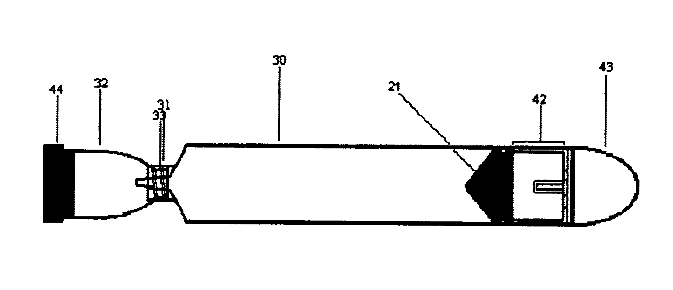
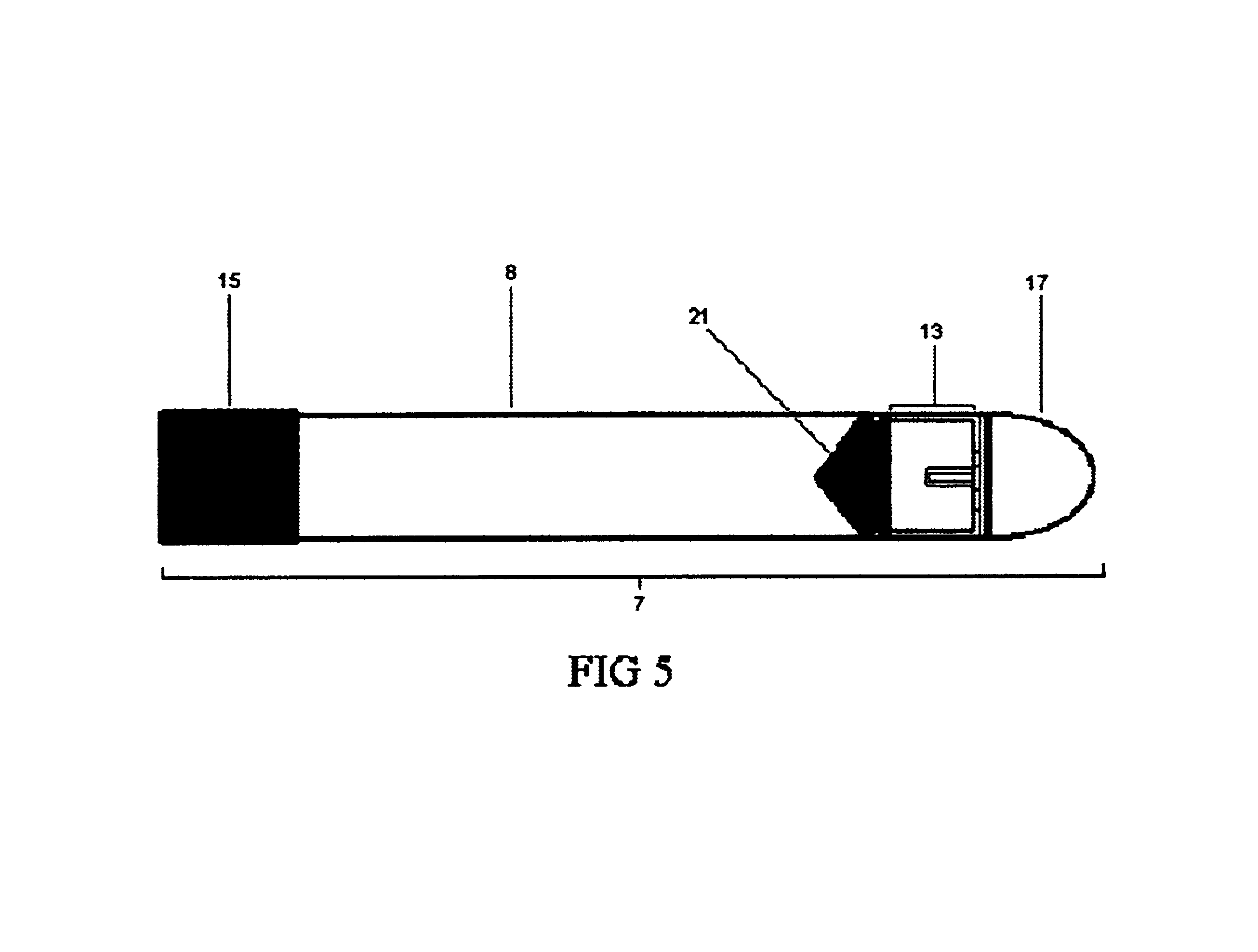
[0080]The vessel plunger 13 should have a cylindrical body having a diameter of roughly 1.3 cm to allow for its entry into the vessel body 8 and to allow it to move freely within the vessel body 8 when not held in place by the plunger lock 17. Continuous with the body at one end will be the rubber seal hold 20. In the first embodiment the rubber seal hold 20 will be a plastic disk over which fits a rubber seal 21, the second seal. This type of attachment is well known in the art of making syringes. The strength of the attachment may also be augmented using an adhesive in order to prevent disengagement of the rubber seal 21 from the hold 20 during assembly. The rubber seal 21 provides the plunger with means for sealing with the vessel body 8. At the other end, the plunger 13 will also contain the plunger grooves 12. As illustrated in FIGS. 9, 10, 11, and 12, the grooves 12 correspond in dimensions and orientation to the body's ridges 11, such that when the grooves 12 and ridges 11 ar...

third embodiment

[0091]A third embodiment for the collection vessel will also be described here. This embodiment is preferred because it uses a transfer needle as opposed to the transfer device 29 in a method of transfer the second body fluid specimen as will be described later.

[0092]The third embodiment comprises: a hollow body having a first and second end; a first seal at said first end; a plunger disposed within said hollow body between said first end and said second end; said plunger providing a second seal; a plunger lock coupled to said plunger; said plunger lock being configured to selectively maintain said plunger at said second end when at least a portion of said hollow body between said first seal and said second seal is at least partially evacuated; an airtight junction that interrupts said hollow body forming a first section and second section; said first section having said first seal; said second section having said plunger and said plunger lock; said plunger lock can further be confi...

fifth embodiment

[0131]A third, fourth, and fifth embodiment of the method for collecting a body fluid specimen is described here, comprising the steps of:[0132]a. preparing the site for puncture using an antiseptic;[0133]b. piercing the prepared puncture site using the fluid collection needle;[0134]c. collecting the first body fluid specimen using the sterile evacuated specimen tube;[0135]d. collecting the second body fluid specimen using an embodiment of the collection vessel;[0136]e. transferring the second body fluid specimen to the evacuated culture vessel using the transfer device.

[0137]As with the first two embodiments, the step of preparing the puncture site may use any one or a combination of the antiseptics listed, but as explained earlier must be capable of aiding in the elimination of an adequate quantity of contaminants. The step of piercing the skin the puncture site may use any of the listed devices for piercing the puncture site. These embodiments also use contaminant redirection as ...


PUM


Abstract

In one aspect of the invention, a method of fluid specimen collection comprises the steps of: preparing a site for puncture using an antiseptic; piercing the prepared puncture site using a fluid collection needle; collecting a first body fluid specimen using a device for collecting the first body fluid specimen; collecting a second body fluid specimen using a device for collecting the second body fluid specimen and; selecting the second body fluid specimen for a diagnostic test of a kind that detects the presence of organisms in the second body fluid specimen. In another aspect of the invention, a vessel for collecting, transporting, and transferring a body fluid specimen comprises: a hollow body having a first and second end; a first seal at said first end; a plunger disposed within said hollow body between said first end and said second end; said plunger providing a second seal; a plunger lock coupled to said plunger; said plunger lock being configured to selectively maintain said plunger at said second end when at least a portion of said hollow body between said first seal and said second seal is at least partially evacuated; said plunger lock can further be configured to release said plunger, thereby allowing said plunger to move toward said first seal within said body.

Description

PARENT CASE TEXT[0001]This application claims benefit of provisional application No. 60 / 263,370 filed Jan. 23, 2001 and provisional application No. 60 / 306,643 filed Jul. 19, 2001, the contents of which are incorporated herein.CROSS REFERENCE TO RELATED APPLICATIONS[0002]N / ASTATEMENT REGARDING FEDERALLY SPONSORED RESEARCH OR DEVELOPMENT[0003]N / AREFERENCE TO A MICROFICHE APPENDIX[0004]N / ADISCUSSION OF THE PRIOR ART[0005]There are a number of known methods for collecting blood / body fluid specimens for culturing. This section discusses prior art methods for collecting blood specimens into evacuated culture vessels. Of these, there are methods that collect directly into the culture vessels and those that collect into an intermediate prior to inoculation of the culture vessels.[0006]The methods of the prior art must deal with two interfering factors in order to avoid false negative and false positive culture results. The first of these is contamination of the blood specimen, which frequen...

Claims


Application Information

Patent Timeline
Patent Type & Authority Patents(United States)
IPC IPC(8): A61B5/15
CPCA61B5/1438A61B5/15003A61B5/150236A61B5/150351A61B5/150389A61B5/150473A61B5/150572A61B5/150732A61B5/154
Inventor STONE, BENJAMIN CURTIS
Owner STONE BENJAMIN CURTIS
Why Eureka
Try Eureka