Photodynamic therapy for selectively closing neovasa in eyeground tissue

a selective closing and neovascular therapy technology, applied in the field of photodynamic therapy, can solve the problems of long-term phototoxicity, poor selectivity to accumulate in the target, and severe damage to visual functions

Inactive Publication Date: 2006-01-10
MEIJI SEIKA KAISHA LTD
View PDF3 Cites 37 Cited by
  • Summary
  • Abstract
  • Description
  • Claims
  • Application Information

AI Technical Summary

Benefits of technology

[0073]Accordingly, the present inventors conclusively consider that, when ICG is intravenously injected at a dose of 0.5 mg / kg to 1 mg / kg to the test monkeys having the experimental CNV in their ocular fundus and then NPe6 is intravenously injected to said monkeys at a dose of 0.5 mg / kg to 10 mg / kg, or desirably at a dose of 0.5 mg / kg to 1 mg / kg as same as the dose of the intravenously injected ICG, either at the same time as the time of the intravenous injection of ICG, or immediately before or immediately after the intravenous injection of ICG, it occurs that both of ICG and NPe6 can concurrently be uptaken into and distributed in the vascular walls of the retinal normal vascular vessels and also in the vascular walls of the CNV in the ocular fundus of the monkeys. The present inventors also consider conclusively that the ICG and NPe6, which have been intravenously injected at the same time or at a substantially same time, are able to bring about the time-dependent change in the concentration of ICG and also the time-dependent change in the concentration of NPe6 within both of the vascular walls of the retinal normal vascular vessels and the vascular walls of the CNV in the ocular fundus of the monkeys, so that the time-dependent change in the concentration of NPe6 can take place concurrently in the same pattern or manner as the time-dependent change in the concentration of ICG in said vascular walls of the retinal normal vascular vessels and of the CNV.

Problems solved by technology

The first-generation photosensitizers for use in PDT had such drawbacks that they have a poor selectivity to accumulate in the target tumor cells while they can readily accumulate in the normal cells, and that the first-generation photosensitizers can additionally involve a long-term phototoxicity in the skin of the patients [see, for example, Cancer Res., Vol. 38, pp.
However, there is nowhere disclosed any experimental Example which to show that any practical application of the method of Richer et al. was made to the field of ophthalmology.
On the other hand, it is known that the neovascularization occurring in the ocular fundus tissue can severely damage the visual functions.
Due to the occurrence of the choroidal neovascularization in the age-related macular degeneration, there are induced subretinal bleeding and subretinal exudation as well as retina detachment and fibrous proliferation, which can bring about a visual deterioration and occasionally blindness.
Furthermore, it is known that when diabetes mellitus has progressed, a proliferative neovascularization is incurred in the retina, resulting in an onset of the proliferative diabetic retinitis and leading sometimes to blindness.
It has been known that the photo-coagulation method made in the above prior art has a drawback that the thermal action of the laser light as employed can destroy even the surrounding normal tissues, whereas the neovascular vessels themselves can be occluded well by the thermal coagulation of them.

Method used


Image

  • Photodynamic therapy for selectively closing neovasa in eyeground tissue

Examples


example 1

for Formulation

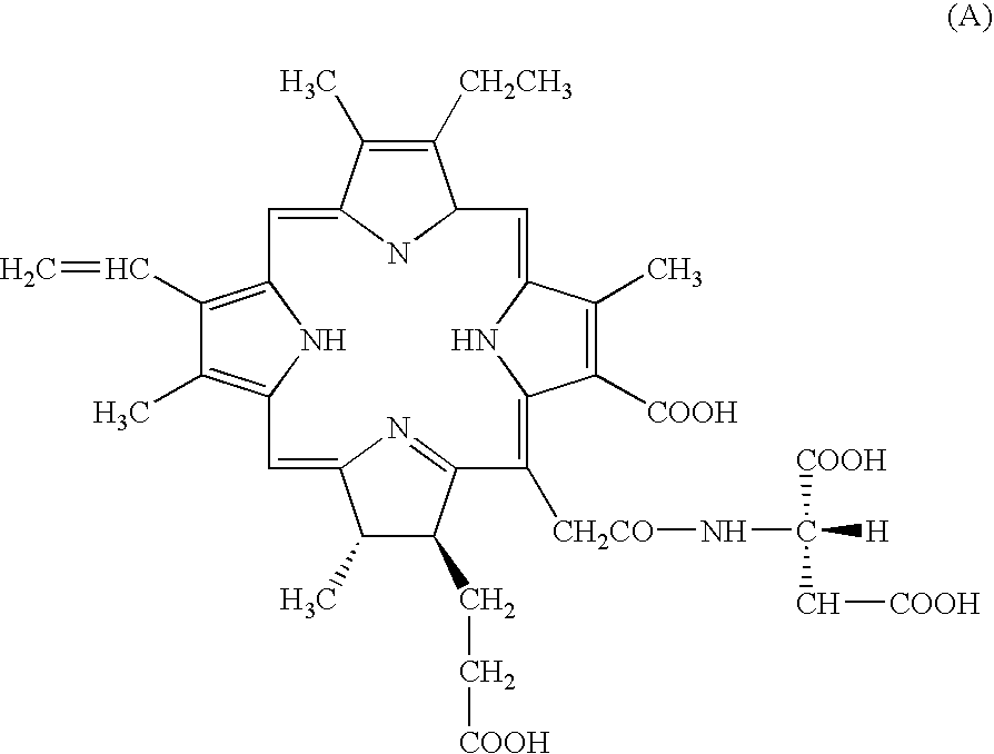
[0145]100 mg of mono-L-aspartyl chlorin e6 tetra-sodium salt was dissolved in 4 ml of distilled water ready for injections. The resulting aqueous solution was then adjusted to pH 7.4. After the aseptic filtration thereof, the resulting sterilized solution was lyophilized in vials. The resulting lyophilized preparation may be dissolved in an appropriate volume of physiological saline, before its use.

example 2

of Formulation

[0146]10 mg / ml of ICG was dissolved in a volume of sterile physiological saline at a concentration of 10 mg / ml. Further, mono-L-aspartyl chlorin e6 tetra-sodium salt was dissolved in a further volume of physiological saline at a concentration of 10 mg / ml. The resultant two aqueous solutions were mixed together in the equal proportions. The resulting solution of ICG and NPe6 is suitable for the intravenous injection of ICG and NPe6 in order to carry out the method of the third aspect of this invention.


PUM

PropertyMeasurementUnit
wavelengthaaaaaaaaaa
irradianceaaaaaaaaaa

Abstract

A method is provided for selectively occluding neovascular vessels formed in the ocular fundus of an eye, which includes intravenously administering a mono-L-aspartyl chlorin compound to a patient; subsequently estimating an appropriate time point when the mono-L-aspartyl chlorin compound has decreased in its concentration or has been eliminated from the retinal normal vascular vessels of the patient but is still remaining at an appropriate concentration in the vascular walls of the neovascular vessels of the ocular fundus; irradiating a laser light at a 664 nm-wavelength which is initiated at the appropriate time; and using the irradiation of the laser light to target lesions comprising the neovascular vessels, at a controlled power.

Description

TECHNICAL FIELD[0001]This invention relates to photodynamic therapy methods for occluding selectively such neovascular vessels as formed in the ocular fundus of eye, without involving any damage or impairment in the normal vascular vessels present in the tissues of the ocular fundus. More specifically, this invention relates to photodynamic therapy methods for occluding exclusively the choroidal neovascular vessels and / or retinal neovascular vessels, without damaging or impairing the retinal normal parenchymal tissue, the retinal normal vascular vessels and / or the choroidal normal vascular vessels of eye, which methods each comprises intravenously administering mono-L-aspartyl chlorin e6 tetra-sodium salt to a mammalian animal having the choroidal neovascular vessels and / or retinal neovascular vessels, and then starting to irradiate a laser light of 664 nm-wavelength to the lesions comprising the neovascular vessels in the ocular fundus at a certain designated fluence of the laser l...

Claims


Application Information

Patent Timeline
Patent Type & Authority Patents(United States)
IPC IPC(8): A61K31/40A61K41/00A61P27/02A61P43/00
CPCA61K31/395A61K41/0071A61K41/0057A61P27/02A61P43/00
Inventor MORI, KEISUKEYONEYA, SHIN
Owner MEIJI SEIKA KAISHA LTD
Why Eureka
Try Eureka