System and method for examining, recording and analyzing dermatological conditions

a dermatological condition and system technology, applied in the field of system and method for examining, recording and analyzing dermatological conditions, can solve the problems of scarring and infection risk, ordinary medical practitioners have difficulty in properly assessing a lesion, and significant waste of resources in the community, so as to reduce the contribution of edge factors

Inactive Publication Date: 2006-01-31
POLARTECHNICS
View PDF50 Cites 197 Cited by
  • Summary
  • Abstract
  • Description
  • Claims
  • Application Information

AI Technical Summary

Benefits of technology

[0032]Thus means of counteracting the TIR effect include: a thick front window material, rendering of the front window edges absorptive and non-radiating to minimise edge contribution to

Problems solved by technology

However, the majority of medical practitioners are not experts in the area of dermatology and each one might see only a few melanoma lesions in a year.
In consequence, the ordinary medical practitioner has difficulty in assessing a lesion properly.
This excess of surgical procedures leads to significant wasted expense to the community, and risks of scarring and infection.
This introduces errors into colour analysis of the ELM images as well as reduces the image contrast.
The absence of a cone means that the image scale is essentially uncontrolled.
It proves to have a significant disadvantage, in that the unsupported skin is allowed to bulge inwards towards the camera.
This bulge may be reduced by reducing the unsupported area, but this is not a realistic approach with a large lesion.
However, the complexity of a lesion makes the identification of even the boundaries between the lesion and the surrounding skin difficult.
This problem is compounded by the obvious fact that human skin colour is widely variable between different individuals and across different races.
Thus it is not possible to specify any particular colour as being “always skin”.
958–964) but with limited success.
The analysis problem is compounded by the fact that the resolution of the images taken with common single-CCD miniature colour TV cameras is not very high (typically, poorer than 0.1 mm on the lesion with a 25 mm field of view), which limits the ability to discriminate fine detail during either on-screen inspection or software-driven image analysis of geometrical features.
However, this technique suffers from a disadvantage in comparison to the use of a 3-CCD camera.
Should this happen there would be a loss of colour registration

Method used


Image

  • System and method for examining, recording and analyzing dermatological conditions
  • System and method for examining, recording and analyzing dermatological conditions
  • System and method for examining, recording and analyzing dermatological conditions

Examples


Embodiment Construction

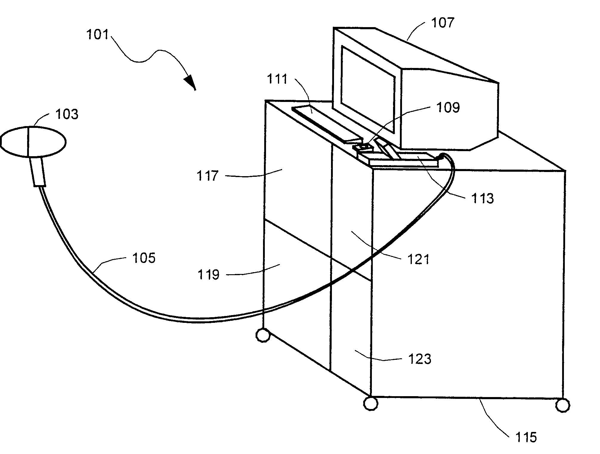
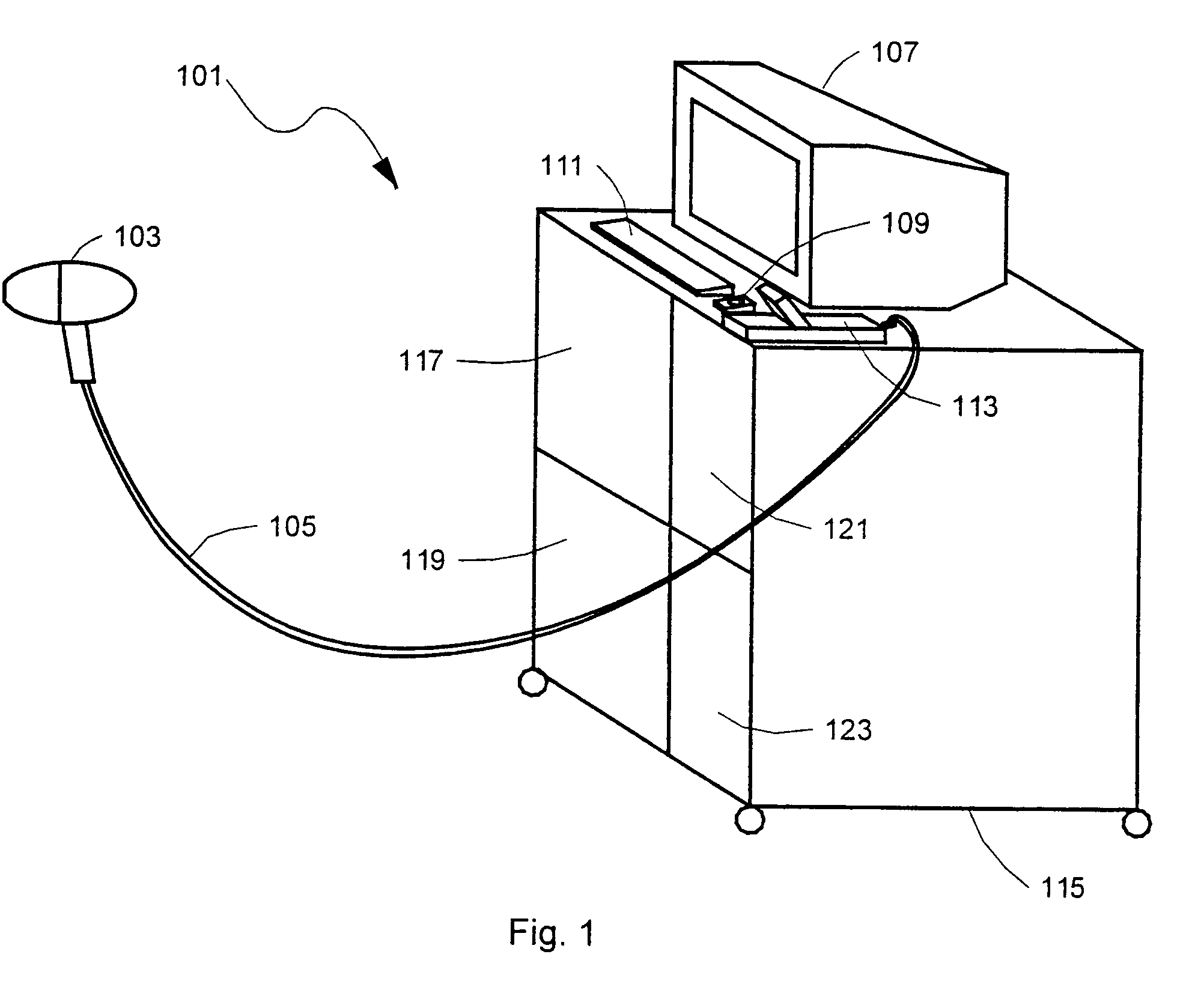
[0075]The preferred embodiment of the invention, illustrated in FIG. 1 and bearing numeral 101, uses video technology and includes a hand-held unit 103; a system trolley 115 containing a computer system 119, a printer 117 connected to the computer system, an illumination source 123 and a video camera controller 121; and, on top of the trolley, a computer monitor 107, a keyboard 111, a mouse or pointing device 109, and a camera cradle 113 for the hand-held unit 103. A flexible cable 105 connects the hand-held unit 103 to the system trolley 115.

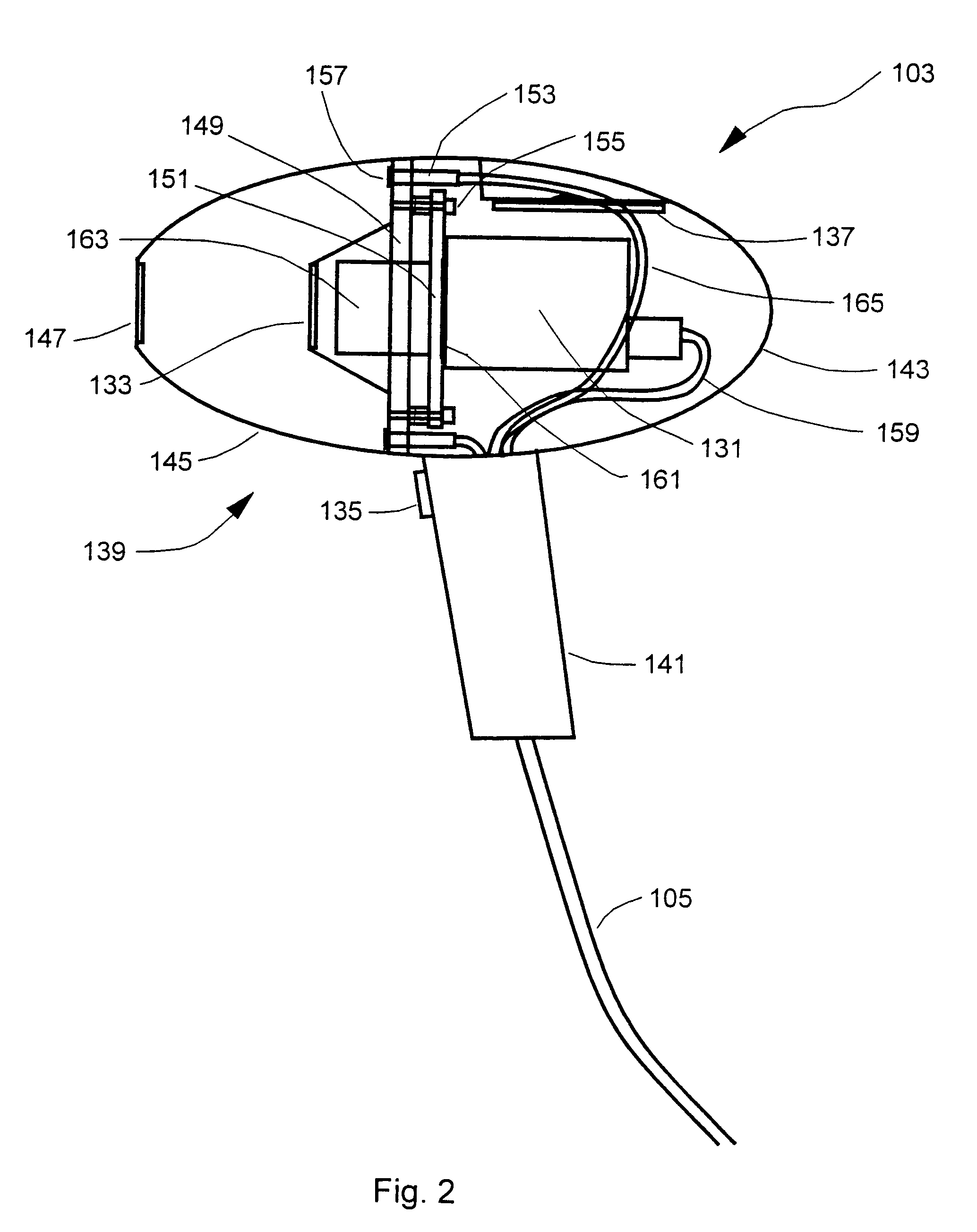
[0076]As illustrated in FIG. 2, the hand-held unit 103 contains a video camera head 131, an optional inner protective cone and window 133, a trigger switch 135, and an optional miniature computer mouse or pointing device 137. The case 139 of the hand-held unit consists of a handle 141, a main body 143, and a removable outer front cone 145 with window 147. Inside the case there is an internal bulkhead 149, and an adjustable camera mount 151 secu...


PUM


Abstract

A system for collecting, storing and displaying dermatological images for the purpose of monitoring and diagnosis of skin conditions and skin cancers, including melanoma. A hand-held unit illuminates a section of the patient's skin, and an imaging device generates imaging signals from light derived from a skin section. Pairs of light output ports in the hand-held unit are arranged such that their intensity distributions overlap at their half-intensity levels so that the resulting summation of their intensities has a flat central region. Three image stores are maintained, one for lesion images, one for “nearby skin” images, and one for reference-white images. The “nearby skin” images are used by the system software to automatically determine the skin/lesion border. The reference white images are used to set the dynamic range of the instrument and to compensate for lighting irregularities. Two images of the same lesion taken at different times may be displayed simultaneously so that changes in the lesion may be determined. The calibration system is designed so that image data taken on any of multiple machines built to the same specification will be corrected back to a common reference standard to ensure absolute accuracy in colour rendition.

Description

[0001]This application claims the benefit of U.S. provisional application No. 60 / 165,072, filed on Nov. 12, 1999.FIELD OF THE INVENTION[0002]This invention relates to an integrated system for the acquisition of high quality medical images of skin, the storage and archiving of those images, the analyses of those images in a patient-oriented database, and the subsequent retrieval of those images and their analyses, and more particularly to such a system for use in the monitoring and diagnosis of skin conditions and skin cancers, including melanoma.BACKGROUND OF THE INVENTION[0003]Malignant melanoma is a form of cancer due to the uncontrolled growth of melanocytic cells just under the surface of the skin. These pigmented cells are responsible for the brown colour in skin and freckles. Malignant melanoma is one of the most aggressive forms of cancer known. The interval between a melanoma site becoming malignant or active and the probable death of the patient in the absence of treatment ...

Claims


Application Information

Patent Timeline
IPC IPC(8): G06K9/00A61B5/00A61B5/103A61B10/00G06T1/00
CPCA61B5/444A61B5/0059
Inventor SKLADNEV, VICTORGUTENEV, ALEXMENZIES, SCOTTTHOMPSON, RICHARDBATRAC, ANDREWBISCHOFF, LEANNECAFFIN, ROGERROWE, STEPHENTALBOT, HUGUESVARVEL, DAVIDWEST, PETER
Owner POLARTECHNICS
Why Eureka
Try Eureka