Method for diagnosis of helicobacter pylori infection

a technology for helicobacter pylori and infection, applied in the field of helicobacter pylori infection diagnosis, can solve the problems of capital equipment and per-patient test cost, inability to prove the reliability of general practitioners' offices, and no test method for i>h, and achieve the effect of simple, rapid and non-invasive diagnostic tes

Inactive Publication Date: 2006-03-21
PHOTONIC BIOSYST
View PDF12 Cites 35 Cited by
  • Summary
  • Abstract
  • Description
  • Claims
  • Application Information

AI Technical Summary

Benefits of technology

[0020]e) comparing the individual's percentage change in post-urea breath ammonia against normal population values wherein H. pylori uninfected individuals display percentage changes below a predefined threshold and H. pylori infected individuals display percentage changes above a given threshold.Alternatively, the follow-on urea administration and percentage change from basal measures can be utilized particularly on those individuals exhibiting intermediate basal results that are not definitive for the subject's H. pylori status as a means to more accurately identify infected individuals.

Problems solved by technology

At present there are no test methods for H. pylori that satisfy the ideal conditions of being non-invasive, rapid, easy to administer, have low capital equipment and per-patient test costs, and capable of being conducted in entirety during a clinician's office visit.
Although the non-invasive antibody-based tests are relatively easy to perform, they have not proved to be reliable in the general practitioner's office.
Furthermore, they incur a blood draw and the costs associated with the blood draw procedure.
Additionally, antibody tests cannot provide a test-of-cure to demonstrate successful antibiotic treatment.
However, to execute these tests, invasive endoscopy procedures are required.
They are therefore neither simple, economical, convenient for the patient, or executable in general clinical practices.
For a number of reasons, it has proven difficult to make measurements of breath ammonia liberated as a by-product of H. pylori urease.
While ammonia readily crosses the stomach and alveolar lining, ammonium ions are not readily absorbed.
Therefore very little ammonia finds its way from the stomach, traffics through the circulatory system, and passes into expired air, consequently making it difficult to measure.
The literature corroborates the difficulty measuring ammonia directly in breath and lack of clinical evidence differentiating H. pylori individuals based on breath measures, such that a simple diagnosis via breath analysis is not expected.
However, he offers no evidence to demonstrate the utility of this maneuver and further, offers no teaching of its clinical utility or basis for deriving diagnoses.
Again however, Katzman's method does not teach diagnosis via ammonia, offering support only for measuring the 13C-labeled CO2 by-product as measured by others (Graham DY et.al., Lancet, 1174–77, May 23, 1987).
Therefore, despite labeled CO2 being measurable in breath and serving diagnostically via the UBT method, it is not to be expected that ammonia would provide a parallel alternative avenue to diagnosis, much less be manifest in any diagnostically useful pattern in the breath.

Method used


Image

  • Method for diagnosis of helicobacter pylori infection
  • Method for diagnosis of helicobacter pylori infection
  • Method for diagnosis of helicobacter pylori infection

Examples


example 1

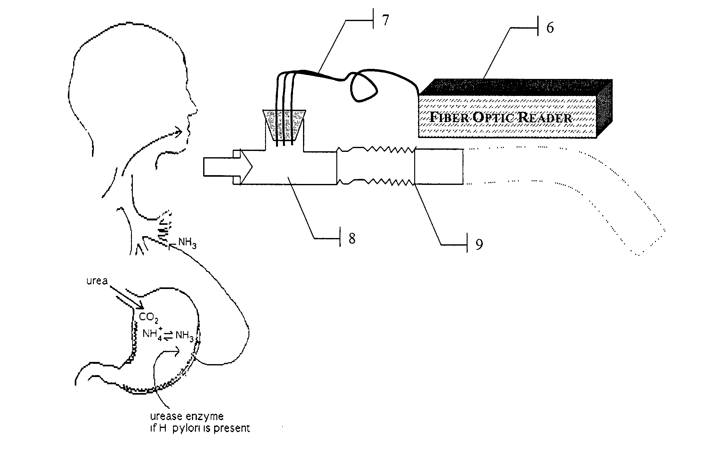
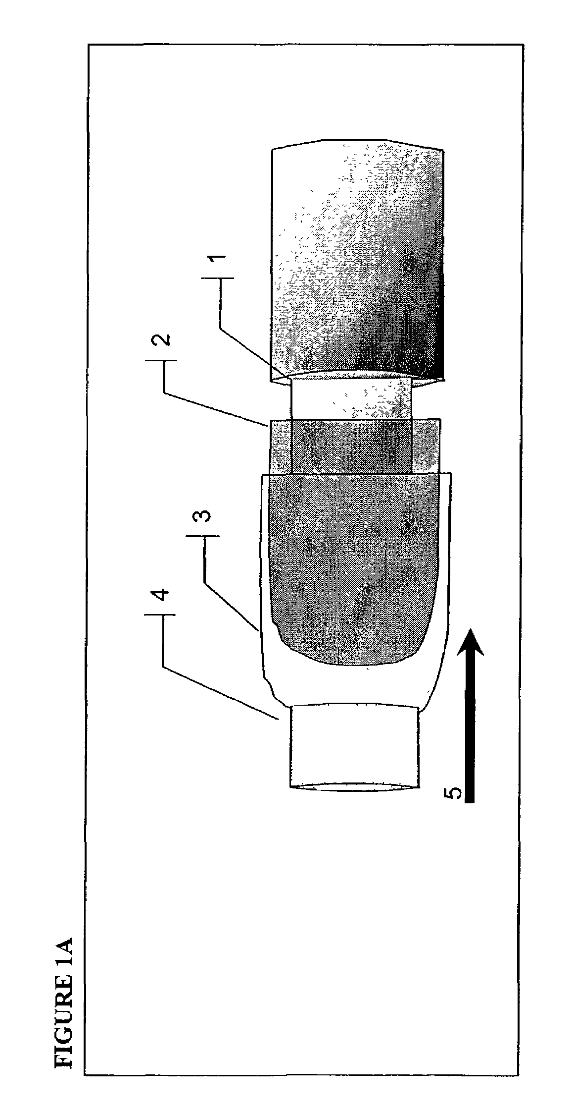
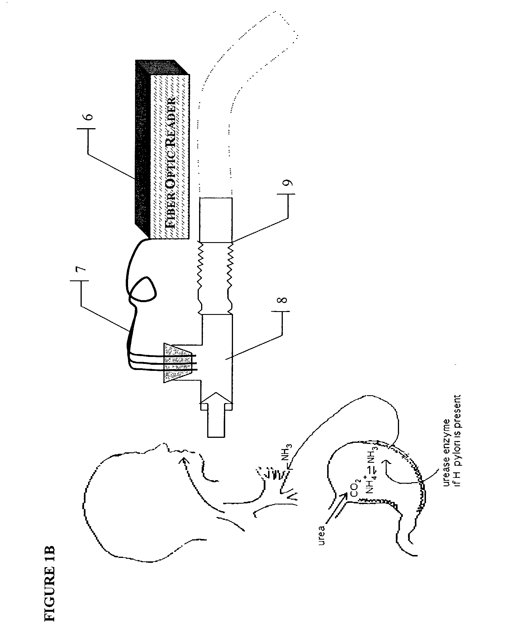
Preparation of Ammonia Sensors for ABT and Optical Sensor Instrumentation

[0046]The following examples describe the preparation of an ammonia sensitive optical sensor useful for the direct determination of breath ammonia measures.

[0047]In one embodiment, the ABT sensor composition is made from an ammonia sensitive indicator dye and a solid phase, for example, a PTFE solid phase in a film form. In one aspect, the sensor compositions are constructed by administering ammonia-sensitive indicator dye(s) in a non-aqueous solvent to a solid-phase PTFE substrate such that the dye is deposited on the solid phase in a form insoluble to aqueous-based solvents. Further, the characteristics of a PTFE film or a porous membrane form are such that it is permeable to gaseous ammonia.

[0048]Optical sensor films for ABT were prepared by dissolving the optical dyes bromocresol-green (BCG) or bromophenol-blue (BPB) in methanol at a concentration of 0.75 mg / L. Other dyes, such as but not limited to, any fl...

example 2

Representative Optical Sensor and Instrument Responses to Ammonia

[0055]To establish the sensor responses to ammonia, a BCG and BPB sensor were connected to appropriate modules and then exposed to 0 ppm, 1 ppm, 4 ppm, 6 ppm and 200 ppm of ammonia in water saturated air. The 200 ppm sample saturates the BPB and BCG dye response ranges and was included only to show a full-range response. The individual channel signal levels at the two wavelengths were recorded. Representative optical signal and Ratio plots for these two sensors are shown in FIGS. 2a–d. As predicted from the BPB and BCG pK's, the BPB sensor demonstrates more of its responsivity in the 0–1 ppm range than the BCG sensor that shows a more extended response over the range of 0–6 ppm.

example 3

Representative H. pylori Positive and Negative Subject Ammonia Breath Test Optical Sensor Responses

[0056]Thirteen volunteers were tested for the presence of H. pylori infection using conventional 14C-urea breath test diagnostic procedures (Ballard Medical, Draper, Utah) in order to classify their clinical status based on current medical practice. Current practice requires subjects fast overnight (typically 8–14 hours) prior to ingesting the 14C-urea capsule and subsequent collection of the subject's breath. A similar fasting regimen was used for the ammonia breath test (ABT). The breath samples were analyzed for the presence of 14C using a scintillation counter. A positive urea breath test was defined as breath 14CO2 excretion greater than 200 dpm, an indeterminate test as breath 14CO2 excretion of 50–200 dpm, and a negative test as 14CO2 excretion less than 50 dpm. Five subjects were found positive for H. pylori and eight were identified as negative for the organism as measured by ...


PUM

PropertyMeasurementUnit
concentrationaaaaaaaaaa
thicknessesaaaaaaaaaa
concentrationsaaaaaaaaaa

Abstract

A rapid, non-invasive breath-test method and device for diagnosing the presence or absence of H. pylori in a subject without administration of isotopic tracers is described. The device consists of a highly sensitive colorimetric ammonia sensor placed in contact with sampled subject breath. The sensor is measured using appropriate reflection spectroscopy instrumentation. The breath-test method consists of measuring a basal ammonia level with the device, administering non-isotopic urea and continuing measurement of the ammonia content in a plurality of consecutive breaths. Diagnostic differences in breath ammonia are identified between H. pylori infected and uninfected individuals.

Description

RELATED APPLICATIONS[0001]This application claims the benefit of the filing of U.S. Ser. No. 60 / 331,275 entitled “Method for Diagnosis of Helicobacter Pylori Infection” filed on Nov. 13, 2001, hereby incorporated by reference.BACKGROUND[0002]The present invention is directed to an effective diagnostic testing for the presence of gastric infection by the microorganism Helicobacter pylori. [0003]Helicobacter pylori is estimated to be responsible for up to 90% of the cases of peptic ulcer disease (PUD) afflicts over 10% of the US population sometime in their lifetime. Estimates for worldwide prevalence of H. pylori infections range from 300 million to over two-thirds of the world's population. H. pylori infection is also associated with 650,000 annual cancer deaths worldwide from gastric adenocarcinoma. The US Communicable Disease Center recommends testing all patients presenting with PUD for diagnosis of H. pylori. [0004]At present there are no test methods for H. pylori that satisfy ...

Claims


Application Information

Patent Timeline
Patent Type & Authority Patents(United States)
IPC IPC(8): A61B5/08G01N1/22A61BA61B5/083G01N31/00G01N33/497
CPCA61B5/083
Inventor HUBBARD, TODD W.PUTNAM, DAVID L.
Owner PHOTONIC BIOSYST
Why Eureka
Try Eureka