
Single nucleotide amplification and detection by polymerase
a single nucleotide and polymerase technology, applied in the field of single nucleotide amplification and detection by polymerase, can solve the problems of difficult detection and characterization of trace amounts of target nucleotides in samples, requirement for strict temperature control, and insufficient quantification due to logarithmic amplification
- Summary
- Abstract
- Description
- Claims
- Application Information
AI Technical Summary
Benefits of technology
Problems solved by technology
Method used
Image

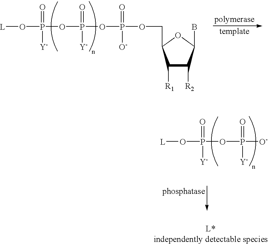
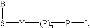
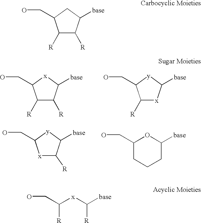
Examples
example 1
Preparation of δ-9H(1,3-dichloro-9,9-dimethylacridin-2-one-7-yl)-dideoxythymidine-5′-tetraphosphate (ddT4P-DDAO)
[0098]ddTTP (100 μl of 80 mM solution) was coevaporated with anhydrous dimethylformamide (DMF, 2×1 ml). To this dicyclohexylcarbodimide (8.3 mg. 5 eq.) was added and the mixture was again coevaporated with anhydrous DMF (1 ml). Residue was taken in anhydrous DMF (1 ml) and reaction was stirred at room temperature overnight. HPLC showed mostly cyclized triphosphate (˜82%). Reaction mixture was concentrated and residue was washed with anhydrous diethyl ether 3×. It was redissolved in anhydrous DMF and concentrated to dryness on rotavap. Residue was taken with DDAO-monophosphate, ammonium salt (5 mg, 1.5 eq.) in 200 μl anhydrous DMF and stirred at 40° C. over the weekend. HPLC showed formation of a new product with desired UV characteristics at 11.96 min. (HPLC Method: 0.30% acetonitrile in 0.1M triethylammonium acetate (pH 7) in 15 min, and 30–50% acetonitrile in 5 min, Nova...
example 2
Preparation of γ (7-Hydroxy-3H-Phenoxazin-3-one)ddGTP (γ-Resorufin-ddGTP)
[0100]ddGTP (125 μl of 86.7 mM solution, 10.8 μmol) was coevaporated with anhydrous DMF (3×0.25 ml). To this, DCC (5 eq.) was added and the mixture was again coevaporated with anhydrous DMF (0.25 ml). Residue was taken in anhydrous DMF (1 ml) and the reaction was stirred at room temperature over a weekend. Resorufin (20 eq.) was coevaporated with anhydrous DMF (2×1 ml) and ddGTP trimetaphosphate from the above cyclization step was added, followed by 20 eq. of triethylamine. After 2 weeks, the reaction mixture was concentrated on a rotavap and the residue was extracted with water (3×2 ml) and filtered. The filtrate was purified on an Xterra RP C18 (19×100 mm) column using 0–30% acetonitrile in 0.1 M triethylammonium bicarbonate (pH 6.7) in 5 column volumes and 30–50% acetonitrile in 1 column volume. The pure fraction was concentrated on a rotavap and coevaporated with methanol (2×5 ml). The residue was dissolved...
example 3
Use of Exonuclease III to Amplify Signal Generated by Incorporation of Nucleotides Labeled on the Terminal Phosphate with Fluorogenic Dyes.
[0102]A 50 μl reaction containing 25 mM Tris HCl, pH 8.05 mM MgCl2, 0.5 mM MnSO4, 40 pmoles ddT4P-DDAO (DDAO-δ-2′,3′-dideoxythymidine-5′-tetraphosphate), 5 pmoles primer (5′GTTTTCCCAGTCACGACGTTGT*A3′ (SEQ ID NO: 1) where * is phosphorothioate linkage) and 10 pmoles template (5′GTCGTTATACAACGTCGTGACTGGGAAAA*ddC3′ (SEQ ID NO: 2) where * is phosphorothioate linkage, ddC indicates a terminal dideoxynucleotide) was annealed by heating to 75° for 4 minutes and cooled to 21° C. Referring now to FIG. 2, to this reaction was added the following: 0.15 units shrimp alkaline phosphatase and 0.5 units exonuclease III (squares) or 0.15 units shrimp alkaline phosphatase, 0.5 units exonuclease III, and 16 units Thermo Sequenase (circles). To a third reaction mixture, 0.15 units shrimp alkaline phosphatase and 16 units Thermo Sequenase were added, then after 10 m...
PUM
| Property | Measurement | Unit |
|---|---|---|
| temperature | aaaaa | aaaaa |
| pH | aaaaa | aaaaa |
| pH | aaaaa | aaaaa |
Abstract
Description
Claims
Application Information