Cancer treatment kits comprising therapeutic conjugates that bind to aminophospholipids

a technology of aminophospholipids and treatment kits, applied in the field of blood vessels and tumor biology, can solve the problems that ps is not likely to be a good candidate for therapeutic intervention, and achieve the effect of large-scale production and easy production

Inactive Publication Date: 2006-06-27
BOARD OF RGT THE UNIV OF TEXAS SYST
View PDF75 Cites 59 Cited by
  • Summary
  • Abstract
  • Description
  • Claims
  • Application Information

AI Technical Summary

Benefits of technology

[0015]The discovery of sufficiently stable PS expression on morphologically intact tumor-associated vascular endothelial cells is important to the targeting nature of the present invention. Should PS translocation to the outer surface of tumor vascular endothelium occur only in dying cells, or should it inevitably trigger cell death, then PS expression would be expected to be transient and PS would not likely be a good candidate target for therapeutic intervention. Surprisingly, the present invention shows that significant stable PS expression occurs in viable endothelial cells in a tumor environment, thus providing ample targeting opportunities.
[0125]In the preparation of immunotoxins, advantages may be achieved through the use of certain linkers. For example, linkers that contain a disulfide bond that is sterically “hindered” are often preferred, due to their greater stability in vivo, thus preventing release of the toxin moiety prior to binding at the site of action. It is generally desired to have a conjugate that will remain intact under conditions found everywhere in the body except the intended site of action, at which point it is desirable that the conjugate have good “release” characteristics.

Problems solved by technology

Should PS translocation to the outer surface of tumor vascular endothelium occur only in dying cells, or should it inevitably trigger cell death, then PS expression would be expected to be transient and PS would not likely be a good candidate target for therapeutic intervention.

Method used


Image

  • Cancer treatment kits comprising therapeutic conjugates that bind to aminophospholipids
  • Cancer treatment kits comprising therapeutic conjugates that bind to aminophospholipids
  • Cancer treatment kits comprising therapeutic conjugates that bind to aminophospholipids

Examples


example i

VCAM-1 Expression on Tumor and Normal Blood Vessels

A. Materials and Methods

1. Materials

[0633]Na125I was obtained from Amersham (Arlington Heights, Ill.). Dulbecco's modified Eagle's tissue culture medium (DMEM) and Dulbecco PBS containing Ca2+ and Mg2+ were obtained from Gibco (Grand Island, N.Y.). Fetal calf serum was obtained from Hyclone (Logan, Utah). O-phenylenediamine, hydrogen peroxide, 3-aminopropyltriethoxy-silane and sterile, endotoxin-free saline (0.9% NaCl in 100 ml of water) were from Sigma (St. Louis, Mo.). SMPT was from Pierce (Rockford, Ill.). Proplex T containing factor VII (74 IU / ml), factor X and factor IX (17 IU / ml) was purchased from Baxter Diagnostics Inc. (McGraw Park, Ill.). Chromogenic substrate, S-2765, for measuring factor Xa proteolytic activity was obtained from Chromogenix (Franklin, Ohio). Purified factor Xa was purchased from American Diagnostica (Greenwich, Conn.). 96 and 48 flat bottom microtiter plates were obtained from Falcon (Becton Dickinson an...

example ii

Localization of Anti-VCAM-1 Antibody In Vivo

A. Methods

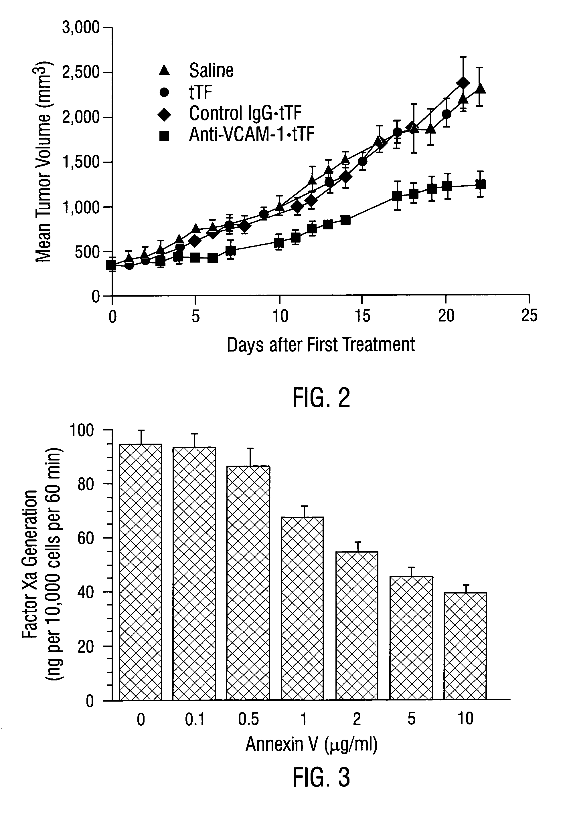
[0645]Male CB 17 SCID mice (Charles River, Wilmington, Mass.) weighing approximately 25 g were injected with 1×10⁷ L540 Hodgkin's lymphoma cells subcutaneously into the right flank. Tumors were allowed to grow to a size of 0.4–0.7 cm3.

[0646]Mice were injected intravenously with 30 μg / 25 g body weight of anti-VCAM-1 antibody, R187 antibody or corresponding coaguligands in 200 μl of saline. Two hours later, animals were anesthetized with metafane and their blood circulation was perfused with heparinized saline as described (Burrows et al, 1992; incorporated herein by reference). The tumor and major organs were removed and snap-frozen in liquid nitrogen.

[0647]Cryostat sections of the tissues were cut and were stained immunohistochemically for the presence of rat IgG or TF. Rat IgG was detected using rabbit anti-rat IgG conjugated to HRP followed by development with carbazole (Fries et al., 1993). Coaguligand was detected using the 1...

example iii

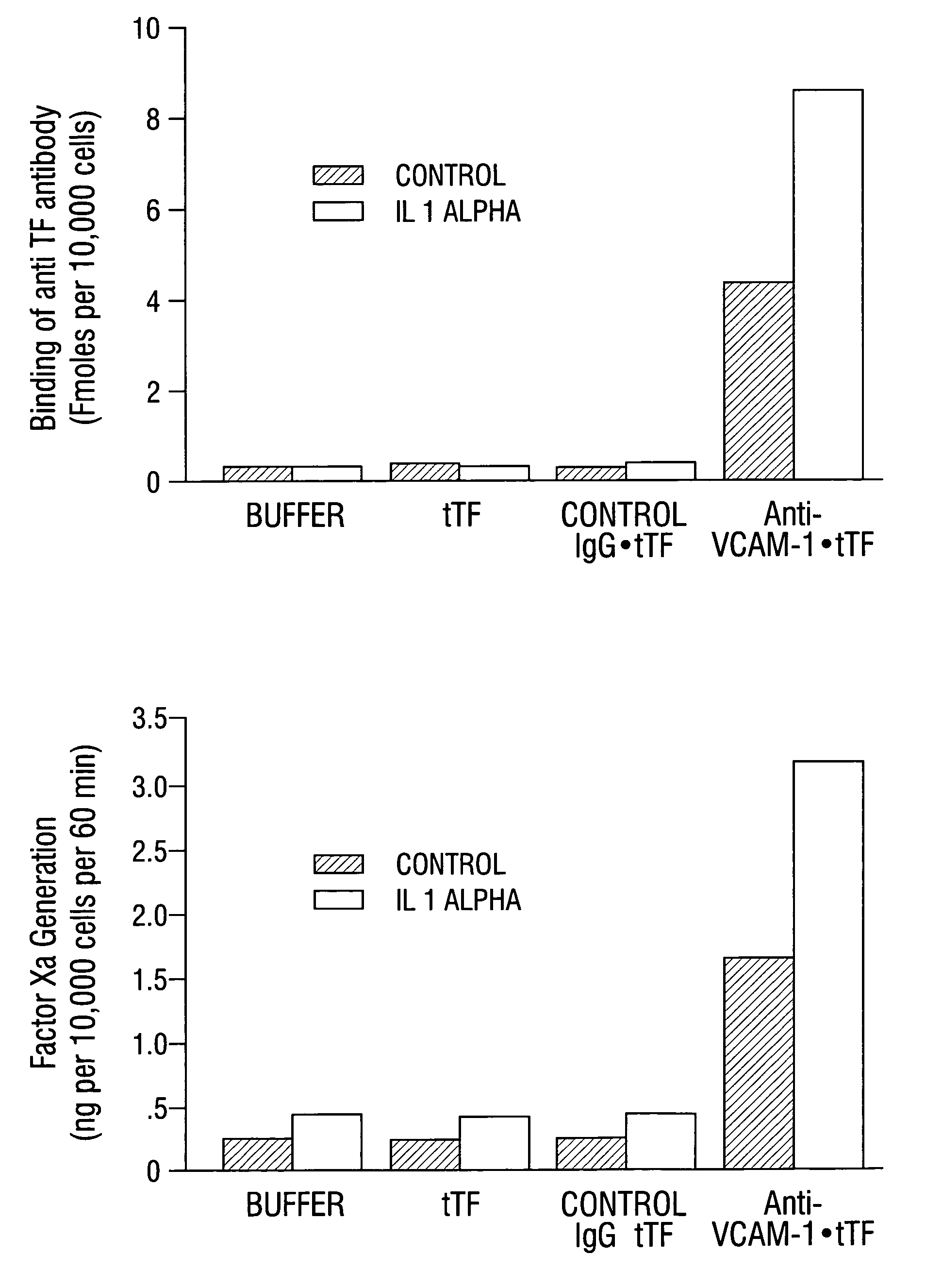
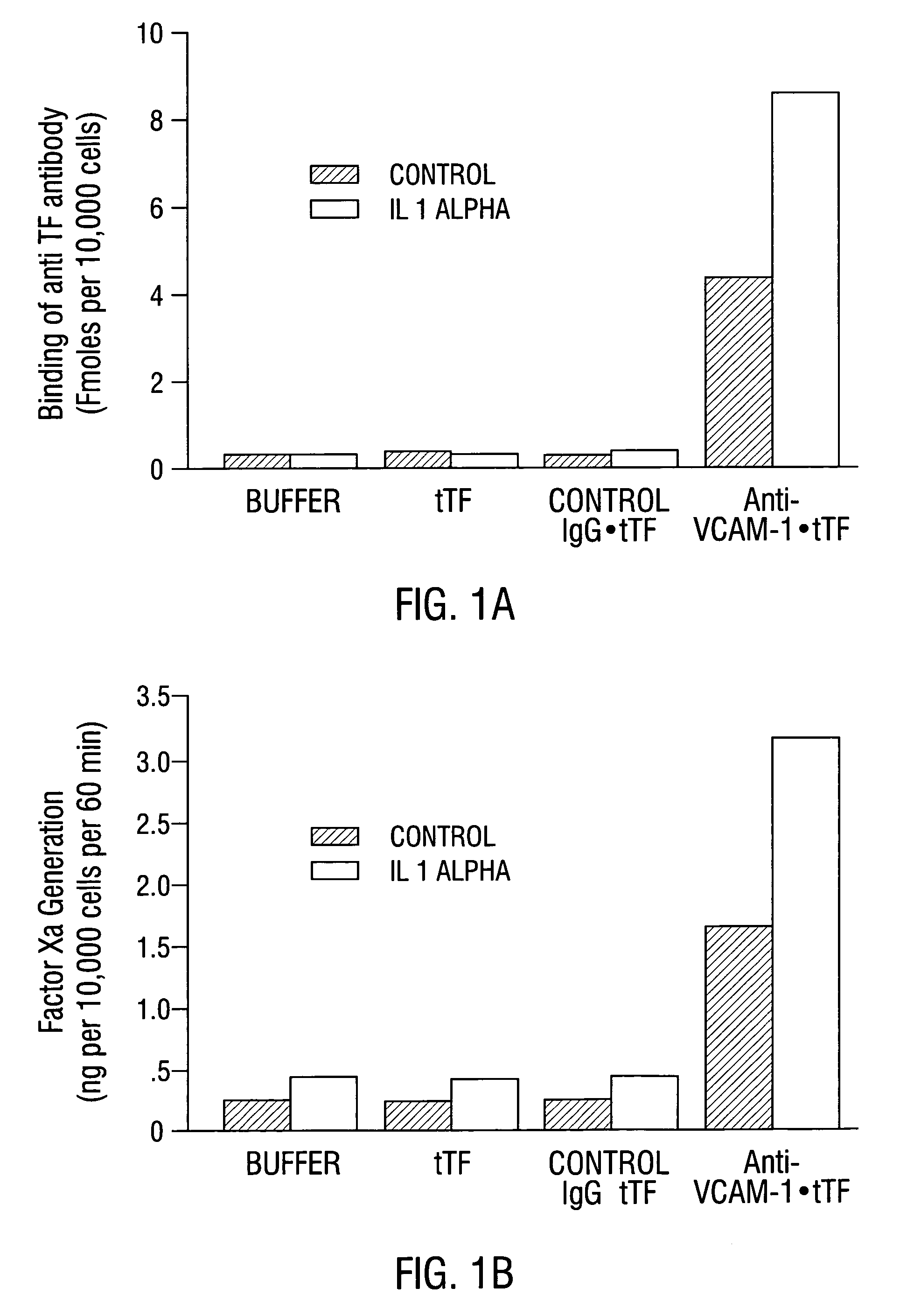
Preparation of Anti-VCAM-1∘tTF Coaguligand

[0650]An anti-VCAM-1∘tTF conjugate or “coaguligand” was prepared as follows. Truncated tissue factor (tTF), with an additional added cysteine introduced at N-terminus (U.S. application Ser. No. 08 / 482,369, incorporated herein by reference), was expressed in E. coli and purified as described by Stone et al. (1995, incorporated herein by reference). After purification, the sulfhydryl group of N′ cysteine-tTF was protected by reaction with Ellman's reagent. The tTF derivative was stored in small volumes at −70° C.

[0651]To prepare the anti-VCAM-1 coaguligand, 5 ml of anti-VCAM-1 antibody IgG (2 mg / ml) in PBS were mixed with 36 μl of SMPT (10 mM) dissolved in dry DMF and incubated at room temperature for 1 h. The mixture was filtered through a column of Sephadex G25 equilibrated in PBS containing 1 mM EDTA. The fractions containing the SMPT-derivatized antibody were concentrated to 4 ml by ultrafiltration in an Amicon cell equipped with a 10,000 ...


PUM

PropertyMeasurementUnit
concentrationsaaaaaaaaaa
volumeaaaaaaaaaa
diameteraaaaaaaaaa

Abstract

Disclosed is the surprising discovery that aminophospholipids, such as phosphatidylserine and phosphatidylethanolamine, are specific, accessible and stable markers of the luminal surface of tumor blood vessels. The present invention thus provides aminophospholipid-targeted diagnostic and therapeutic constructs for use in tumor intervention. Antibody-therapeutic agent conjugates and constructs that bind to aminophospholipids are particularly provided, as are methods of specifically delivering therapeutic agents, including toxins and coagulants, to the stably-expressed aminophospholipids of tumor blood vessels, thereby inducing thrombosis, necrosis and tumor regression.

Description

[0001]The present application claims priority to first provisional application Ser. No. 60 / 092,589, filed Jul. 13, 1998, and second provisional application Ser. No. 60 / 110,600, filed Dec. 2, 1998, the entire text and figures of which applications are incorporated herein by reference without disclaimer.[0002]The U.S. Government owns rights in the present invention pursuant to grant numbers 1RO1CA74951-01 and 5RO1CA54168-05 from the National Institutes of Health.BACKGROUND OF THE INVENTION[0003]1. Field of the Invention[0004]The present invention relates generally to the fields of blood vessels and tumor biology. More particularly, it embodies the surprising findings that aminophospholipids, such as phosphatidylserine and phosphatidylethanolamine, are accessible, stable and specific markers of tumor vasculature. The invention thus provides therapeutic constructs and conjugates that bind to aminophospholipids for use in delivering toxins and coagulants to tumor blood vessels and for in...

Claims


Application Information

Patent Timeline
Patent Type & Authority Patents(United States)
IPC IPC(8): A61K39/395A61K38/00A61K45/00A61K47/48A61K49/00A61K49/04A61K51/10A61P35/00C07K16/28C12P21/08
CPCA01K2217/05A61K39/395A61K51/1027C07K16/2836C07K2317/24C07K2317/31C07K2319/00A61K47/6835A61K47/6849A61P35/00A61P9/00A61K2300/00A61K49/04A61N5/10
Inventor THORPE, PHILIP E.RAN, SOPHIABREKKEN, ROLF A.
Owner BOARD OF RGT THE UNIV OF TEXAS SYST
Why Eureka
Try Eureka