Method and screening candidate compounds for drug against tumor

a candidate compound and tumor technology, applied in the field of tumor drug screening, can solve the problems of unfulfilled identification of mutations and mast cell leukemic cell lines

Inactive Publication Date: 2006-10-24
CHUGAI PHARMA CO LTD
View PDF1 Cites 9 Cited by
  • Summary
  • Abstract
  • Description
  • Claims
  • Application Information

AI Technical Summary

Benefits of technology

[0051]To eliminate the effects of nonselective cytotoxic substances in the screening system in which cell proliferation is used as an index, the parent cells are cultured in the presence of IL-3 and a test sample is added to the culture to determine the effects of the test sample on IL-3-dependent cell-proliferation in parallel with the experiment above.

Problems solved by technology

However, mutations in c-kit gene have been found in the mast cell leukemic cell lines, which mutations have not been fully identified in clinical samples (Tsujimura, T. et al., 1994, Blood, 83:2619–2626; Kitayama, H., 1996, Blood, 88: 995–1004; Tsujimura, Y. et al., 1996, Blood, 87: 273–283).

Method used


Image

  • Method and screening candidate compounds for drug against tumor
  • Method and screening candidate compounds for drug against tumor
  • Method and screening candidate compounds for drug against tumor

Examples


example 1

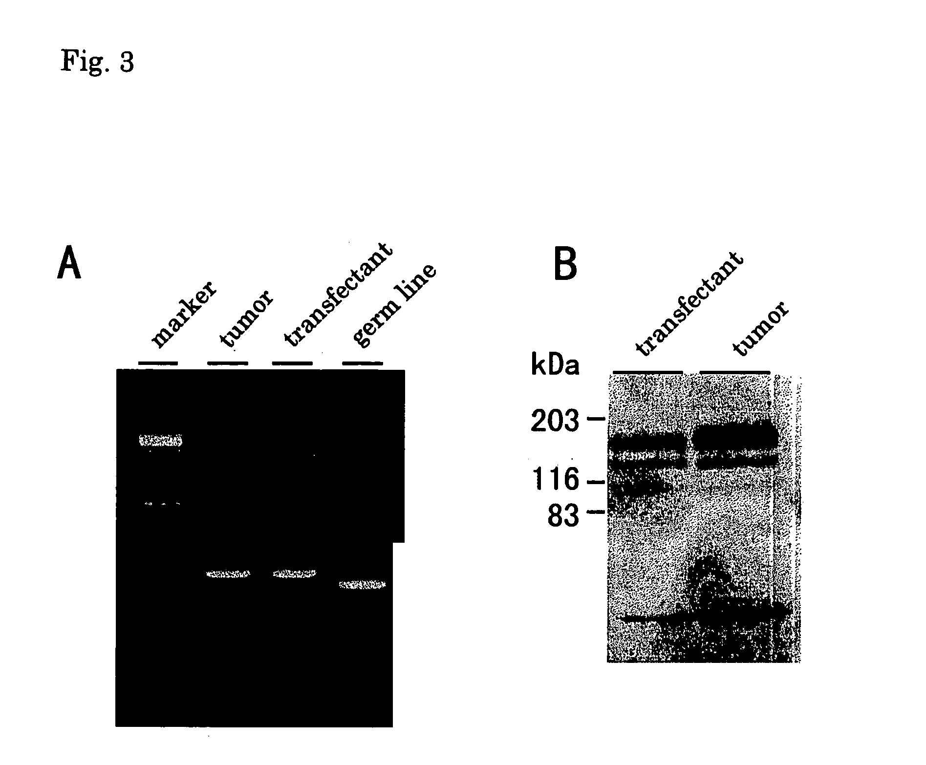
Detection of FLT3 / ITD in Leukemia Cells

[0059]High molecular weight DNA was isolated from leukemia cells, and a DNA fragment containing the JM domain of the FLT3 protein was amplified by PCR according to the method described in Kiyoi, H., Leukemia 11: 1447–1452, 1997. The bands that differed from the band of the wild-type in size were excised from agarose gel and purified with Qiaex gel extraction kit (Qiagen), followed by cloning into pMOSBlue T vector (Amersham) according to the manufacturer's instruction. Ten colonies of the recombinants were cultured on the LB medium, and the plasmid DNA was prepared with QIAprep spin plasmid miniprep kit (QIAGEN). Nucleotide sequences of these clones were confirmed by sequencing. Expression of the FLT3 mRNA was confirmed by RT-PCR according to the method described in Kiyoi, H., Leukemia 11: 1447–1452, 1997. The bands that differed from the band of the wild type in size were cloned according to the method described above, and their nucleotide seq...

example 2

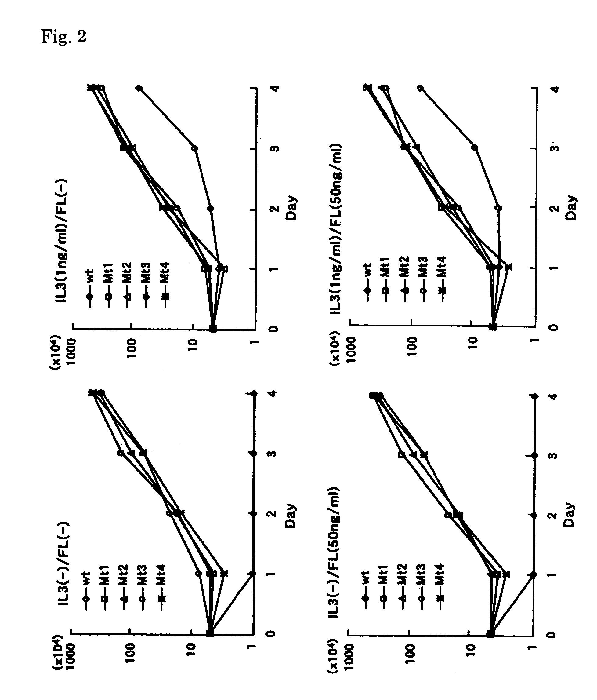
Introduction of an FLT3 / ITD Expression Vector into Blood Cells

[0063]Total RNA was extracted from leukemia cells and was used for cDNA synthesis. The cDNA synthesized was used as a template to amplify the MunI-EcoRV fragment, which contains the tandem repeat region found in mutants FTL3 cDNA, by RT-PCR. MunI-F primer (SEQ ID NO: 9 / 5′-CAACAATTGGTGTTTGTCTCCTCTT-3′) and EcRV-R primer (SEQ ID NO: 10 / 5′-CATGATATCTCGAGCCAATCCAAAG-3′) were used for the amplification. The amplified fragments were cleaved with MunI and EcoRV (Boehringer-Mannheim-Yamanouchi), resolved on agarose gel, and purified according to the aforementioned method. Expression vector pCDHF3 (a gift from Dr. Olivier Rosnet), which carries a full-length wild type FLT3 cDNA (Rosnet, O. et al., Blood 82:1110–1119; Accession No. S64785), was cleaved with MunI and EcoRV, and the purified FLT3 / ITD fragment was inserted into the vector. Four mutants of FLT3 / ITD (Mt1, Mt2, Mt3 and Mt4) were used. Nucleotide sequences of the mutated ...

example 3

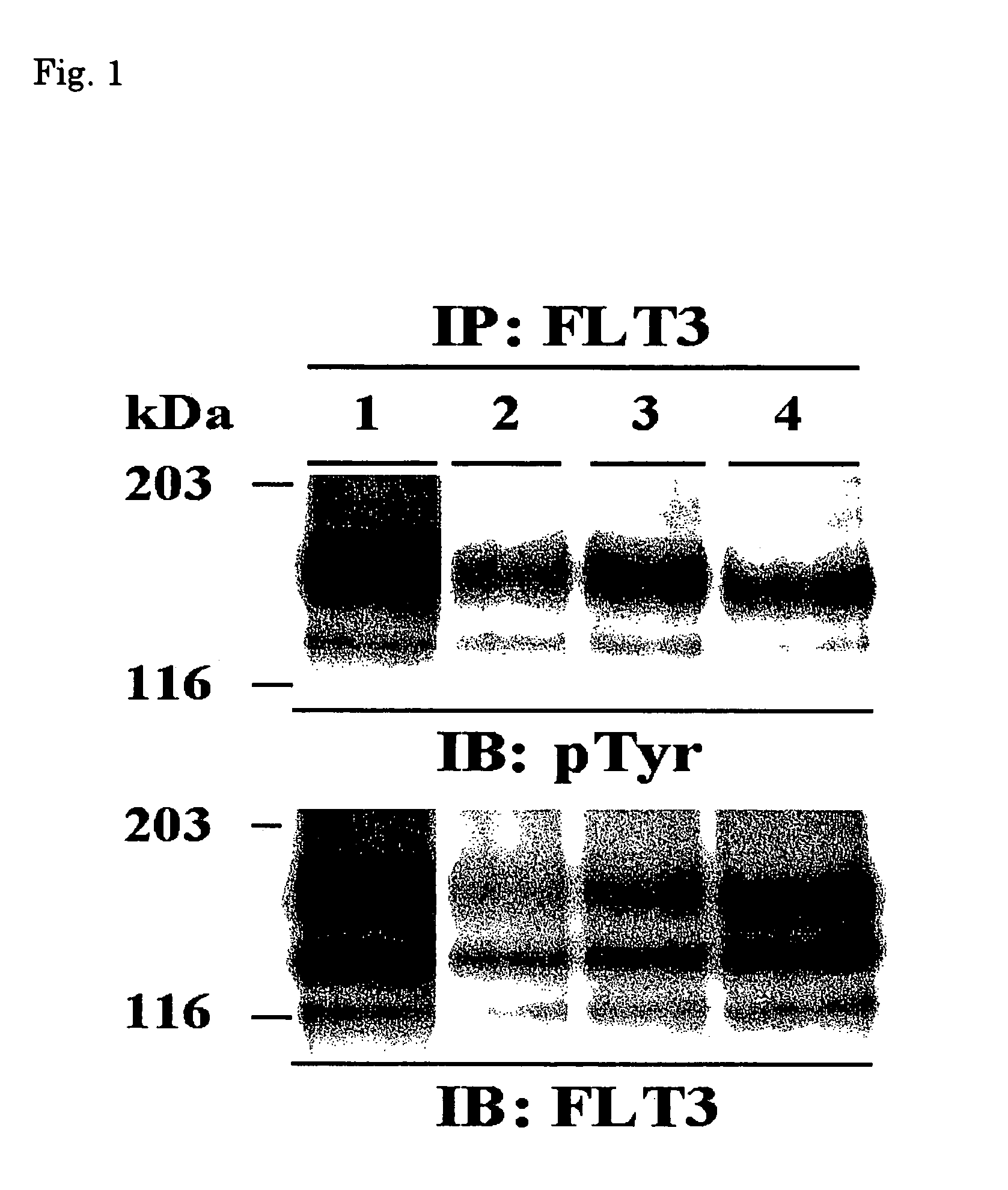
Tyrosine-Phosphorylation of the FLT3 Molecules in the Transfectants

[0066](1) The transfectants were cultured in RPMI 1640 medium supplemented with 10% FCS (GIBCO), and then centrifuged at 1000 rpm for 5 minutes to recover 2×10⁷ cells. The cell pellets were washed with PBS, dissolved in lysis buffer (20 mM Tris-HCl, pH 7.5, 150 mM NaCl, 2 mM EDTA, Nonidet P-40, 50 mM NaF, 10 mg / ml aprotinin, 10 mg / ml leupeptin, 1 mM Na3VO4, 50 mM Na2MoO4, 1 mM phenylmethylsulfonyl fluoride (PMSF)), allowed to stand for 1 hour at 4° C., then centrifuged at 15000 rpm for 30 minutes. Rabbit anti-human FLT3 antibody (Santa Cruz Biotechnology, Santa Cruz, Calif., USA) was added to the supernatants and the mixture was stirred for 2 hours at 4° C. After adding Protein A / G Plus agarose (Santa Cruz), the resulting mixture was stirred for 2 hours at 4° C. and washed with the lysis buffer three times. The pellets were then dissolved in sample loading buffer (0.125 M Tris-HCl, pH 6.8, 10% 2-mercaptoethanol, 4% S...


PUM

PropertyMeasurementUnit
pHaaaaaaaaaa
pHaaaaaaaaaa
lengthaaaaaaaaaa

Abstract

Investigation on the frequency of FLT3/ITD found in various blood cancers has revealed that the frequency is high in acute myeloblastic leukemia in particular. Studies on the effects of FLT3/ITD in the blood cell lines revealed that the tyrosine residues in FLT3/ITD is constitutively phosphorylated in these cell lines and that blood cells into which FLT3/ITD is introduced show IL-3 independent proliferation. Moreover, the blood cells into which FLT3/ITD is introduced are found to be capable of forming tumors and inhibit cell differentiation. The inventors have found that it is possible to screen for pharmaceutical compounds against tumors by using inhibition of these FLT3/ITD functions as an index.

Description

TECHNICAL FIELD[0001]The present invention relates to a method for screening a candidate compound for a drug against tumors, in particular, blood cancers. More specifically, it relates to a method for screening a compound that inhibits the function of FLT3 / ITD in animal cells, including blood cell lines.BACKGROUND ART[0002]FTL3, as well as KIT, FMS and PDGFR, is a protein of a class III receptor tyrosine kinase (RTK), and is presumed to be involved in the hematopoietic system (Rosnet, O. et al., 1991, Genomics 9:380–385; Rosnet, O. et al., 1991, Oncogene, 6:1641–1650; Matthews, W. et al., 1991, Cell, 65: 1143–1152; Rosnet, O. et al., 1993, Blood, 82: 1110–1119). Structurally, RTK has an extracellular region containing five immunoglobulin-like domains, one juxtamembrane region (JM domain), two tyrosine domains (TK1 and TK2) intervened by a kinase insert domain (KI domain), and the C-terminal domain. FLT3 is strongly expressed in the hematopoietic stem cells as well as the brain, plac...

Claims


Application Information

Patent Timeline
Patent Type & Authority Patents(United States)
IPC IPC(8): C12Q1/00G01N33/53G01N33/567A01N37/18A61K35/14G01N33/50G01N33/574
CPCG01N33/5011G01N33/57426A61P35/00
Inventor KIYOI, HITOSHINAOE, TOMOKITOWATARI, MASAYUKIKITAMURA, TOSHIO
Owner CHUGAI PHARMA CO LTD
Why Eureka
Try Eureka