System and method for magnetic-resonance-guided electrophysiologic and ablation procedures

a magnetic resonance and electrophysiologic technology, applied in the field of electrophysiologic diagnostic and therapeutic procedures, can solve the problems of inability to determine the optimal approach, inability to achieve the optimal approach, so as to improve the safety and accuracy of electrophysiologic procedures, improve the resolution and reliability, and simplify the production of continuous linear lesions

Inactive Publication Date: 2006-12-26
THE JOHNS HOPKINS UNIVERSITY SCHOOL OF MEDICINE
View PDF41 Cites 233 Cited by
  • Summary
  • Abstract
  • Description
  • Claims
  • Application Information

AI Technical Summary

Benefits of technology

[0023]It is a further object of the invention to provide a system and method for imaging ablation lesions with increased resolution and reliability.
[0024]The invention provides a system and method for using magnetic resonance imaging to increase the safety and accuracy of electrophysiologic procedures. The system in its preferred embodiment provides an invasive combined electrophysiology and imaging antenna catheter which includes an RF antenna for receiving magnetic resonance signals and diagnostic electrodes for receiving electrical potentials. The combined electrophysiology and imaging antenna catheter is used in combination with a magnetic resonance imaging scanner to guide and provide visualization during electrophysiologic diagnostic or therapeutic procedures. The invention is particularly applicable to catheter ablation of atrial and ventricular arrhythmias. In embodiments which are useful for catheter ablation, the combined electrophysiology and imaging antenna catheter may further include an ablation tip, and such embodiment may be used as an intracardiac device to both deliver energy to selected areas of tissue and visualize the resulting ablation lesions, thereby greatly simplifying production of continuous linear lesions. Additionally, the ablation electrode can be used as an active tracking device that receives signal from the body coil excitation. Gradient echoes are then generated along three orthogonal axes to frequency encode the location of the coil and thus provide the three-dimensional space coordinates of the electrode tip. These numeric coordinates can then be used to control the imaging plane of the scanner, thereby allowing accurate imaging slices to be automatically prescribed though the anatomic target for RF therapy. The invention further includes embodiments useful for guiding electrophysiologic diagnostic and therapeutic procedures other than ablation. Imaging of ablation lesions may be further enhanced by use of MR contrast agents. The antenna utilized in the combined electrophysiology and imaging catheter for receiving MR signals is preferably of the coaxial or “loopless” type that utilizes a helical whip. High-resolution images from the antenna may be combined with low-resolution images from surface coils of the MR scanner to produce a composite image. The invention further provides a system for eliminating the pickup of RF energy in which intracardiac wires are detuned, by for example low-pass filters, so that they become very inefficient antennas. An RF filtering system is provided for suppressing the MR imaging signal while not attenuating the RF ablative current. Steering means may be provided for steering the invasive catheter under MR guidance. Lastly, the invention provides a method and system for acquisition of high-density electroanatomic data using a specially designed multi-electrode catheter and the MRI scanner. This will be achieved by using an active tracking system that allows the location of each electrode to be determined.

Problems solved by technology

They represent the most frequently encountered tachycardias, account for the most morbidity and mortality, and, despite much progress, remain therapeutic challenges.
These symptoms, side effects of drugs, and most importantly, thromboembolic complications in the brain (leading to approximately 75,000 strokes per year), make atrial fibrillation a formidable challenge.
The optimal approach is uncertain.
A major disadvantage of antiarrhythmic therapy is the induction of sustained, and sometimes lethal, arrhythmias (proarrhythmia) in up to 10% of patients.
The substantial incidence of thromboembolic strokes makes chronic anticoagulation important, but bleeding complications are not unusual, and anticoagulation cannot be used in all patients.
Medical management of atrial fibrillation, therefore, is inadequate.
Despite these encouraging results, this procedure has not gained widespread acceptance because of the long duration of recovery and risks associated with cardiac surgery.
One of the main limitations of the procedure is the difficulty associated with creating and confirming the presence of continuous linear lesions in the atrium.
This difficulty contributes significantly to the long procedure durations discussed above.
Since x-ray shadows are the superposition of contributions from many structures, and since the discrimination of different soft tissues is not great, it is often very difficult to determine exactly where the catheter is within the heart.
In addition, the boarders of the heart are generally not accurately defined, so it is generally not possible to know if the catheter has penetrated the wall of the heart.
The system does not obviate the need for initial placement using x-ray fluoroscopy, and cannot directly image ablated tissue.
While MRI may provide the visual guidance necessary for creating and confirming linear lesions, it has been assumed that electrical wires implanted in a patient can act as antennas to pick up radio-frequency energy in an MR system and conduct that energy to the patient, thereby causing tissue injury.
However, these systems are not suitable for use in the heart.
There is no provision in the disclosed probe for measuring electrical signals; and, it is unclear how much resolution the probe provides.

Method used


Image

  • System and method for magnetic-resonance-guided electrophysiologic and ablation procedures
  • System and method for magnetic-resonance-guided electrophysiologic and ablation procedures
  • System and method for magnetic-resonance-guided electrophysiologic and ablation procedures

Examples


Embodiment Construction

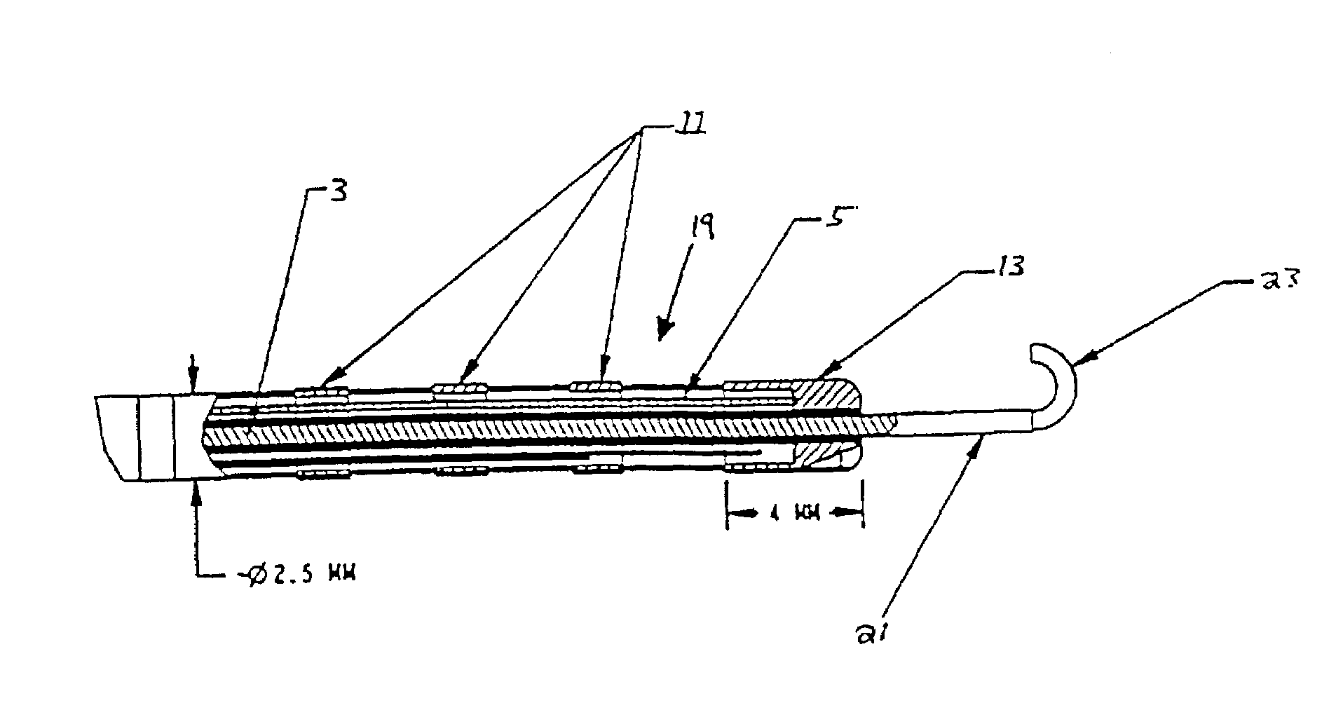
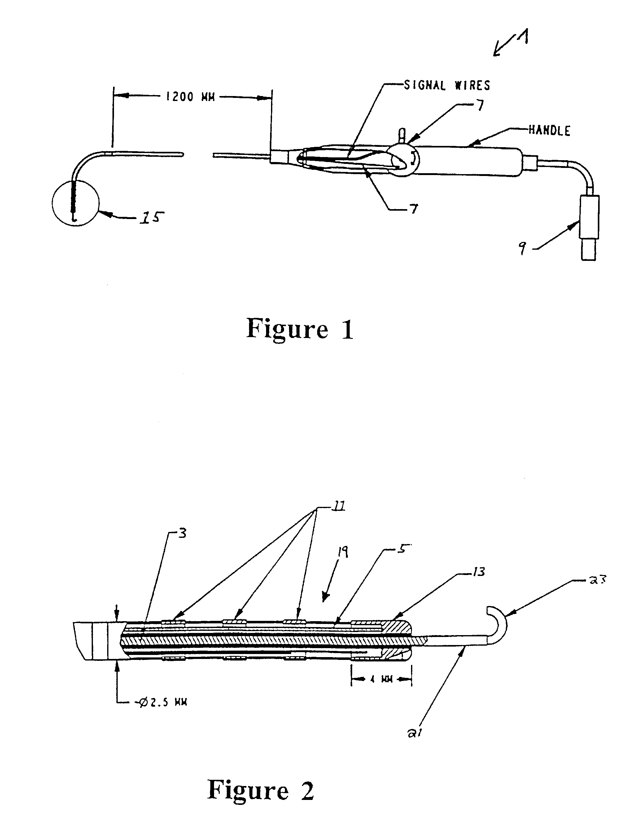
[0033]The invention in its preferred embodiment uses MR imaging to allow catheters to be placed without radiation, and provides very accurate localization of catheter tips in 3-dimensional space. With current MRI scanners, resolution is limited by the distance the RF coil is from the volume of tissue being imaged. RF from any particular imaging volume is picked up by the surface coil. The gradients select a volume inside the body for imaging, but the coil outside the body picks up the signal from the volume. The farther the surface coil is from the imaging volume, the more noise will be present.

[0034]In accordance with a preferred embodiment of the invention, an intracardiac receiving coil / antenna is used so that the receiving coil / antenna is closer to the imaging volume (lesions), thereby reducing noise, increasing signal, and improving resolution where it is needed most.

[0035]In a first embodiment of the invention, MRI is used to facilitate catheter ablation of atrial fibrillation...


PUM


Abstract

A system and method for using magnetic resonance imaging to increase the accuracy of electrophysiologic procedures is disclosed. The system in its preferred embodiment provides an invasive combined electrophysiology and imaging antenna catheter which includes an RF antenna for receiving magnetic resonance signals and diagnostic electrodes for receiving electrical potentials. The combined electrophysiology and imaging antenna catheter is used in combination with a magnetic resonance imaging scanner to guide and provide visualization during electrophysiologic diagnostic or therapeutic procedures. The invention is particularly applicable to catheter ablation, e.g., ablation of atrial fibrillation. In embodiments which are useful for catheter ablation, the combined electrophysiology and imaging antenna catheter may further include an ablation tip, and such embodiment may be used as an intracardiac device to both deliver energy to selected areas of tissue and visualize the resulting ablation lesions, thereby greatly simplifying production of continuous linear lesions. The invention further includes embodiments useful for guiding electrophysiologic diagnostic and therapeutic procedures other than ablation. Imaging of ablation lesions may be further enhanced by use of MR contrast agents. The antenna utilized in the combined electrophysiology and imaging catheter for receiving MR signals is preferably of the coaxial or “loopless” type. High-resolution images from the antenna may be combined with low-resolution images from surface coils of the MR scanner to produce a composite image. The invention further provides a system for eliminating the pickup of RF energy in which intracardiac wires are detuned by filtering so that they become very inefficient antennas. An RF filtering system is provided for suppressing the MR imaging signal while not attenuating the RF ablative current. Steering means may be provided for steering the invasive catheter under MR guidance. Other ablative methods can be used such as laser, ultrasound, and low temperatures.

Description

[0001]This application is a divisional application of and claims priority to U.S. patent application Ser. No. 09 / 428,990, filed Oct. 29, 1999, now U.S. Pat. No. 6,701,176 and claims the benefit of U.S. Provisional Patent Application No. 60 / 106,965 filed Nov. 4, 1998, the entire disclosure of which is incorporated herein by reference.BACKGROUND OF THE INVENTION[0002]1. Field of the Invention[0003]The invention relates in general to ablation and electrophysiologic diagnostic and therapeutic procedures, and in particular to systems and methods for guiding and providing visualization during such procedures.[0004]2. Related Art[0005]Atrial fibrillation and ventricular tachyarrhythmias occurring in patients with structurally abnormal hearts are of great concern in contemporary cardiology. They represent the most frequently encountered tachycardias, account for the most morbidity and mortality, and, despite much progress, remain therapeutic challenges.[0006]Atrial fibrillation affects a la...

Claims


Application Information

Patent Timeline
Patent Type & Authority Patents(United States)
IPC IPC(8): A61B5/055A61B17/00A61B19/00G01R33/28
CPCA61B5/055A61B5/415A61B5/418A61B18/1492A61B19/52G01R33/285G01R33/34084A61B2017/00039A61B2017/00053A61B2019/5236G01R33/4808G01R33/5601A61B90/36A61B2090/374
Inventor HALPERIN, HENRY R.BERGER, RONALD D.ATALAR, ERGINMCVEIGH, ELLIOT R.LARDO, ALBERTCALKINS, HUGHLIMA, JOAO
Owner THE JOHNS HOPKINS UNIVERSITY SCHOOL OF MEDICINE
Why Eureka
Try Eureka