Materials and methods for management of hyperacute rejection in human xenotransplantation

a technology for xenotransplantation and hyperacute rejection, which is applied in the field of xenotransplantation, can solve the problems of increasing the financial cost of dialysis in australia and new zealand, and increasing the cost of dialysis. , to achieve the effect of reducing or reducing reducing or eliminating the hyperacute rejection respons

Inactive Publication Date: 2007-04-10
BRESAGEN +1
View PDF14 Cites 11 Cited by
  • Summary
  • Abstract
  • Description
  • Claims
  • Application Information

AI Technical Summary

Benefits of technology

[0027]The invention further includes a method for eliminating or reducing hyperacute rejection of non-primate mammalian cells by human serum, comprising adding, to the human serum, a physiologically acceptable amount of galactose or a saccharide in which the terminal carbohydrate is an a galactose linked at position 1, prior to exposure of the human serum to the non-primate cells. The amount of galactose or saccharide added is sufficient to reduce

Problems solved by technology

It is widely acknowledged that there is an acute, worldwide shortage of human organs for transplantation.
In Japan the imbalance between supply and demand is even greater due to religious prohibitions on the use of organs from cadaveric donors.
The direct financial cost of dialysis in Australia and New Zealand is approximately $A45,000/patient/year.
In addition, indirect costs due to unemployment and sickness are higher in dialysis patients and the social costs are considerable.
These statistics indicate that a) transplantation is the optimal therapy for end stage renal failure; b) there is an undersupply of donor kidneys; and c) present strategies aimed at increasing the transplant rate have been less than successful.
There are, in addition, serious shortages of other transplantable organs including hearts, livers, lungs and

Method used


Image

  • Materials and methods for management of hyperacute rejection in human xenotransplantation
  • Materials and methods for management of hyperacute rejection in human xenotransplantation
  • Materials and methods for management of hyperacute rejection in human xenotransplantation

Examples


example 1

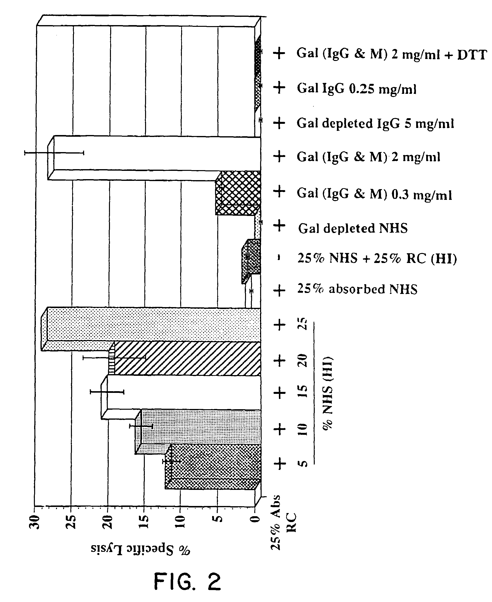
Affinity Purification of Human Anti-GAL Antibodies

[0102]Anti-GAL antibodies were purified from normal heat inactivated AB serum (from CS1 Parkville, Victoria, Australia) using the following sets of procedures.

A. Preparation of Total Anti-GAL (IgG+IgM) Antibodies

The following procedures are performed at 4° C.

[0103]1. Desalt 15–30 ml serum (in 3 ml batches) by passage through a pre-equilibrated (20 ml application buffer: 20 mM K2HPO4, 30 mM NaCl, pH 8) Econo Pac 10DG (Bio-Rad, Richmond, USA) column. Alternatively, for large scale preparations, desalt by dialysis exhaustively against application buffer.[0104]2. Wash column with 4 ml aliquots of application buffer. Collect and pool column eluates.[0105]3. Apply pooled desalted serum to a pre-equilibrated (20 ml application buffer) Synsorb 115 (galactosyl-galactose; Chembiomed, Alberta, Canada) or D(+) Melibiose-Agarose (Sigma) affinity column (5 ml–50 ml depending on the yield required).[0106]4. Collect run-through (partially anti-GAL-d...

example 2

Reactivity of IgG and IgM Anti-GAL Antibodies and Depleted Serum with Porcine Cells and Tissues

I. Cells

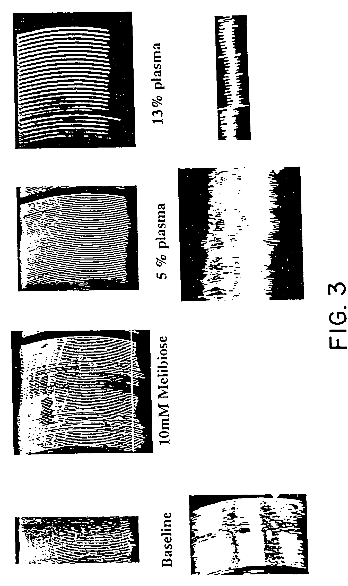
[0124]Reactivity of IgG and IgM anti-GAL antibodies was assessed using either porcine aortic endothelial cells (prepared by the inventors as described below) or porcine epithelial cell line LLC PK1 (PK1), obtained from the American Type Culture Collection (ATCC), Accession No. CRL1392.

A. Isolation and Culture of Porcine Aortic Endothelial Cells (PAE's)

[0125]Pigs were blood typed (using human typing reagents) to identify “O-type” pigs, i.e, pigs unreactive with antibodies to A or B human red blood cell antigens. Aortas were excised from “O-type” pigs, then transported from the abattoir to the laboratory on ice. PAE's were isolated by collagenase treatment as described by Gimbrone et al., J. Cell Biol. 60: 673–84 (1974). PAE's were cultured in RPMI medium containing 10% fetal calf serum (FCS), supplemented with 150 g / ml endothelial cell supplement (Sigma) and 50 g / ml heparin (Sigma)....

example 3

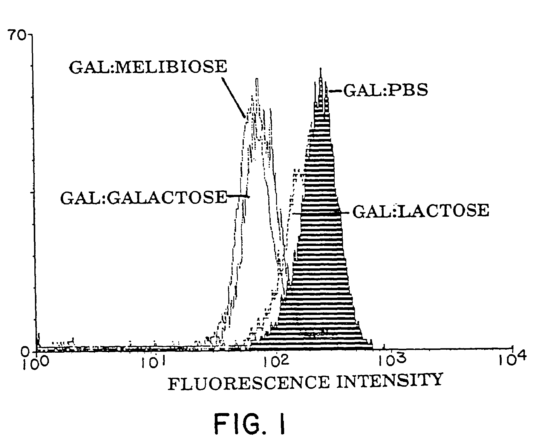
Hemagglutination of Pig RBC by Human Serum: Sugar Inhibition Studies

[0162]The methods used to investigate the hemagglutination of pig red blood cells (RBC's) by human serum was adapted from the methods described by Galili, J. Exp. Med. 160: 1579–81 (1984) and Severson, Immunol. 96: 785–789 (1966).

I. Methods

[0163]A. Media / Solution Preparation[0164]1. Human Serum Albumin (HSA) (CSL, Melbourne, Australia) (5 mg / ml) was dissolved in PBS, filter sterilized, and stored at 4° C.[0165]2. Preparation of sugars:[0166] 1 M stock solutions of sugar were prepared by dissolving the amount indicated in 100 ml of PBS. Sodium azide was added (0.02%) and solutions stored at 4° C.

[0167]

α-Lactose (4-O-β-D.galactopyranosyl-α-D-glucose36.0 gD(+)galactose18.0 gStachyose (α-D-gal-[1->6]-α-D-Glc-[1->2]-β-D-Fru)66.6 gMelibiose (6-O-α-D-galactopyranosyl-D-glucose)34.2 gSucrose (α-D-Glucopyranosyl β-D-fructofuranoside)34.2 gD-(+)-Glucose18.0 gα-D-(+)-Fucose (6-Deoxy-D-galactopyranose)16.4 g

[0168]All sugars wer...


PUM


Abstract

Human pre-formed xenoantibodies play an important role in the hyperacute rejection response in human xenotransplantation. Disclosed are materials and methods for removing or neutralizing such antibodies. Also disclosed are materials and methods for reducing or eliminating the epitopes in the donor organs that are recognized by such antibodies. Such epitopes are formed as the result of activity by the enzyme α-1,3 galactosyltransferase. The porcine gene encoding α-1,3 galactosyltransferase is disclosed, as are materials and methods for inactivating (“knocking out”) the α-1,3 galactosyltransferase gene in mammalian cells and embryos. Included are nucleic acid constructs useful for inactivating the α-1,3 galactosyltransferase gene in a target cell. Also disclosed is a novel leukemia inhibitory factor (T-LIF) that is useful for maintenance of embryonic stem cells and primordial germ cells in culture.

Description

CROSS REFERENCE TO RELATED APPLICATIONS[0001]This application claims benefit of priority, and is a continuation, of U.S. application Ser. No. 08 / 984,900, filed Dec. 4, 1997, now U.S. Pat. No. 6,849,448, which claims benefit of priority, and is a divisional, of U.S. application Ser. No. 08 / 378,617, filed Jan. 26, 1995, now U.S. Pat. No. 5,849,991, issued Dec. 15, 1998, which claims benefit of priority, and is a continuation-in-part, of U.S. application Ser. No. 08 / 188,607, filed Jan. 27, 1994, now abandoned.FIELD OF THE INVENTION[0002]This invention relates generally to the field of xenotransplantation. In particular this invention relates to methods and materials for reduction or elimination of the hyperacute rejection response in humans. More particularly, this invention relates to methods for treating human serum to reduce or eliminate hyperacute rejection. This invention also relates to methods and materials for generating non-human organs lacking or having reduced α 1,3 galactos...

Claims


Application Information

Patent Timeline
IPC IPC(8): A01N63/00C07H21/04C12N15/00C12N15/63C12N15/87A01K67/027A61K31/70A61K38/00C07K1/22C07K14/47C07K14/54C07K16/40C07K16/44C12N5/10C12N9/10C12N15/09C12N15/54C12N15/85
CPCA01K67/0276A61K31/70A61K31/7004C07K14/54C07K16/40C12N9/1051C12N15/8509A01K2217/075A01K2227/105A01K2227/108A01K2267/02A01K2267/025A61K38/00A01K2267/03C12N2800/30
Inventor D'APICE, ANTHONY J. F.PEARSE, MARTIN J.ROBINS, ALLAN J.CRAWFORD, ROBERT J.RATHJEN, PETER D.
Owner BRESAGEN
Why Eureka
Try Eureka