
Method for treatment of biological tissues to mitigate post-implantation calcification and thrombosis

Inactive Publication Date: 2007-05-08
EDWARDS LIFESCIENCES CORP
View PDF44 Cites 62 Cited by
  • Summary
  • Abstract
  • Description
  • Claims
  • Application Information

AI Technical Summary

Benefits of technology

[0019]5. TERMINAL STERILIZATION—subjecting the biological tissue to a sterilant (e.g., a liquid sterilant such as 0.2–2.0% by weight glutaraldehyde solution) for a sterilization time period. A 0.625% glutaraldehyde solution may be used in combination with heat (i.e. warming above room temperature but below a temperature which would cause damage to the tissue of the bioprosthesis), as the sterilant. Alternatively, a suitable sterilant solution may comprise an osmotically balanced aqueous solution alone or in combination with a non-contacting source of sterilization (e.g., radiation, electron beam, UV, or other similar expedient), or include an aqueous solution of glutaraldehyde in combination with the above-described DSC mixture or some components of such DSC mixture. In instances where a 0.625% glutaraldehyde solution is used as the sterilant, the sterilization time period may be 1–6 days at 37 degrees C. or 1–2 days at 50 degrees C.). This terminal sterilization step may be performed after packaging of the bioprosthesis in its final container, thereby eliminating the need for any subsequent handling of the bioprosthesis until the time of implantation.

Problems solved by technology

One problem which has been associated with the implantation of bioprosthetic materials is that the connective tissue proteins (i.e., collagen and elastin) within these materials can become calcified following implantation within the body.
Such calcification can result in undesirable stiffening or degradation of the bioprosthesis.

Method used


Image

  • Method for treatment of biological tissues to mitigate post-implantation calcification and thrombosis
  • Method for treatment of biological tissues to mitigate post-implantation calcification and thrombosis
  • Method for treatment of biological tissues to mitigate post-implantation calcification and thrombosis

Examples


Embodiment Construction

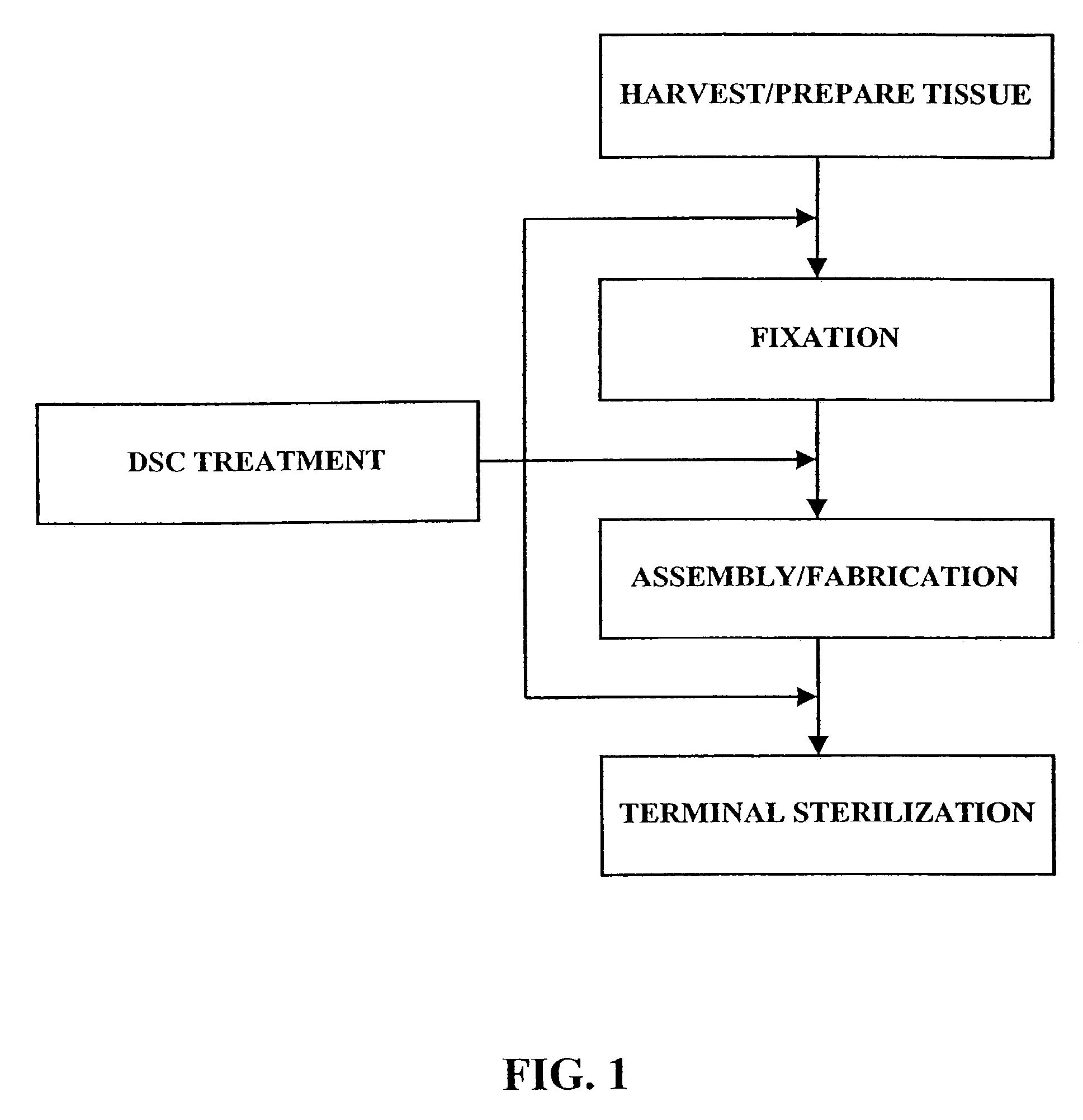
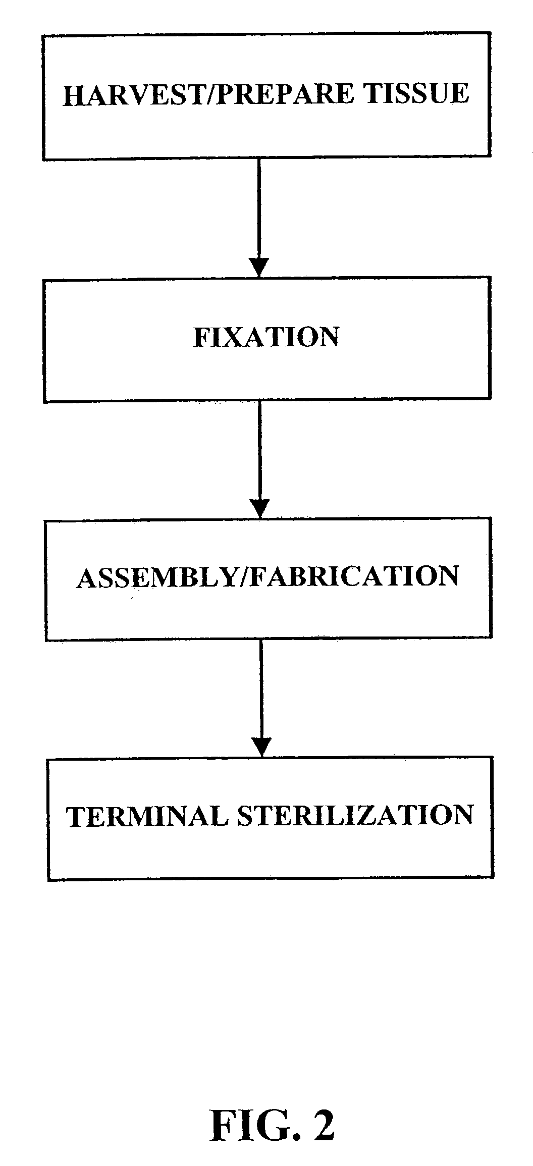
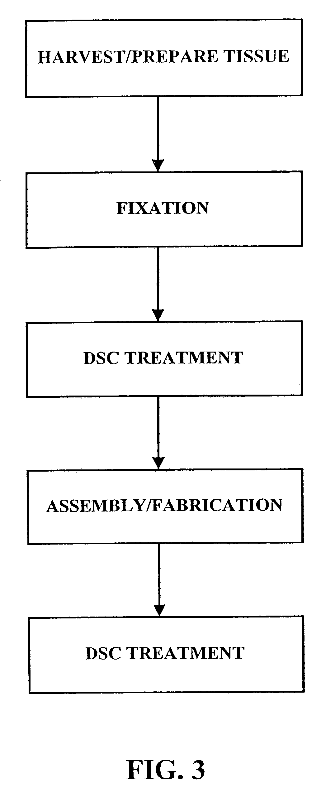
[0028]As shown in FIG. 1, the fixation / sterilization method of the present invention generally comprises six (6) steps as follows:

Step 1: Harvest / Prepare Biological Tissue

[0029]The desired biological tissue is harvested (i.e., surgically removed or cut away from its host animal). Thereafter, it is typically, trimmed or cut to size and washed with sterile water, balanced salt solution, saline or other suitable washing solution.

Step 2: Fixation of Biological Tissue

[0030]The biological tissue is then contacted with a crosslinking agent, such as an aldehyde (e.g., formaldehyde, glutaraldehyde, dialdehyde starch), polyglycidyl either (e.g., Denacol 810), diisocyanates, photooxidation, or carbodiimide(s)) to crosslink the connective tissue proteins present within the tissue. Due to the long standing use and experience with glutaraldehyde, a presently preferred fixative for use in this step is a solution of 0.2–2.0% by weight glutaraldehyde. For example, the biological tissue may be immers...


PUM

PropertyMeasurementUnit
Temperatureaaaaaaaaaa

Abstract

A method of treating a biological tissue including contacting the biological tissue with an aqueous sterilizing solution, and maintaining the aqueous sterilizing solution at a temperature of about 50° C. for a time period of about 1 to 2 days. The method of treating a biological tissue may be utilized as a terminal sterilization step in a method for fixation of biological tissues, and bioprosthetic devices may be prepared by such fixation method. The fixation method may include the steps of A) fixing the tissue, B) treating the tissue with a mixture of i) a denaturant, ii) a surfactant and iii) a crosslinking agent, C) fabricating or forming the bioprosthesis (e.g., forming the tissue and attaching any non-biological components thereto) and D) subjecting the bioprosthesis to the terminal sterilization method. The aqueous sterilizing solution may be glutaraldehyde of about 0.625 weight percent buffered to a pH of about 7.4.

Description

[0001]The present application is a continuation of U.S. application Ser. No. 09 / 828,650, filed on Apr. 6, 2001, now U.S. Pat. No. 6,547,827, which is a continuation of U.S. application Ser. No. 09 / 157,546, filed Sep. 21, 1998, “now U.S. Pat. No. 6,214,054.FIELD OF THE INVENTION[0002]The invention pertains generally to medical method / devices and more particularly to a method for fixing (e.g., tanning or crosslinking) and sterilizing biological tissue to I) decrease the fixed tissue's propensity for post-implantation calcification and ii) decrease the thrombogenicity of the fixed tissue.BACKGROUND OF THE INVENTION[0003]Various types of implantable medical devices may be formed wholly or partially of biological tissue that has been chemically “fixed” or preserved. The technique used for chemical fixation of biological tissues typically requires exposure of the biological tissue to one or more chemical agents that are capable of forming cross-linkages between connective tissue protein m...

Claims


Application Information

Patent Timeline
IPC IPC(8): A61L2/00A61L27/36A61L27/00A61L33/00A61L33/12
CPCA61L27/3604A61L27/3641A61L27/3645A61L27/3683A61L27/3687A61L27/3691A61L33/12A61L2400/02A61L2430/40Y10S623/901Y10S623/92Y10S623/915Y10S623/917A61L2/0088A61P19/00
Inventor CARPENTIER, ALAIN F.CARPENTIER, SOPHIECUNANAN, CRYSTAL M.QUINTERO, LILLIAN J.HELMUS, MICHAEL N.LOSHBAUGH, CHRISTINESARNER, H. CHRIS
Owner EDWARDS LIFESCIENCES CORP
Learn More