

Coronavirus isolated from humans
a human coronavirus and human genome technology, applied in the field of newly isolated human coronavirus, can solve the problems of hampered past efforts to develop rapid diagnostics and vaccines for human coronavirus infection, coronaviruses generally do not grow well in cell culture,
- Summary
- Abstract
- Description
- Claims
- Application Information
AI Technical Summary
Benefits of technology
Problems solved by technology
Method used
Image

Examples
example 1
Isolation and Characterization of SARS-CoV
Virus Isolation and Ultrastructural Characterization
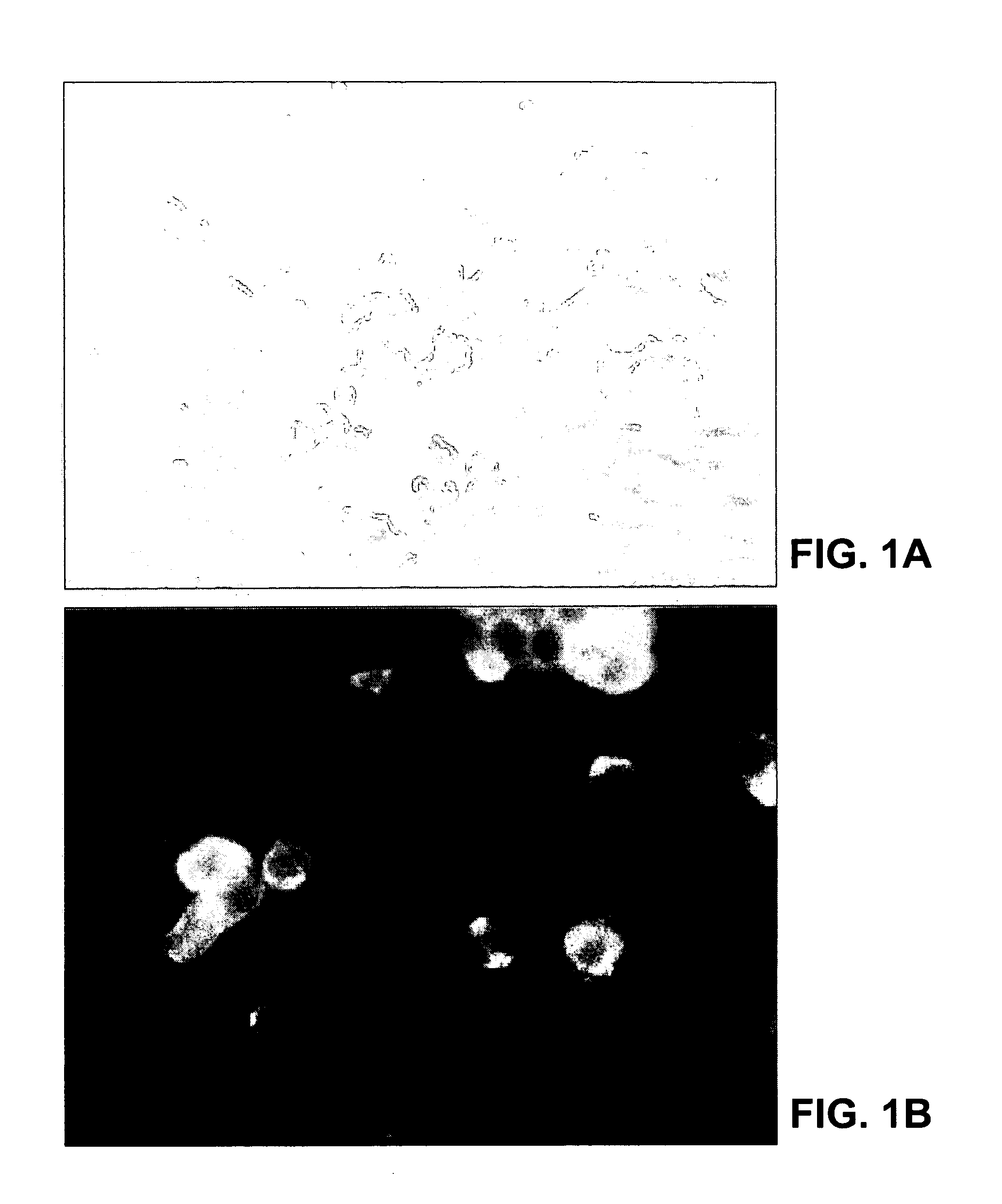
[0165]This example describes the original isolation and characterization of a new human coronavirus from patients with SARS.
[0166]A variety of clinical specimens (blood, serum, material from oropharyngeal swabs or washings, material from nasopharyngeal swabs, and tissues of major organs collected at autopsy) from patients meeting the case definition of SARS were sent to the Centers for Disease Control and Prevention (CDC) as part of the etiologic investigation of SARS. These samples were inoculated onto a number of continuous cell lines, including Vero E6, NCI-H292, MDCK, LLC-MK2, and B95-8 cells, and into suckling ICR mice by the intracranial and intraperitoneal routes. All cultures were observed daily for CPE. Maintenance medium was replenished at day seven, and cultures were terminated fourteen days after inoculation. Any cultures exhibiting identifiable CPE were subjected to several pro...
example 2
Detection of SARS-CoV in a Subject
[0184]This example demonstrates the detection of SARS-CoV in patient specimens using SARS-CoV-specific primers.
[0185]The SARS-specific primers Cor-p-F2 (SEQ ID NO: 13), Cor-p-F3 (SEQ ID NO: 14) and Cor-p-R1 (SEQ ID NO: 15) were used to test patient specimens for SARS. One primer for each set was 5′-end-labeled with 6-FAM to facilitate GeneScan analysis. One-step amplification reactions were performed with the Access RT-PCR System (Promega, Madison, Wis.) as described by Falsey et al., J. Infect. Dis. 87:785–90, 2003. Positive and negative RT-PCR controls, containing standardized viral RNA extracts, and nuclease-free water were included in each run. Amplified 6-FAM-labeled products were analyzed by capillary electrophoresis on an ABI 3100 Prism Genetic Analyzer with GeneScan software (version 3.1.2; Applied Biosystems; Foster City, Calif.). Specimens were considered positive for SARS-CoV if the amplification products were within one nucleotide of the...
example 3
Immunohistochemical and Histopathological Analysis, and Electron-Microscopical Analysis of Bronchoalveolar Lavage Fluid
[0186]This example illustrates immunohistochemical, histopathological and electron-microscopical analysis of Vero E6 cells infected with the SARS-CoV and tissue samples from SARS patients.
[0187]Formalin-fixed, paraffin-embedded Vero E6 cells infected with the SARS-CoV and tissues obtained from patients with SARS were stained with hematoxylin and eosin and various immunohistochemical stains. Immunohistochemical assays were based on a method described previously for hantavirus (Zaki et al., Amer. J. Pathol. 146:552–79, 1995). Briefly, 4-μm sections were deparaffinized, rehydrated, and digested in Proteinase K for 15 minutes. Slides were then incubated for 60 minutes at room temperature with monoclonal antibodies, polyclonal antiserum or ascitic fluids derived from animal species with reactivities to various known coronaviruses, and with a convalescent-phase serum spec...
PUM
| Property | Measurement | Unit |
|---|---|---|
| diameter | aaaaa | aaaaa |
| diameter | aaaaa | aaaaa |
| concentration | aaaaa | aaaaa |
Abstract
Description
Claims
Application Information