

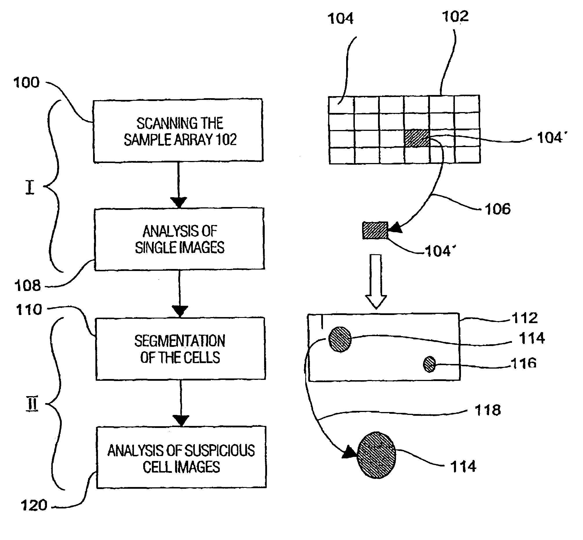
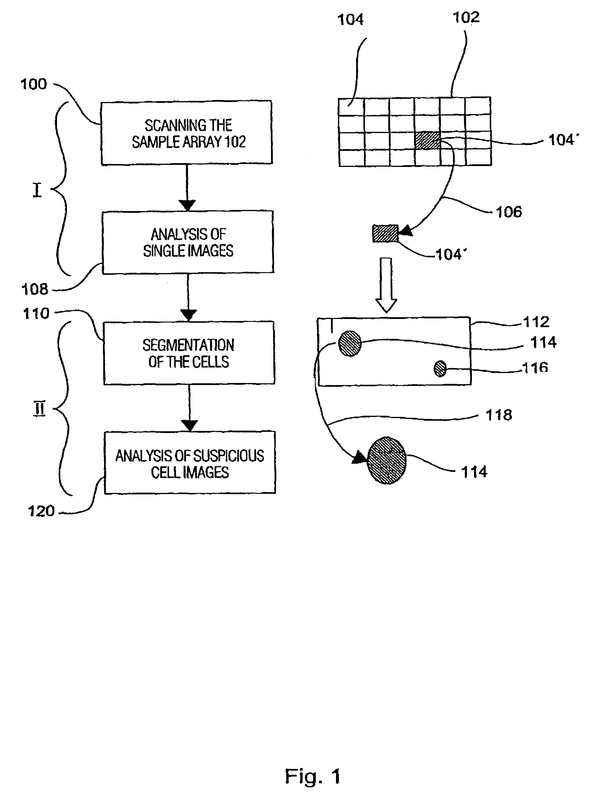
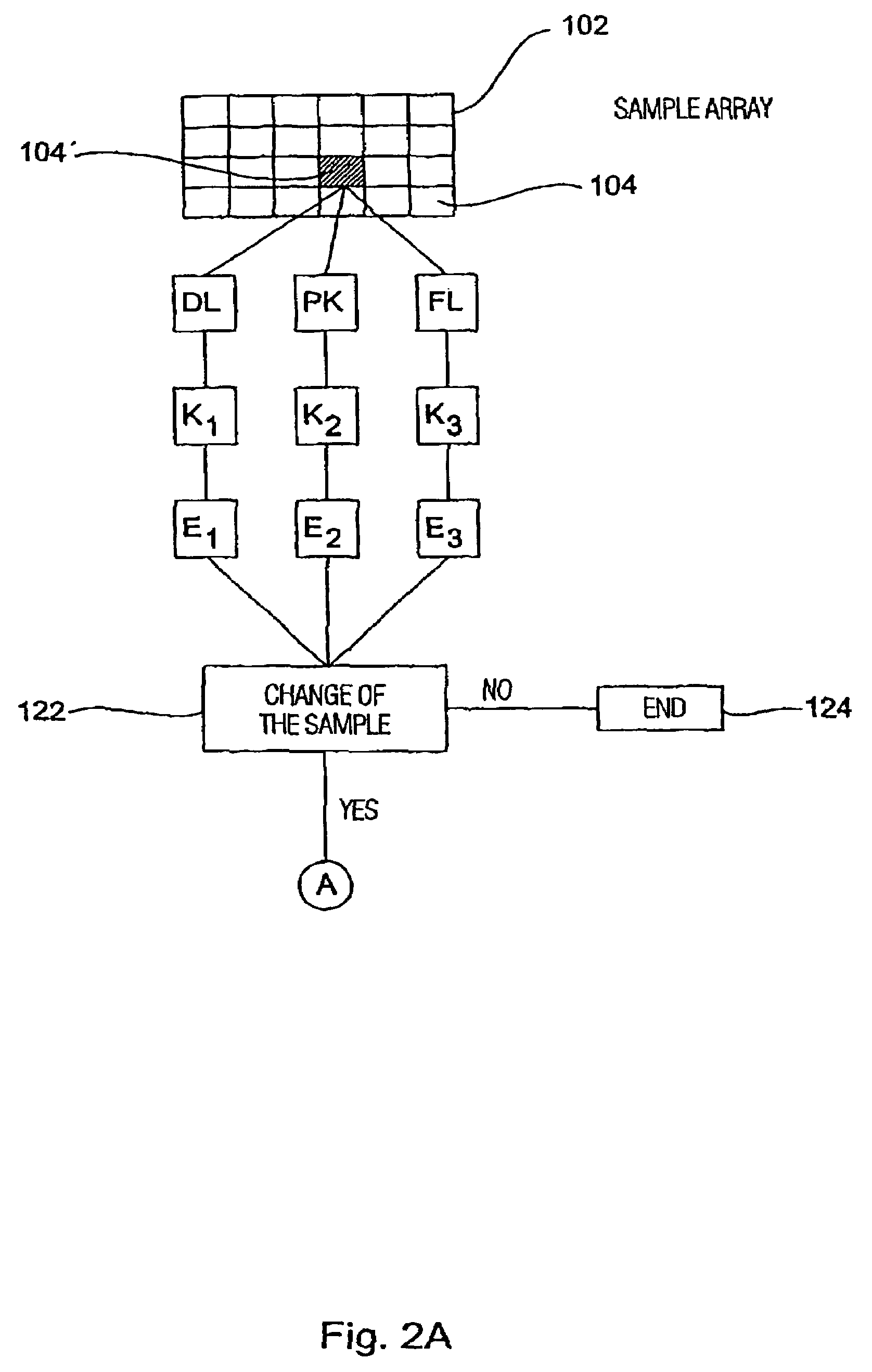
Method for analyzing a biological sample
a biological sample and analysis method technology, applied in the field of biological sample analysis, can solve the problems of inability to achieve a decisive breakthrough in treatment success, inability to secure the progression assessment of tissue changes with the diagnostic methods currently applied, and inability to achieve the required classification security, etc., to achieve high-quality reproducible preparation
- Summary
- Abstract
- Description
- Claims
- Application Information
AI Technical Summary
Benefits of technology
Problems solved by technology
Method used
Image

Examples
Embodiment Construction
[0030]Before preferred embodiments of the present invention are subsequently set forth in greater detail, general aspects of the invention are again explained in the following.
[0031]One possibility for the recognition of tumor cells is to detect their oxidative DNA damage. The tumor cells always have higher oxidative DNA damage than cells of the corresponding normal tissue. This can be seen from a series of publications on the content of the mutagenic base damage 8-oxaguanine and other oxidative DNA change in tumor and normal tissues of the gastro-intestinal tract and of the lung, prostate, brain, ovaries, mamma, etc., such as in Olinski et al. (1992) FEBS Lett. 309, 193-198; Jaruga et al. (1994) FEBS Lett. 341, 59-64; Musarrat et al. (1996) Eur. J. Cancer 32A, 1209-1214; Malins et al. (1996) Proc. Natl. Acad Sci. USA 93, 2557-2563; Matsui et al (2000) Cancer Lett. 151, 87-95. Even in leukemia cells increased 8-oxoguanine levels have been found (Senturker et al. (1997) FEBS Lett., 4...
PUM
| Property | Measurement | Unit |
|---|---|---|
| fluorescence | aaaaa | aaaaa |
| phase contrast | aaaaa | aaaaa |
| shape | aaaaa | aaaaa |
Abstract
Description
Claims
Application Information