
LC/MS method of analyzing high molecular weight proteins

a high molecular weight protein and ms technology, applied in chemical/physical processes, peptides, particle separator tube details, etc., can solve the problems of ineffective application of mass spectrometry for the analysis of higher molecular weight proteins, limited application of high performance liquid chromatography (hplc) readily adaptable for the separation and analysis of low molecular weight species, and insufficiently achieving the applicability of mass spectrometry for the analysis of higher molecular

Inactive Publication Date: 2008-02-12
AMGEN INC
View PDF29 Cites 34 Cited by
  • Summary
  • Abstract
  • Description
  • Claims
  • Application Information

AI Technical Summary

Benefits of technology

This approach enables clear identification of protein conformation and post-translational modifications, improving the resolution and accuracy of protein analysis for proteins above 90 kDa, including antibodies, by reducing peak broadening and enhancing the detection of molecular weight and structural integrity.

Problems solved by technology

Purification techniques such as high performance liquid chromatography (HPLC) that are readily adaptable for the separation and analysis of low molecular weight species often have limited application when being used to separate protein species larger than 50 kDa.
Nevertheless, there remains a significant problem in RP chromatography of large proteins in that a single protein generally produces broad, asymmetrical and even multiple chromatographic peaks at near-room temperatures.
While mass spectrometry has been used for the analysis of small protein fragments of less than 90 kDa, the applicability of mass spectrometry for the analysis of higher molecular weight proteins has not been effectively achieved.
However, these techniques remain inadequate for analysis of antibodies and other large conformationally complex proteins.
This is due to an insufficient resolution resulting from the confounding effects of the large numbers of very similar peptides that result from variable protein glycosylation and enzymatic digests of moderately sized glycoproteins.
Nonetheless, the authors acknowledged that more work needs to be done to improve the power of this approach, and that high-yield concentration steps will be required due to extensive carbohydrate heterogeneity.
Thus, useful information from the LC-ESI-MS or MALDI-TOF techniques can only be obtained from analysis of intact proteins that are relatively small, e.g., less than 10 kDa.
Although the peptide mapping can be used to obtain the more detailed information, this method consumes a lot of time for sample preparation and data interpretation and becomes very complicated when molecular weight of protein increases.
However, many biologically-relevant proteins have higher molecular weights and further, have post-translational modifications that can confound their analyses in mass spectrometry and / or HPLC.

Method used


Image

  • LC/MS method of analyzing high molecular weight proteins
  • LC/MS method of analyzing high molecular weight proteins
  • LC/MS method of analyzing high molecular weight proteins

Examples


example 1

Analysis of Glycosylation Profiles

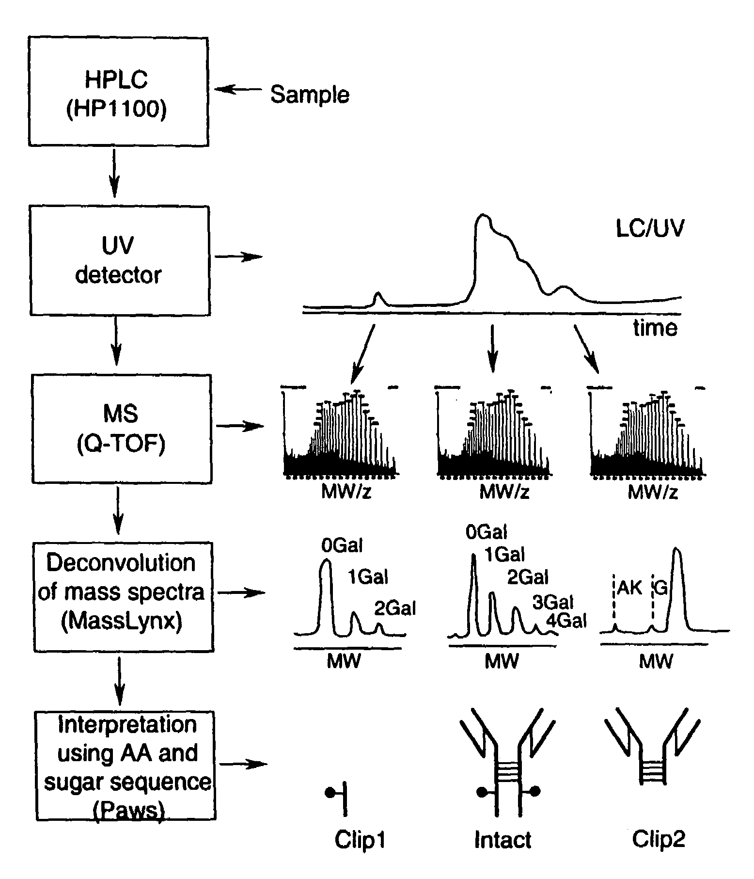
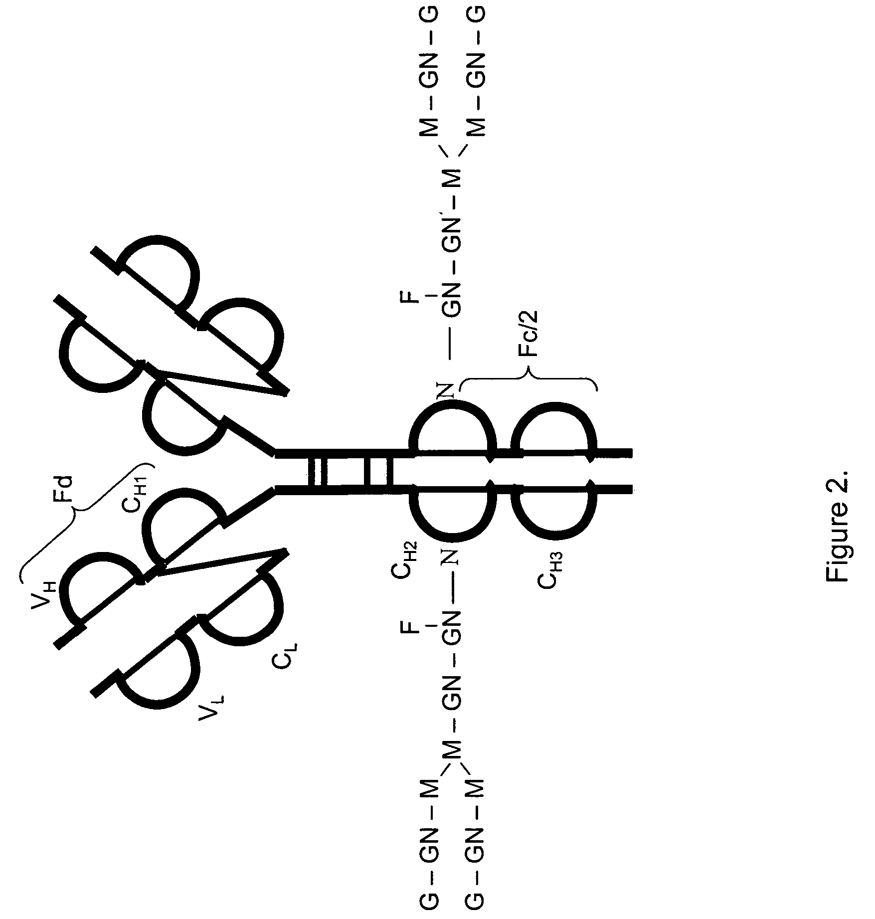
[0117]FIG. 2 shows a structure of an IgG2 antibody with attached oligosaccharides. Table 3 presents a range of oligosaccharide structures possible for most antibodies, their molecular weight values and abbreviaton codes. FIG. 3 shows a UV chromatogram of the IgG2 antibody and the deconvoluted electrospray ionization mass spectrum of this antibody with the glycosylation profile. The mass spectrometric profile in FIG. 3B indicates that the most abundant glycosylation form of IgG2 is missing all galactose residues. FIG. 4 is a structure of IgG1 antibody. FIG. 5 shows UV chromatogram of the IgG1 and the deconvoluted electrospray ionization mass spectrum of this antibody with its glycosylation profile. The glycosylation profile of this antibody is more complicated than for IgG2 antibody, because the IgG1 has an additional N-glycosylation in the VH domain. These additional glycans also have biantennary structure bit with terminal sialic acid residues. In ...

example 2

Identification of Cleavage Sites

[0120]Polypeptide bond cleavage is one of the common degradation pathways in proteins, which may take place during manufacturing and delivery of pharmaceuticals such antibodies. It can be caused by metal ions or the presence of residual proteases after purification. Identification of the clipping sites remains an important task because it allows detection and removal of the cause of degradation. FIG. 6 shows the UV chromatogram of IgG2 antibody, which may be contaminated with a protease, after 8 weeks at 37° C. versus a control sample. Peaks 1 and 9 represent cleavage products of the IgG2. The mass spectrum for the main component of IgG2 eluting as peaks 5-7 is shown in FIG. 3. The main component (peaks 5-7) of both the degraded and control samples has the same MW value within the error margin. The masses for peak 1 and 9 cleavage products were identified from the mass spectra presented in FIGS. 7 and 8. Identified clipping sites are between residues ...

example 3

Identification of Dimers

[0122]Formation of dimers, aggregates, and associated fragments is another pathway of protein degradation especially in formulation solutions with high protein concentrations. A degraded sample of a second IgG1 antibody was separated by the reversed phase HPLC to separate a main peak and a dimer. The measured MW of the monomer was 149525 Da, as compared to theoretical calculated value of 149522 (+20 ppm). The measured MW of the dimer was 299090 Da, as compared to a theoretical calculated value of 299050 Da (130 ppm). See FIGS. 10 and 11 for more details. This example shows the utility of the method for quick identification of protein aggregates.


PUM

PropertyMeasurementUnit
temperatureaaaaaaaaaa
molecular weightaaaaaaaaaa

Abstract

The present invention is directed to a novel method of analysis of high molecular weight proteins. More specifically, it is directed to a novel reversed-phase LC / MS method of analysis of high molecular weight proteins including antibodies.

Description

[0001]The present application claims the benefit of priority under 35 U.S.C. §119 of U.S. Provisional Patent Application No. 60 / 538,982 filed Jan. 23, 2004 and U.S. Provisional Patent Application No. 60 / 548,302 filed Feb. 27, 2004. The entire text of each of the foregoing applications is incorporated herein by reference.BACKGROUND[0002]1. Field of the Invention[0003]The present invention is generally directed to LC / MS analysis of high molecular weight proteins, including antibodies and antibody conjugates or fragments.[0004]2. Background of the Related Art[0005]Purification techniques such as high performance liquid chromatography (HPLC) that are readily adaptable for the separation and analysis of low molecular weight species often have limited application when being used to separate protein species larger than 50 kDa. In addition, the characterization and / or consistency of manufacture of a given protein often relies on peptide mapping in order to monitor the amino acid sequence an...

Claims


Application Information

Patent Timeline
Patent Type & Authority Patents(United States)
IPC IPC(8): B01D15/08C07K1/02C07K1/04C07K1/20C07K16/06C12N15/10G01N30/02G01N30/30G01N30/34G01N30/46G01N30/54G01N30/72G01N30/88G01N33/558H01J49/04
CPCC07K1/023C07K1/047G01N30/30G01N30/34G01N30/7266G01N30/02G01N30/54G01N2030/3007G01N2030/3076G01N2030/8813B01D15/325
Inventor DILLON, THOMASBONDARENKO, PAVELPIPES, GARYRICCI, MARGARETREHDER, DOUGLASKLEEMANN, GERD
Owner AMGEN INC
Learn More