Magnetic resonance imaging agents for the detection of physiological agents
- Summary
- Abstract
- Description
- Claims
- Application Information
AI Technical Summary
Benefits of technology
Problems solved by technology
Method used
Image
Examples
example 1
Synthesis and Characterization of Galactose-DOTA Derivative
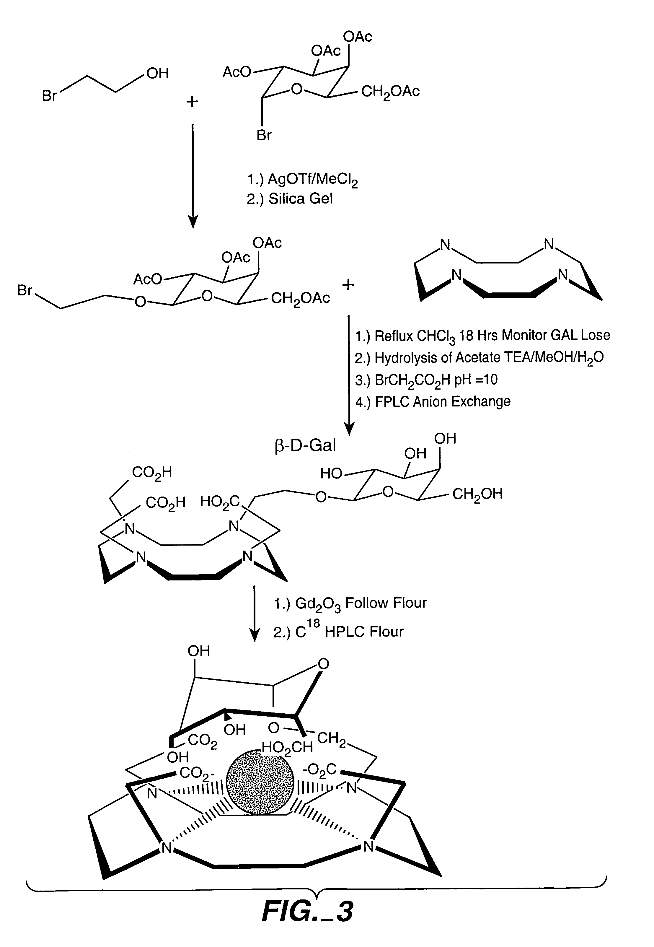
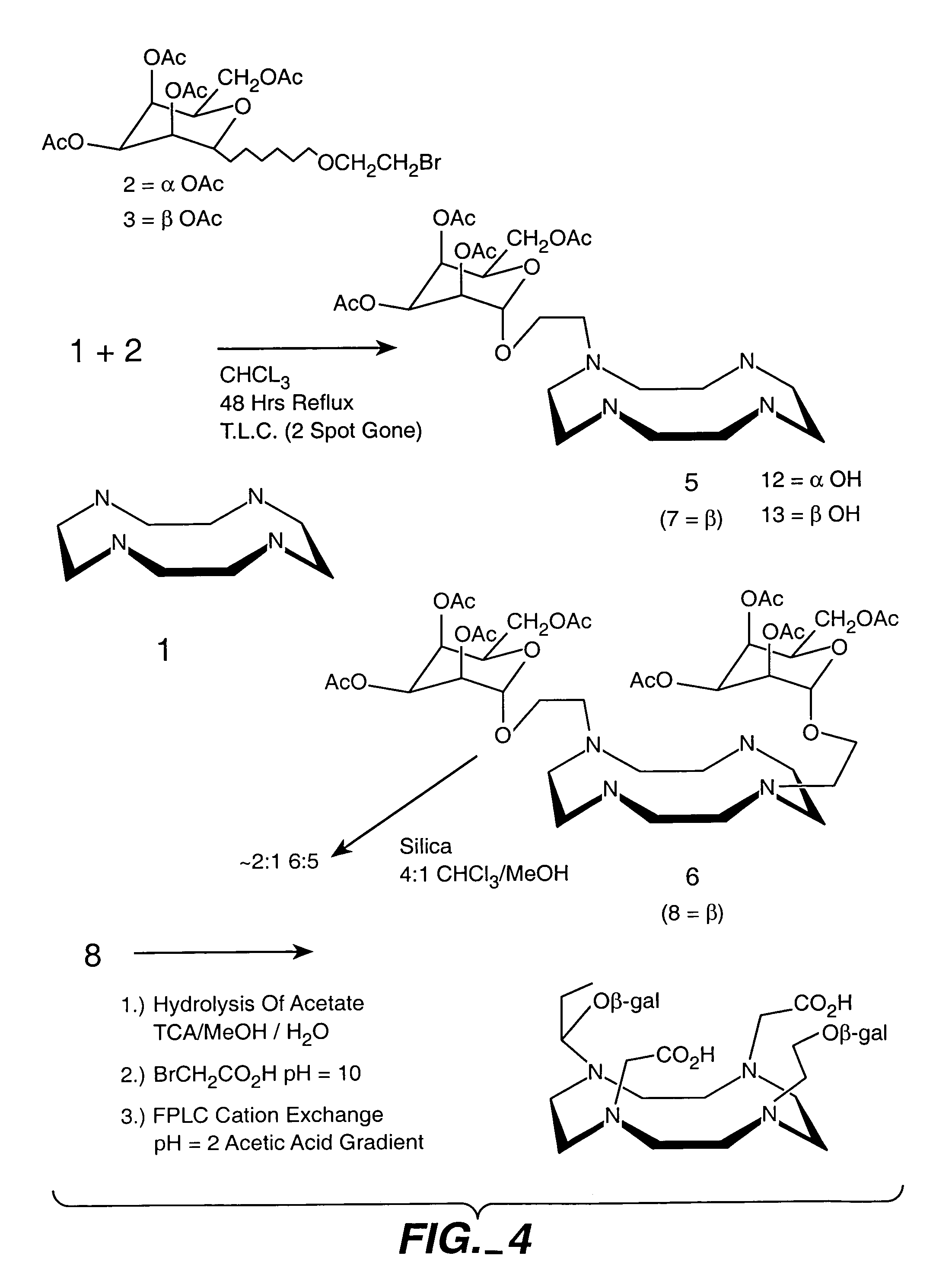
[0201]Synthesis of Do3a-hydroxyethyl-beta-galactose Gadolinium complex (FIG. 4). Acetyl protected bromo-galactose (Aldrich) was reacted with bromoethanol. Difference ratios of the alpha- and beta-bromoethyl ether of the acetylgalactose were obtained in good yield. The isomers were separated using silica gel chromatography and their assignments were made by hydrolyzing the acetyl protecting groups and comparing the proton NMR coupling constants to known compounds. Recently an x-ray structure was done confirming these assignments (data not shown).
[0202]The beta-isomer was reacted with cyclen at reflux in chloroform with monitoring of the reaction by TLC. Hydrolysis of the acetates was achieved with TEA / MCOH / H2O overnight, and the solvent was removed under low vacuum. The resulting product was reacted directly with bromoacetic acid and then maintained at pH 10-10.5 until the pH remained constant. The possible products all would...
example 2
Synthesis of BAPTA-DTPA and BAPTA-DOTA Derivatives
[0210]Two representative synthetic schemes are shown for the synthesis of a BAPTA-DTPA derivative in FIGS. 7 and 8. In FIG. 7 (the preferred method), structure I was prepared by modification of published procedures (Tsien et al., supra) and coupled to hexamethylenediamine using NaCNBrH3 in dry methanol. The ratio of reactants used was 6:1:0.6 (diamine:BAPTA aldehyde:NaCNBrH3). The reaction was quenched with the addition of concentrated HCl and the product purified by HPLC (II). This material was reacted with the mono (or bis) anhydride of DTPA with the protecting groups left on the BAPTA until after the Gd(III)Cl3 or Gd2O3 was added (elevated pH, heat). The final product was purified by ion-exchange HPLC.
[0211]In FIG. 8, the monoanhydride of DTPA was prepared and reacted with a bisalkylamine (e.g. NH2(CH2)6(NH2). This material was purified by ion-exchange HPLC and placed in a round bottom flask equipped with argon inlet and pressure ...
example 3
The Use of R Groups to Increase Signal
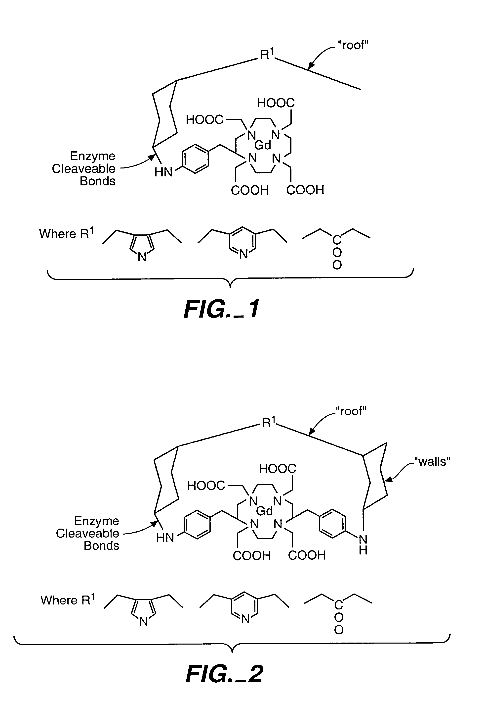
[0212]The Example 1 compound exhibits an enhancement of roughly 20% upon exposure to the target analyte, in this case β-galactosidase. In order to increase the MR contrast enhancement, our intention was to further decrease the access of bulk water to the Gd(III) site by stabilizing the position of the galactopyranose unit on top of the macrocyclic framework. Several studies dealing with intramolecular dynamic processes in tetraazacarboxyclic macrocycles were recently reported (see Kang et al., Inorg. Chem. 36:2912 (1993); Aime et al., Inorg. Chem. 36:2095 (1997); Pittet et al., J. Am. Chem. Soc. Dalton Trans. 1997, 895-900; Spirlet et al., J. Am. Chem. Soc. Dalton Trans. 1997, 497-500, all of which are incorporated by reference. This work demonstrated that introducing α-methyl groups to the ethylenic groups of carboxylic arms increases the rigidity of the amino-carboxylate macrocyclic framework. We therefore added sterically bulky α-methyl group...
PUM
 Abstract
Description
Claims
Application Information
 - R&D
- Intellectual Property
- Life Sciences
- Materials
- Tech Scout
- Unparalleled Data Quality
- Higher Quality Content
- 60% Fewer Hallucinations
Browse by: Latest US Patents, China's latest patents, Technical Efficacy Thesaurus, Application Domain, Technology Topic, Popular Technical Reports.
© 2025 PatSnap. All rights reserved.Legal|Privacy policy|Modern Slavery Act Transparency Statement|Sitemap|About US| Contact US: help@patsnap.com


