
Isolation of neural stem cells using gangliosides and other surface markers

Inactive Publication Date: 2008-09-02
CHILDRENS HOSPITAL OF ORANGE COUNTY +1
View PDF5 Cites 17 Cited by
  • Summary
  • Abstract
  • Description
  • Claims
  • Application Information

AI Technical Summary

Benefits of technology

[0020]A further embodiment is a method for controlling excessive proliferation of neural transplants, by: administering an agent which reduces the proliferation of neural transplant cells selected from the group consisting of: antibodies to neural stem cell markers, antisense oligonucleotides for neural stem cell markers, and antagonists of neural stem cell markers. In one embodiment, the neural stem cell markers are selected from the group consisting of: GD2, TAPA-1,

Problems solved by technology

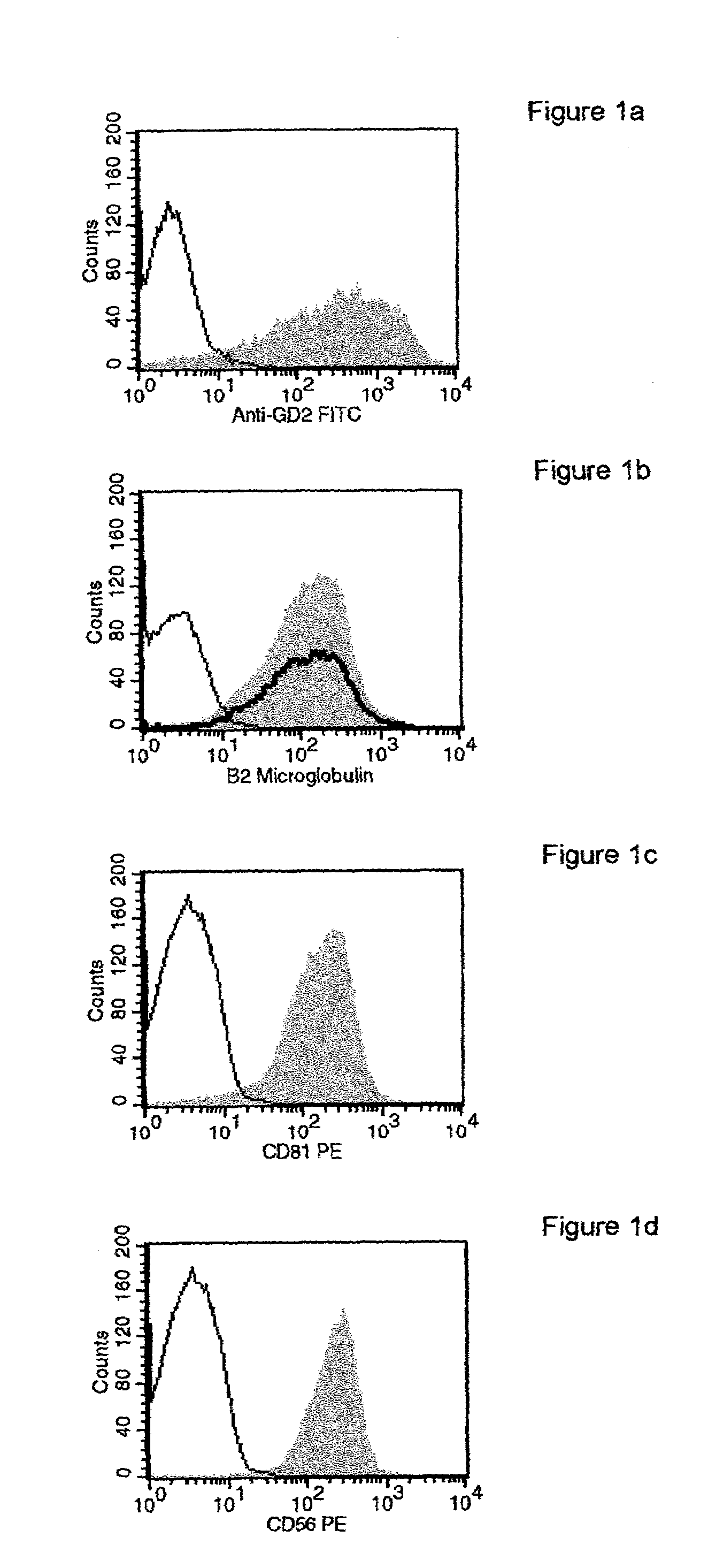
Previous methods for the isolation of neural stem cells (NSCs) using the cell marker CD133 have proved problematic, therefore, a method was developed which allows neural stem cells, human or otherwise, to be enriched without reference to CD133.
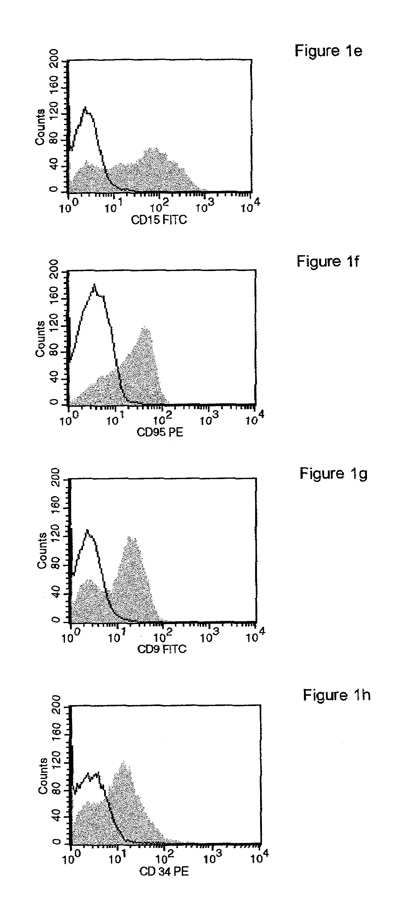
Furthermore, in contrast to previous studies (Uchida, et al, 2000) it was shown that these neural progenitors are CD34+ and CD133−, suggesting that the previous method of identifying NSCs was flawed.

Method used


Image

  • Isolation of neural stem cells using gangliosides and other surface markers
  • Isolation of neural stem cells using gangliosides and other surface markers
  • Isolation of neural stem cells using gangliosides and other surface markers

Examples


example 1

Identification of Positive and Negative Neural Stem Cell Markers in Human Neural Stem Cells

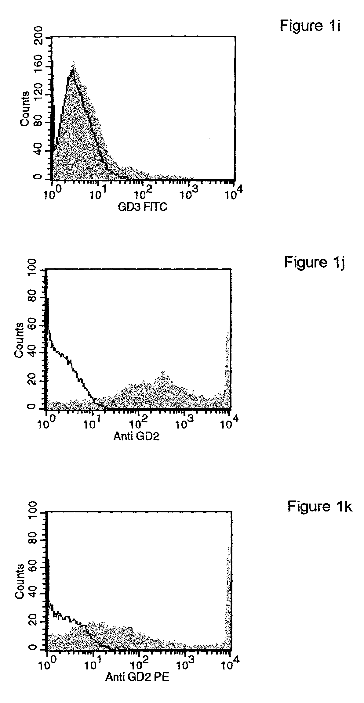
[0067]Initially, a human neuronal progenitor cell line was obtained from Michael Young, Ph.D. who obtained it from Clonetics, (Walkersville, Md., catalog number CC-2599). These cells had been obtained from donated prenatal tissue and tested negative for a range of infectious pathogens. Cells were obtained frozen and subsequently grown in tissue culture using a defined medium consisting of DMEM / F12 high glucose, N2 Supplement (Life Technologies), bFGF (20-25 ng / ml) and EGF (20-25 ng / ml), and L-glutamine. Cells were characterized as multipotent neural progenitor cells based on the ability to propagate over many passages, expression of nestin and Ki-67, proto-neuronal morphology, as well as the ability to differentiate into neurons and glia (Mizumoto, et al., 2001; Young, et al., unpublished data).

[0068]For the present study, cells were initially grown as neurospheres until plentiful, then sphere...

example 2

Identification of Neural Retinal Cell Markers

[0077]Stem cells from the neural retina of GFP-transgenic mice were found to express the markers previously shown for brain-derived stem cells. In FIG. 2 the expression of GD2 ganglioside, CD15, and the tetraspanins CD9 and CD81, are shown using flow cytometry.

example 3

Identification of Neural Cell Markers Associated with Differentiation

[0078]The human neuronal progenitor cells (hNSCs) in Example 1 were treated with Fetal calf serum (FCS) to induce differentiation and some of the markers were reexamined. FIG. 3 depicts the influence of these differentiating conditions on the expression of target molecules by hNSCs. In each case the target molecule is shown as solid gray, the isotype control with a fine black outline. The bold outline indicates the profile of the target molecule after hNSCs were cultured in fetal bovine serum (FBS). FIG. 3a shows that CD34 expression increased under these conditions, FIG. 3b shows that CD15 expression fell to control levels, FIG. 3c shows that GD2 ganglioside expression decreased by an order of magnitude, FIG. 3d shows that CD9 expression fell to a lesser degree.

[0079]The expression of certain NSC-specific markers decreased during differentiation, suggesting that these markers are strong markers of “true” neural st...


PUM

PropertyMeasurementUnit
Lengthaaaaaaaaaa
Affinityaaaaaaaaaa

Abstract

During the growth and study of NSCs, a range of molecules present on the surface of multipotent neural stem and progenitor cells (NSCs) were identified. These markers were identified using a number of human and murine neural stem cell lines, including retinal stem cells (RSCs). The NSC-specific markers identified included gene products as well as non-protein molecules and sugar epitopes not directly coded in the genome. Together with surface markers which were determined to be absent from the surface of hNSCs, the molecules described herein provide a means to enrich for neural stem cells, or neural progenitor subpopulations, particularly using combinatorial cell sorting strategies. These same molecules also represent targets for pharmacological manipulation of NSC populations and subpopulations, both in vivo and ex vivo. Furthermore, these molecules provide potential targets for therapeutic manipulation of other neural precursor-related cell types including malignant cell types as well as diseases originating from, or preferentially affecting, various uncommitted or replication-competent cell types.

Description

RELATED APPLICATION[0001]This application claims priority under 35 U.S.C. § 119(e) to U.S. Provisional Application 60 / 285,407, filed Apr. 20, 2001.FIELD OF THE INVENTION[0002]The present invention relates generally to the isolation of neural stem cells. More specifically, the present invention relates to a method of using gangliosides and other markers to isolate neural stem cells from the central nervous system, including brain, spinal cord, and retina.BACKGROUND OF THE INVENTION[0003]Cell transplantation has over the last two decades emerged as a promising approach for restoration of function in neurodegenerative diseases, in particular Parkinson's and Huntington's disease. Clinical trials have so far focused on the use of implants of embryonic mesencephalic tissue containing already fate-committed dopaminergic neuroblasts with the capacity to develop into fully mature dopamine neurons in their new location in the host brain. A major limitation of the fetal cell transplantation pr...

Claims


Application Information

Patent Timeline
IPC IPC(8): C12N5/00C12N5/06C12N5/08A61K35/12C07K16/28C12N5/02C12N5/077C12N5/0797G01N33/574
CPCC07K16/28G01N33/57484G01N33/57407C12N5/0623A61K35/12C12N2503/00
Inventor KLASSEN, HENRYSCHWARTZ, MICHAELYOUNG, MICHAEL J.
Owner CHILDRENS HOSPITAL OF ORANGE COUNTY
Learn More