Controlled high efficiency lesion formation using high intensity ultrasound

a high-efficiency, controlled technology, applied in the field of endoscopic, can solve the problems of significant trauma to patients, excessive bleeding and pain, and significant cost, and achieve the effect of efficient treatment of uterine fibroids

Inactive Publication Date: 2008-12-30
OTSUKA MEDICAL DEVICES
View PDF24 Cites 95 Cited by
  • Summary
  • Abstract
  • Description
  • Claims
  • Application Information

AI Technical Summary

Benefits of technology

[0026]The present invention is directed to a method and apparatus for efficiently treating uterine fibroids and other diseases with high intensity u

Problems solved by technology

Fibroids may cause excessive bleeding and pain.
There are more than 80,000 abdominal myomectomies each year in the U.S. These surgical procedures cause significant trauma to the patients and result in significant costs.
Consequently, patients need several days of hospital stay and suffer from the prolonged recovery.
The mechanical damage to the fibroid and the uterus can cause bleeding during the treatment and adhesions after the treatment.
Suturing the damage in the uterus is very difficult in the laparoscopic MIS procedure.
Also, most of these alternative treatments are time consuming and technically challenging.
The fibroids then starve and die due to lack of a blood supply.
The embolization procedure may cause severe pain in the first few days after the treatment.
Other disadvantages of UAE may include long X-ray radiation exposure during the procedure and other long-term potential adverse effects.
One disadvantage of the

Method used


Image

  • Controlled high efficiency lesion formation using high intensity ultrasound
  • Controlled high efficiency lesion formation using high intensity ultrasound
  • Controlled high efficiency lesion formation using high intensity ultrasound

Examples


Embodiment Construction

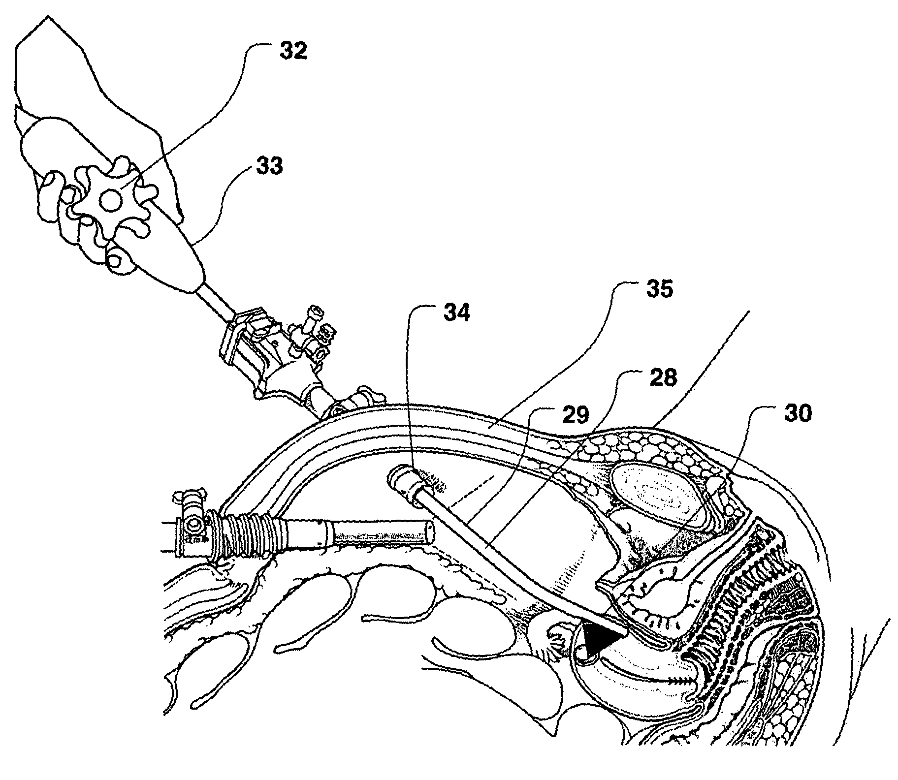
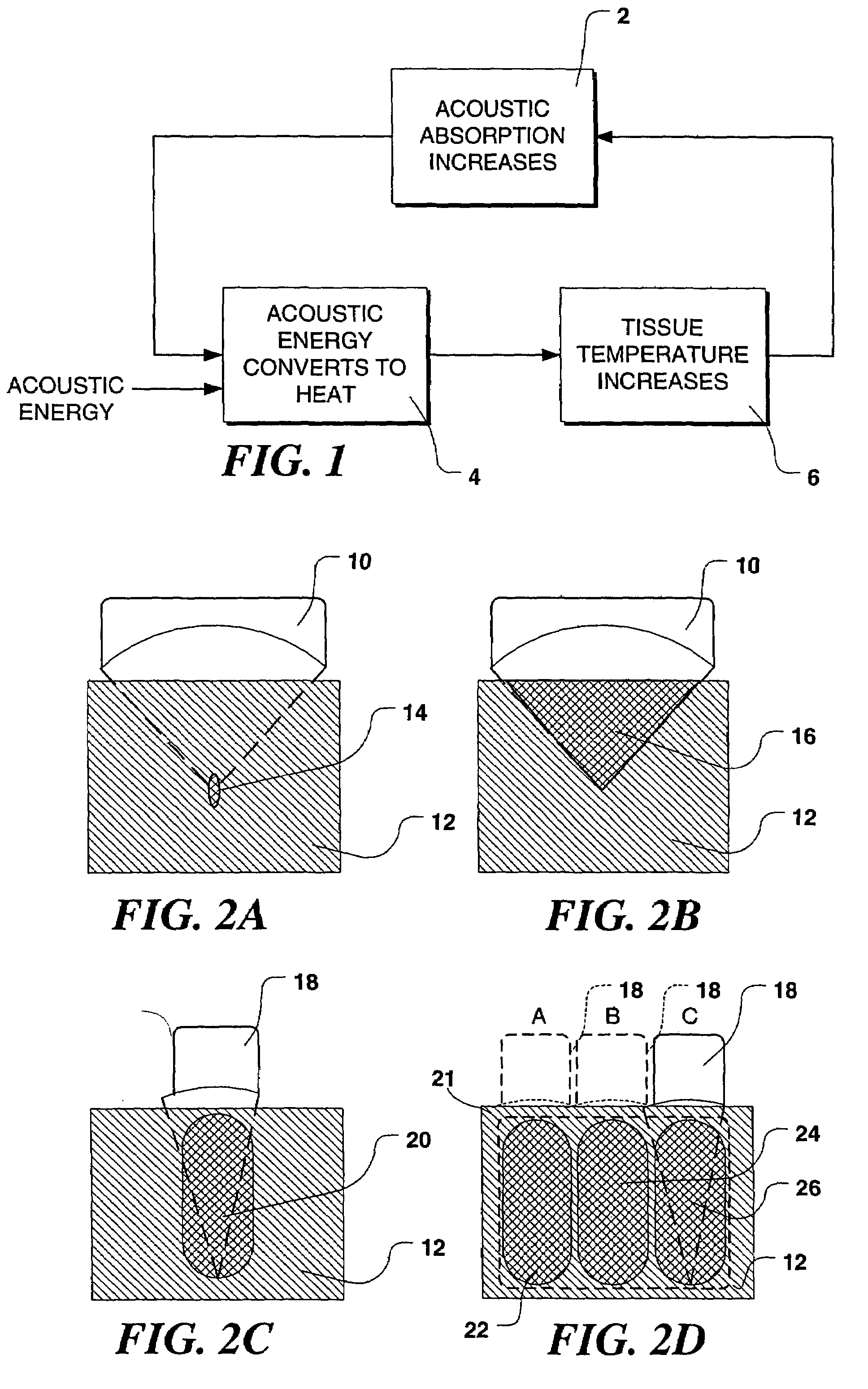
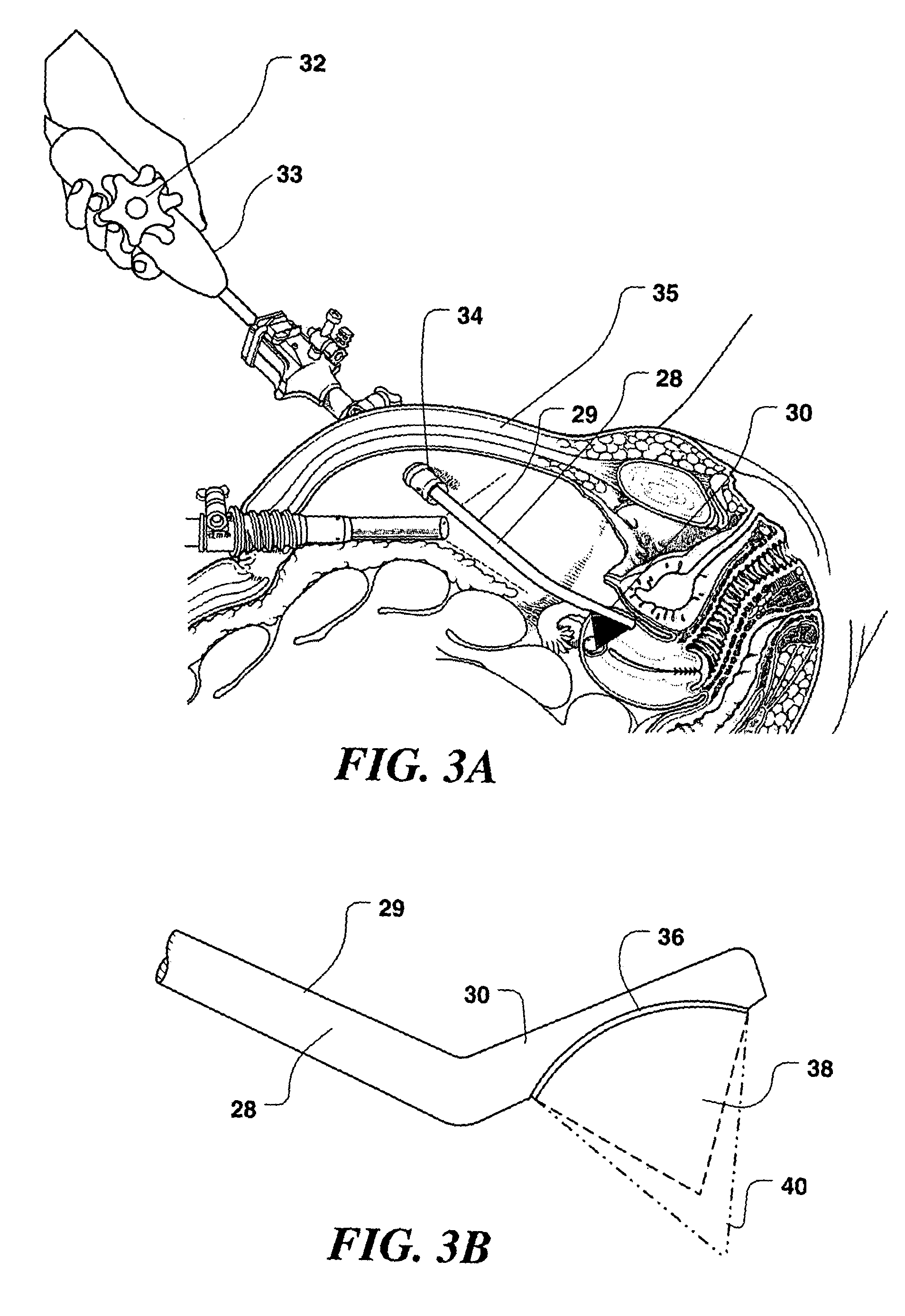
[0047]In the following description of the present invention, its application in treating uterine fibroid tumors is discuss in some detail. However, it should be emphasized that the device and methods described herein may also be used to apply ultrasound therapy treatment to other organ systems, lesions, and disease states. The therapy delivered may be thermal ablation, where a temperature rise is established to a level at which tissues are no longer viable; mechanical ablation, where cavitation is employed as the primary ablative means; or may achieve hemostasis wherein bleeding or blood flow in intact organs is arrested. Such applications of the present invention may be accomplished in open, invasive surgery, by way of established minimally invasive techniques (for example, by way of body entry through one or more small incisions or punctures), or in some cases, noninvasively, through the skin surface or through the linings of body cavities such as the rectum, vagina, or esophagus....


PUM


Abstract

An ultrasound system used for both imaging and delivery high intensity ultrasound energy therapy to treatment sites and a method for treating tumors and other undesired tissue within a patient's body with an ultrasound device. The ultrasound device has an ultrasound transducer array disposed on a distal end of an elongate, relatively thin shaft. In one form of the invention, the transducer array is disposed within a liquid-filled elastomeric material that more effectively couples ultrasound energy into the tumor, that is directly contacted with the device. Using the device in a continuous wave mode, a necrotic zone of tissue having a desired size and shape (e.g., a necrotic volume selected to interrupt a blood supply to a tumor) can be created by controlling at least one of the f-number, duration, intensity, and direction of the ultrasound energy administered. This method speeds the therapy and avoids continuously pausing to enable intervening normal tissue to cool.

Description

FIELD OF THE INVENTION[0001]This invention relates generally to methods and apparatus for efficiently heating biological tissues with high intensity ultrasound for therapeutic purposes, and in particular, to endoscopic devices for applying ultrasound energy to uterine fibroids and other pathologic tissues that are inside body or organ cavities, to destroy the tumor or the diseased tissue.BACKGROUND OF THE INVENTION[0002]Fibroids are benign tumors in women's uteri. There are different types of fibroids, including submucosal, which are inside the uterine cavity; intramural, which are in the uterine wall; and subserosal, which are outside the uterus. Fibroids may cause excessive bleeding and pain. For symptomatic fibroids, surgery is the predominate treatment. Every year in the U.S., there are more than 200,000 cases of fibroid-caused hysterectomies. To preserve the uterus, the patient may choose myomectomy, which removes the fibroids only. There are more than 80,000 abdominal myomecto...

Claims


Application Information

Patent Timeline
IPC IPC(8): A61H1/00A61H5/00A61B17/42A61B19/00A61N7/02
CPCA61B8/12A61B8/4281A61B8/546A61B8/445A61N7/02A61N7/00A61B2017/4216A61N7/022A61N2007/0052A61B2019/5276A61B2090/378
Inventor WENG, LEEPEROZEK, DAVID M.ZHANG, JIMIN
Owner OTSUKA MEDICAL DEVICES
Why Eureka
Try Eureka