Peyer's patch and/or M-cell targeting ligands
a technology of m-cell and m-cell, applied in the field of targeting ligands, can solve the problem that no human m-cell marker has been identified as a target for the delivery of vaccines and/or other drugs through the m-cell
- Summary
- Abstract
- Description
- Claims
- Application Information
AI Technical Summary
Benefits of technology
Problems solved by technology
Method used
Image
Examples
example 1
[0134]The Screening Process: Biopanning In Vivo
[0135]A Phage Display Peptide Library (12-mer at 1.5×10¹¹ pfu) was inoculated intraduodenally into a rat loop model (n=5) Blood samples were taken from the rat loop at time periods of 0, 30, 60, 90 and 120 minutes. The animals were then sacrificed and the loops excised. From these loops, Peyer's patch and non-Peyer's patch tissues were isolated, washed and homogenized. The bacteriophage present in the tissue samples were amplified in E. Coli and isolated by polyethylene glycol (PEG) precipitation. Peyer's patch specific phage were titred and selected for use in subsequent screening cycles.
[0136]Four screening cycles were completed and phage titres (in pfu / ml) were obtained at cycle 4.
[0137]
TABLE 4Phage titres (pfu / ml) in crude tissuehomogenates (rat loop model; cycle 4)Rat No.Peyer's Patch11.0 × 10625.6 × 10534.0 × 10342.6 × 10654.6 × 104
example 2
[0138]Phage Binding Studies
[0139]The phage pools obtained in Example 1 were plated out on LB agar plates with top agar and phage clones were selected for evaluation by an ELISA analysis of binding to Peyer's patch tissue from various species, along with Caco-2 and IEC-6 cell models.
[0140]The ELISA was run with 5 μg / ml of phage homogenates, Blocking buffer: 1% BSA-TBS, wash buffer: TBS-Tween (0.05%), anti-M13 biotin conjugate (Research Diagnostics RDI-PRO61597) at a 1:5000 dilution, ExtrAvidin Alkaline Phosphatase (Sigma E-2636) at a 1:5000 dilution and pNPP substrate.
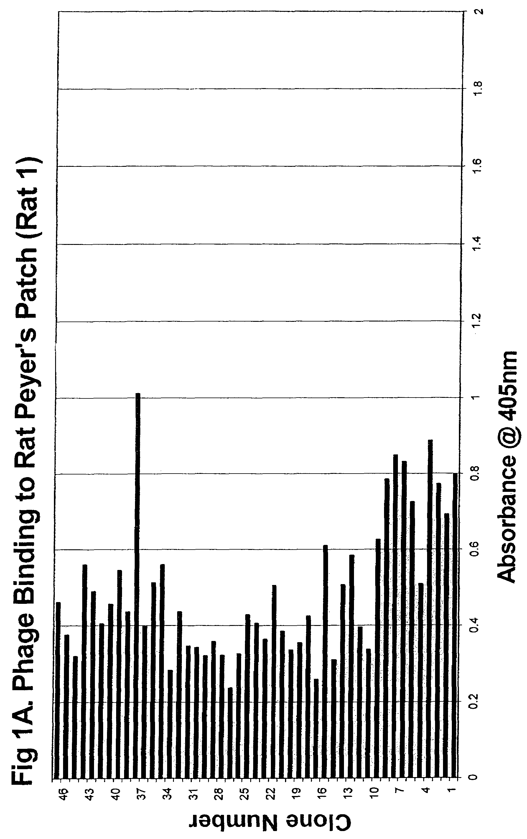
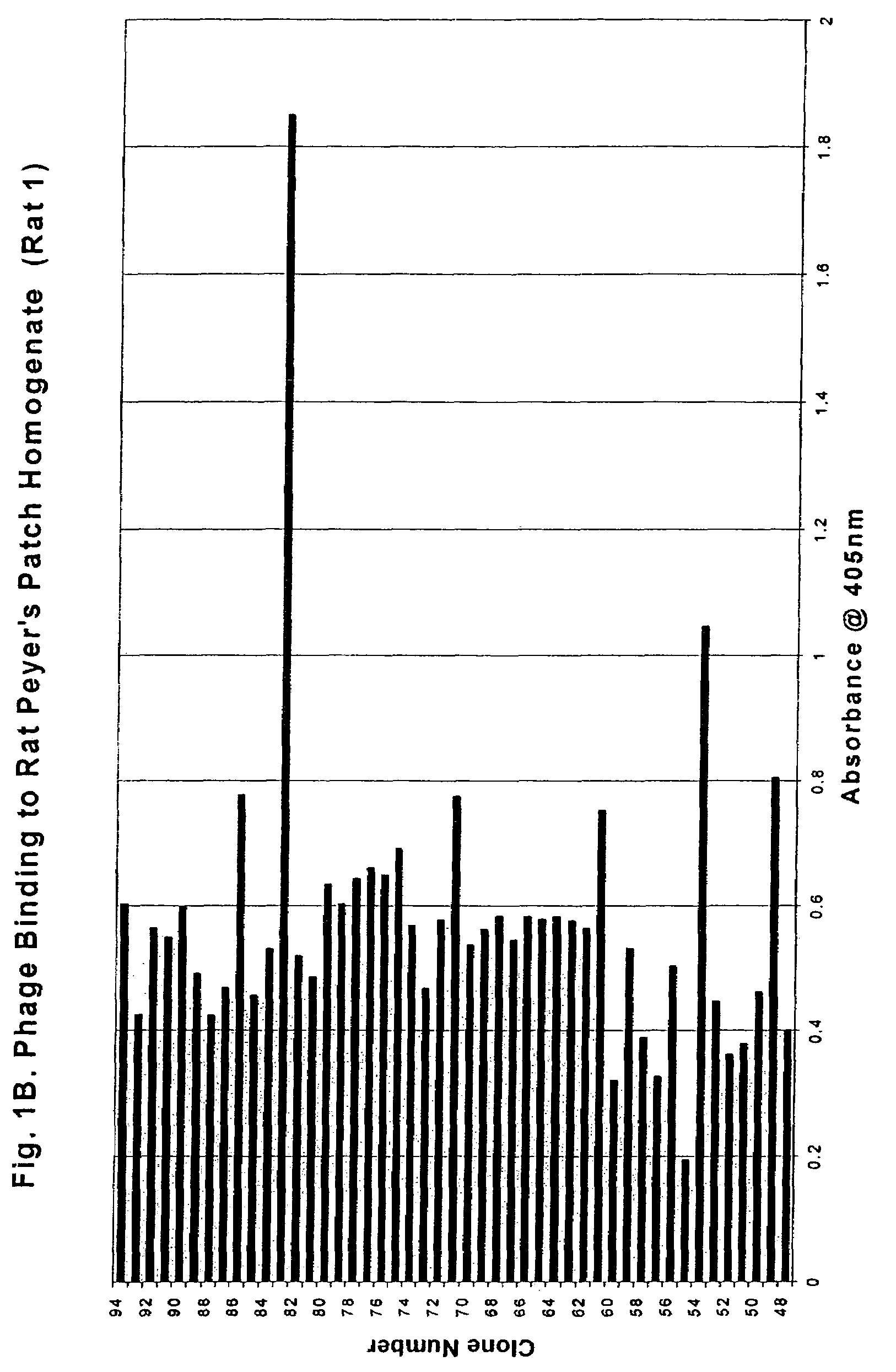
[0141]Five hundred clones (Rats 1-5 from Cycle 4) were subsequently assayed using the above method. (See FIGS. 1A-5 for binding profiles). The clones exhibited a broad range of activity with concentration-dependent binding clearly detectable for all high-binding clones.
[0142]
TABLE 5Table of phage clone numbers & SEQ ID NOs.No. of copies ofSEQ ID ofeachcorrespondingSEQ ID NO:Amino Acid Sequenceclone isolatedClone No.DNA ...
example 3
[0143]1. Specificity Determination: Analysis of Phage Binding to Rat Small Intestine and / or Peyer's Patch
[0144]A Biotin-ExtrAvidin Alkaline Phosphatase assay was established for high throughput screening of the phage clones. The initial screens identified 55 out of the 500 clones as high-binding clones (an absorbency reading of >0.75).
[0145]The rat tissue homogenates were prepared by harvesting rat GI and Peyer's patch and storing them on ice until needed, or 1-2 hours. The tissue was then put into homogenization buffer (250 mM Sucrose, 12 mM Tris, 16 mM EDTA) with protease inhibitor cocktail. A hand-held homogenizer was used to break up the tissue for 3-4 minutes. The contents of the homogenizer were then transferred to microfuge tubes and spun at 1500 rpm for 1 minute. The supernatant was taken off and measured for protein content using the Bio-Rad Assay. Specificity studies were then run to allow differentiation between Peyer's patch specific and non-specific binding properties. ...
PUM
 Abstract
Description
Claims
Application Information
 - R&D
- Intellectual Property
- Life Sciences
- Materials
- Tech Scout
- Unparalleled Data Quality
- Higher Quality Content
- 60% Fewer Hallucinations
Browse by: Latest US Patents, China's latest patents, Technical Efficacy Thesaurus, Application Domain, Technology Topic, Popular Technical Reports.
© 2025 PatSnap. All rights reserved.Legal|Privacy policy|Modern Slavery Act Transparency Statement|Sitemap|About US| Contact US: help@patsnap.com


