
Generation of virus-like particles and use as panfilovirus vaccine

a technology of virus-like particles and vaccines, which is applied in the direction of antibody medical ingredients, peptide sources, peptides, etc., can solve the problems of limited knowledge of the mechanism of pathogenicity, no vaccines or therapeutics available to prevent or treat filovirus infections, and hampered efforts to develop therapeutics against ebola and marburg

Inactive Publication Date: 2010-03-23
UNITED STATES OF AMERICA THE AS REPRESENTED BY THE SEC OF THE ARMY
View PDF5 Cites 16 Cited by
  • Summary
  • Abstract
  • Description
  • Claims
  • Application Information

AI Technical Summary

Benefits of technology

"The present invention is about a method for generating genome-free filovirus virus-like particles (VLPs) that resemble native virus. The VLPs can be used as a research tool and as a vaccine or delivery vehicle for therapeutic or diagnostic agents. The invention also provides a method for inducing an immune response to a filovirus, a vaccine for blocking filovirus entry into cells, and a method for evaluating the effectiveness of agents or chemicals that can block filovirus entry. The invention also describes the use of noninfectious filovirus VLPs in an in vitro assay for testing the efficacy of potential agents to inhibit or reduce filovirus entry into cells or budding from cells."

Problems solved by technology

Although natural outbreaks have been geographically restricted so far, limited knowledge of the mechanisms of pathogenicity, potential of aerosol transmission (Jaax et al., 1995, Lancet 346, no.
8991-8992, 1669), unknown natural reservoir, and lack of immunological and pharmacological therapeutic measures, pose a challenge to classification of the public health threat of Marburg and Ebola viruses.
Currently, there are no vaccines or therapeutics available to prevent or treat filovirus infections.
Unfortunately, questions remain about many of the vaccine strategies used thus far, including acceptable vaccine doses, safety considerations, the impact of prior immunity to the vaccine vector, and the ability of these vaccine strategies to cross-protect against multiple strains of EBOV and MARV (Hart, M. K., 2003, Vaccine research efforts for filoviruses.
Efforts to develop therapeutics against Ebola and Marburg have been hampered, in part, by poor understanding of the process of filovirus entry and budding at the molecular level.

Method used


Image

  • Generation of virus-like particles and use as panfilovirus vaccine
  • Generation of virus-like particles and use as panfilovirus vaccine
  • Generation of virus-like particles and use as panfilovirus vaccine

Examples


example 1

[0100]Association of Filovirus Glycoproteins with Lipid Rafts.

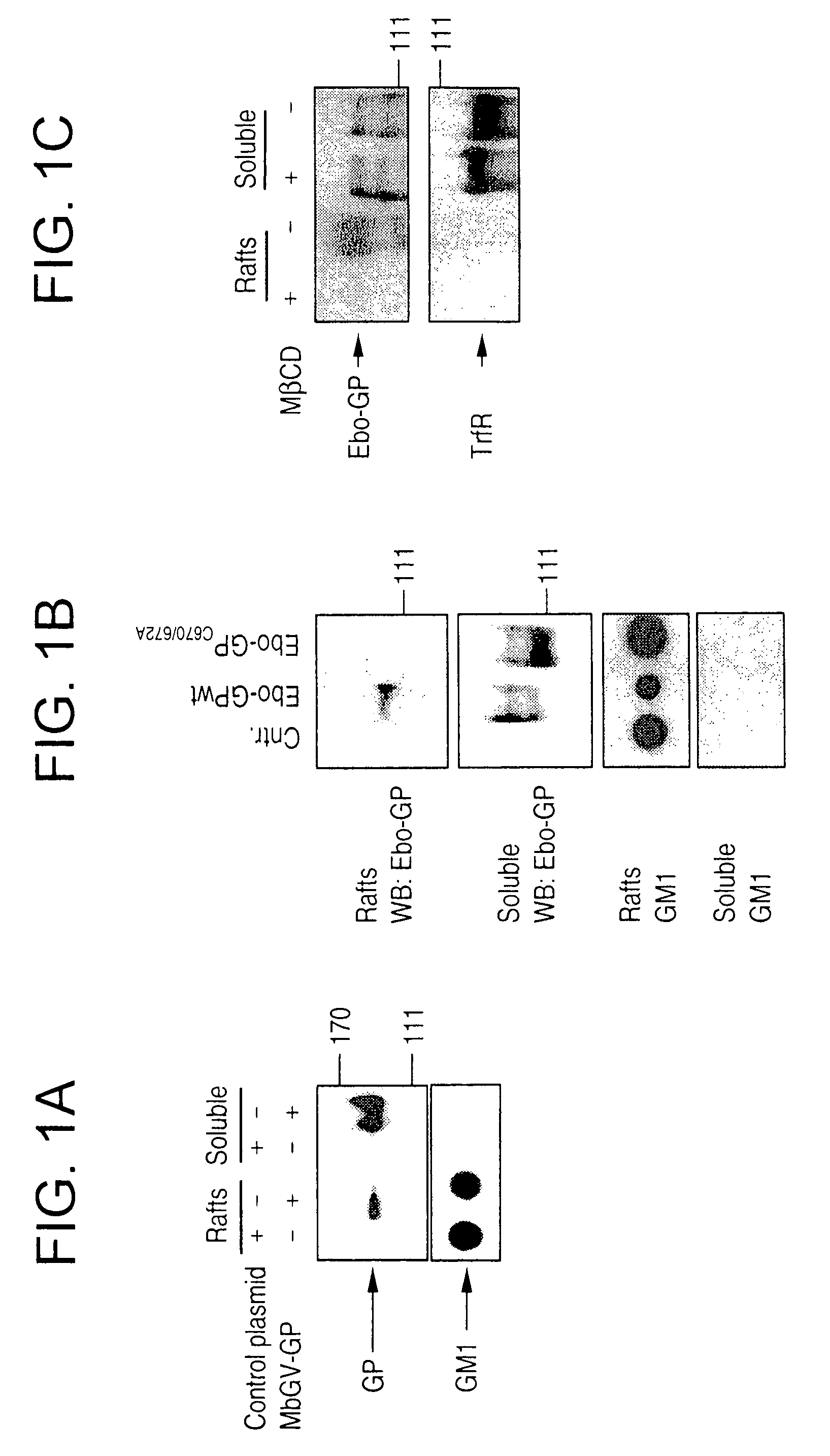
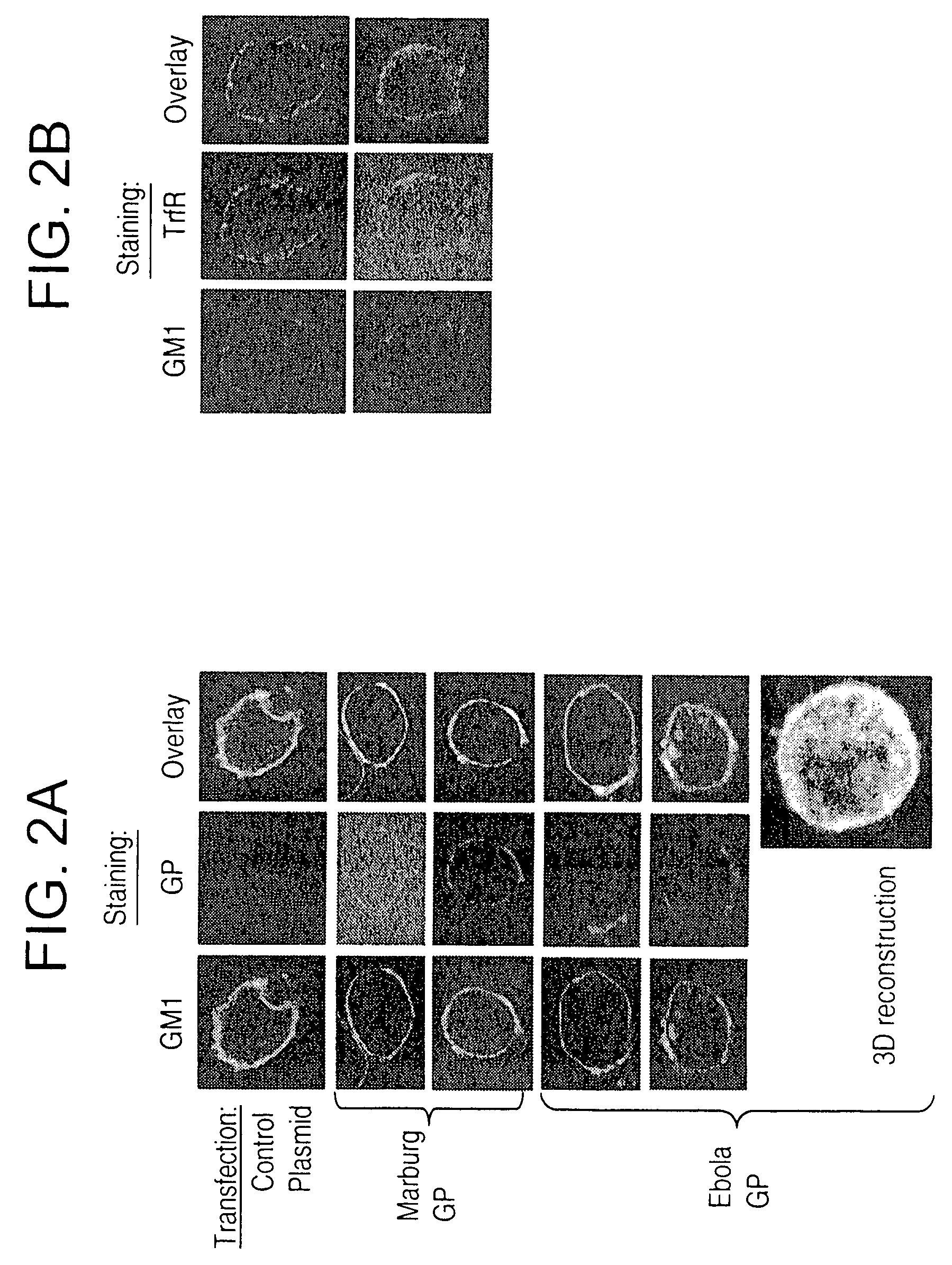
[0101]Targeting of membrane-spanning proteins to lipid rafts is commonly governed by dual acylation of cysteine residues at the cytosolic end of the transmembrane domains (Rousso et al, 2000, supra; Zhang et al., 1998, Immunity 9, 239). The filovirus envelope glycoproteins (GP) contain such acylation signals in their transmembrane domains (Feldmann and Klenk, 1996, supra) and palmitoylation of Ebola GP has been recently reported (Ito et al., 2001, J. virol. 75, 1576). By transient expression of the filovirus envelope glycoproteins in 293T cells and subsequent extraction of rafts by sucrose gradient ultracentrifugation (Aman and Ravichandran, 2000, supra), we examined whether these glycoproteins localize to lipid rafts. As shown in FIG. 1 (A and B), a significant fraction of Ebola and Marburg GPs were found to reside in rafts. In contrast, an Ebola GP, mutated at cysteine residues 670 and 672 (EbO-GPC670 / 672A), the putativ...

example 2

[0102]Filoviral Proteins Associate with Lipid Rafts in Cells Infected with Live Virus.

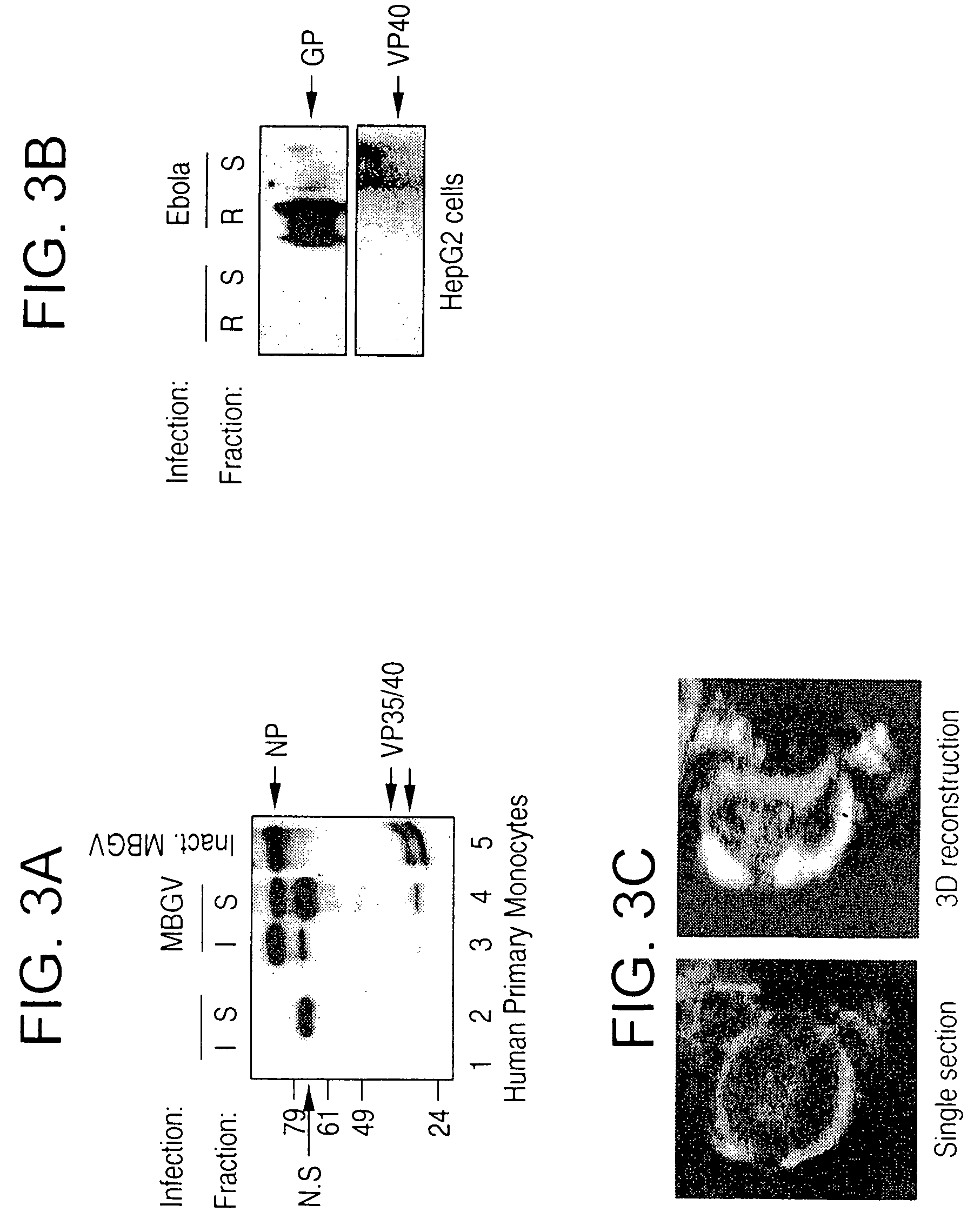
[0103]Two of the primary target cells of filoviruses are monocyte / macrophages and hepatocytes (Feldman and Klenk, 1996, supra). Thus, to examine the localization of EBOV and MBGV proteins with respect to lipid rafts during infection with live virus, primary human monocytes, HepG2 hepatocytes, and also Vero-E6 cells (commonly used to propagate filoviruses) were used as target cells. Human monocytes were infected with the Musoke strain of MBGV, after 24 h detergent-insoluble and detergent-soluble fractions were separated by centrifugation (Rousso et al., 2000, supra). As shown in FIG. 3A, a major fraction of viral proteins was detected in the detergent-insoluble fraction (I) 24 hours after infection. We then performed similar experiments with HepG2 cells, infected with EBOV-Zaire95 and prepared lipid rafts by sucrose gradient ultracentrifugation. Similar to Marburg, Ebola VP40 and GP were detected ma...

example 3

[0104]Ebola and Marburg Virions Incorporate the Raft Molecule GM1 During Budding

[0105]To determine whether the virus was released through lipid rafts, we analyzed EBOV from culture supernatants of infected cells for the presence of the raft marker GM1. Enveloped viruses bud as virions surrounded by the portion of the plasma membrane at which assembly takes place (Simons and Garoff, 1980, J. Gen. Virol. 50, 1). Release of virions through lipid rafts would therefore result in incorporation of raft-associated molecules in the viral envelope, thus identifying virus budding from the rafts. As shown in FIG. 4A, EBOV immunoprecipitated with anti-Ebola GP antibody from supernatant of infected Vero-E6 cells contained readily detectable levels of GM1. We also analyzed inactivated Marburg virus that had been purified by ultracentrifugation for the incorporation of GM1 and demonstrated that GM1 was detectable in MBGV (FIG. 4B, lower panel). In contrast, the raft-excluded protein TrfR was not in...


PUM

PropertyMeasurementUnit
optical densityaaaaaaaaaa
lengthaaaaaaaaaa
diameteraaaaaaaaaa

Abstract

In this application are described filovirus-like particles for both Ebola and Marburg and their use as a diagnostic and therapeutic agent as well as a filovirus vaccine. Also described is the association of Ebola and Marburg with lipid rafts during assembly and budding, and the requirement of functional rafts for entry of filoviruses into cells.

Description

[0001]This application is a continuation-in-part application of U.S. application Ser. No. 10 / 289,839 filed on Nov. 7, 2002 now abandoned, which claims the benefit of priority under 35 U.S.C. 119(e) from U.S. Application Ser. No. 60 / 338,936 filed on Nov. 7, 2001, now expired. This application also claims benefit of priority under 35 U.S.C. 119(e) from U.S. Application Ser. No. 60 / 562,800 and 60 / 562,801 filed on Apr. 13, 2004, all of which are herein incorporated by reference in their entirety.INTRODUCTION[0002]The filoviruses Ebola (EBOV) and Marburg (MBGV) are two of the most pathogenic viruses in humans and non-human primates (Feldman and Klenk, 1996, Adv. Virus Res. 47, 1), which cause a severe hemorrhagic fever (Johnson et al., 1997, Lancet 1, no. 8011, P. 569). The mortality rates associated with infections of Ebola or Marburg virus are up to 90% (Feldman and Klenk, 1996, supra; Johnson et al., 1997, supra). Although natural outbreaks have been geographically restricted so far, ...

Claims


Application Information

Patent Timeline
Patent Type & Authority Patents(United States)
IPC IPC(8): A61K39/12C12N5/10C12N7/00C12Q1/70A61K39/295C07K14/08C12N7/04
CPCA61K39/12C07K14/005C12N7/00A61K2039/5258C12N2760/14122C12N2810/60C12N2760/14145C12N2760/14222C12N2760/14223C12N2760/14245C12N2760/14123C12N2760/14134C12N2760/14234
Inventor BAVARI, SINAAMAN, M. JAVADSCHMALJOHN, ALAN L.WARFIELD, KELLY L.SWENSON, DANA
Owner UNITED STATES OF AMERICA THE AS REPRESENTED BY THE SEC OF THE ARMY
Learn More