Three-dimensional fabrication of biocompatible structures in anatomical shapes and dimensions for tissue engineering and organ replacement
a biocompatible structure and three-dimensional fabrication technology, applied in biochemistry equipment, biochemistry equipment and processes, general culture methods, etc., can solve the problems of compromising the health of the organ recipient, and affecting the quality of life of the organ. significant developments in connection with many internal, physical structures, especially those of hollow and epithelial organs, have been limited
- Summary
- Abstract
- Description
- Claims
- Application Information
AI Technical Summary
Benefits of technology
Problems solved by technology
Method used
Image
Examples
example 1
Characterization of Pore Sizes Fabricated by Three-Dimensional Printing
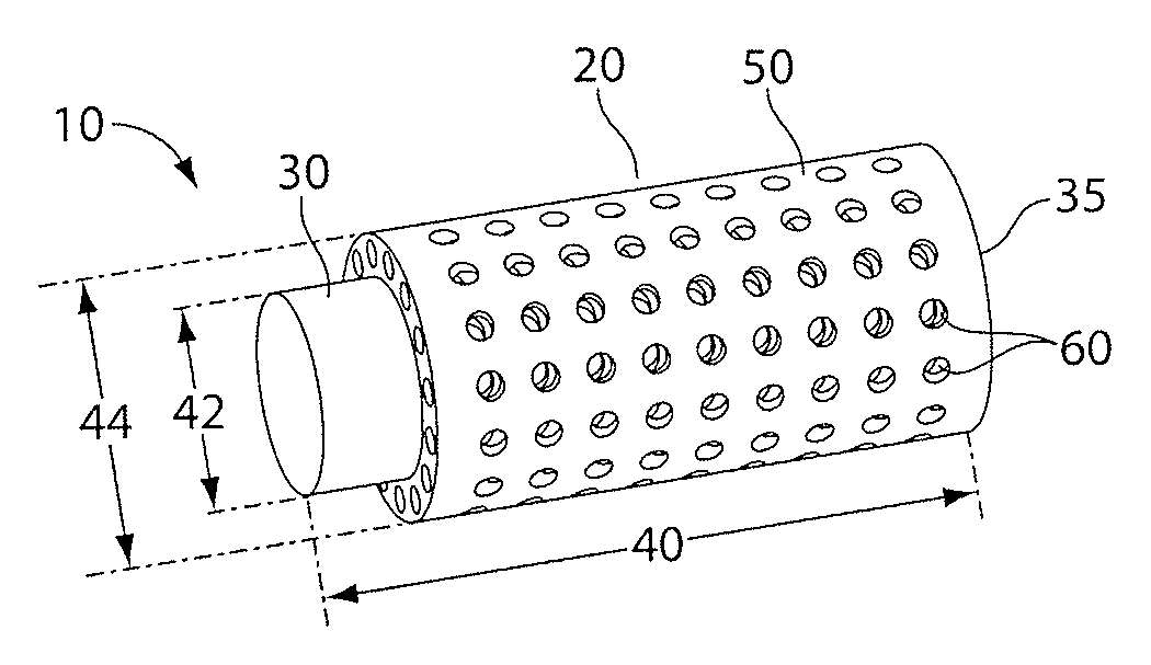
[0116]For the formation of structures that can allow the transport of components, such as soluble factors between portions of the structure, pores can be designed and printed on the structures. To characterize the minimum pore size obtainable with the Eden 260 RPT, a 500 μm thick stencil was designed with pores sizes ranging from 100 μm to 1000 μm, in intervals of 100 μm, and printed (FIG. 3). After the stencil had been printed, the bulk of the support material was peeled. Next, the stencil was rinse with de-ionized water and immersed in 25% tetra methyl ammonium hydroxide (TMAH) solution and the pore sizes were measured over time (FIG. 4). Complete removal of sacrificial material on the super structure was typically achieved within 5 hours of TMAH immersion.
example 2
Characterization of Surface Roughness of Structures Fabricated by Three-Dimensional Printing
[0117]Roughness of surfaces of structures fabricated using the EDEN 260 RPT were measured with an AlphaStep profiler. The surfaces had a RMS roughness of 3 microns and waviness of 40 microns (peak to peak), as shown in FIG. 16. Typical sizes of cells seeded onto surfaces of printed structures ranged from 10-30 μm in diameter.
[0118]To examine the influence of surface roughness on cell culture, endothelial and tubule epithelial cells were seeded onto printed semi-tubular structures. Madin-Darby Canine Kidney cells (MDCK) were obtained from American Type Culture Collection (Rockville, USA). The endothelial cell type (HMEC-1) was provided by the National Center for Infectious Diseases (Atlanta, USA). All cell types were cultured in DMEM (Invitrogen Singapore, Singapore) with 10% fetal calf serum (Invitrogen Singapore, Singapore) and 1% antibiotic-antimycotic solution (Invitrogen Singapore, Singap...
example 3
Reducing Surface Roughness by Coating Surfaces with Materials
[0119]Synthetic materials, cyanoacrylate and a polysulphone / PMMA solution, were used to coat surfaces of structures made by three-dimensional printing to reduce surface roughness. Using printed tubular structures as described above, the tubes were filled with cyanoacrylate and spun at 2500 RPM for 1 min. The tubes were then left to stand for 2 hours to allow the solvent in the cyanacrylate to evaporate. The tubes was sliced open with a scalpel and the internal surface of the pipe was examined with SEM (FIGS. 18A and 18B) and a profilometer scan (FIG. 19). From both the SEM micrograph and the profilometer scan, it was observed that surface roughness as well as the waviness of printed structure was significantly reduced by coating the surface with these materials.
[0120]Experiments to reduce surface roughness were also performed by coating collagen type 1 and fibronectin on the surface of a printed material in the shape of a ...
PUM
| Property | Measurement | Unit |
|---|---|---|
| thickness | aaaaa | aaaaa |
| inner diameter | aaaaa | aaaaa |
| pore size | aaaaa | aaaaa |
Abstract
Description
Claims
Application Information
 - R&D
- Intellectual Property
- Life Sciences
- Materials
- Tech Scout
- Unparalleled Data Quality
- Higher Quality Content
- 60% Fewer Hallucinations
Browse by: Latest US Patents, China's latest patents, Technical Efficacy Thesaurus, Application Domain, Technology Topic, Popular Technical Reports.
© 2025 PatSnap. All rights reserved.Legal|Privacy policy|Modern Slavery Act Transparency Statement|Sitemap|About US| Contact US: help@patsnap.com


