Methods and devices for orthovoltage ocular radiotherapy and treatment planning

a technology of which is applied in the field of methods and devices for orthovoltage ocular radiotherapy and treatment planning, can solve the problems of limited, if any, verification of the orientation of the eye, lack of efficacy in those studies, and substantial irradiation of non-targeted structures, so as to reduce eye motion

Active Publication Date: 2010-09-21
CARL ZEISS MEDITEC INC
View PDF206 Cites 61 Cited by
  • Summary
  • Abstract
  • Description
  • Claims
  • Application Information

AI Technical Summary

Problems solved by technology

In addition, during treatment, there was limited, if any, verification of the orientation of the eye relative to the pre-operative CT scan or verification of maintenance of eye orientation during treatment.
Due the penetrating nature of the MeV radiation and the beam width, among other things, prior art treatment beam 6 results in substantial irradiation of the non-targeted structures.
In addition, these prior art attempt at applying radiation to the macula did not consider eye movements or eye position.
Such fractionation and minimalist dosing and planning schemes likely lead to the lack of efficacy in those studies.
Another significant limitation of the treatments in prior art external beam trials, there was no accounting for eye movement and position during the treatment.
However, the eye movement and the center of rotation vary from patient to patient, and it is difficult to know the exact dose applied to the macula.
In addition, eye location and movement were not controlled in this study.
Such complications likely negated any benefit of the therapy.
Although relatively sophisticated, the system 800 may be limited to the ophthalmic region and therefore takes advantage of specific imaging equipment only available for the eye.
Adjustment of the power on the power supply will result in a decrease in the peak voltage of x-rays, limiting the amount of higher energy photons.
Note that further increases in polar angle may present inconvenience or discomfort with respect to the range of eyelid retraction, or by interference with the beam by tissues adjacent the eye.
It has been found that the average shift values shown lead to surprisingly small errors in the population studied, a maximum error of 0.20 mm in the horizontal direction and 0.08 mm in the vertical direction.
The heat generated by the anode is the major limiting factor in the ultimate flux which can be achieved by the x-ray source.
However, its dose uniformity is significantly reduced within the macula target.
On the other hand, a real or at least conceptual hazard of high degree-of-freedom robotic systems employing energy beam treatment, is the large possible range of beam paths (e.g., upon a control system failure), and associated risk issues, regulatory complexity, and high end-user installation and site modification costs.
However, the da Vinci is not autonomous and requires an expert surgeon to move its arms.
However, the linear accelerator which moves around the patient is over 1 ton in weight and cannot move close enough to the patient to deliver beams of X-ray to the eye.
Furthermore, the system does not include an eye stabilization system to allow for alignment relative to the eye.
In addition, a real or at least conceptual hazard of high degree-of-freedom robotic systems employing energy beam treatment, is the large possible range of beam paths (e.g., upon a control system failure), and associated risk issues, regulatory complexity, and high end-user installation and site modification costs.
Because each treatment dose is relatively short and applied over a small distance, the robot can sacrifice speed and travel distance for smaller size.
In one preferred embodiment, negative pressure applied to the eye, for example, a negative pressure of 20-50 mm Hg, is effective to stabilize the position of the eye on the device, that is, substantially prevent movement of the eye with respect to the device, but by itself is not sufficient to hold the eye-contact device on the eye.
Gating may be triggered by error or failure conditions such as loss of eye tracking by the system, loss of limbus tracking, or other system-based failure deemed justification for interruption of radiation treatment, e.g., due to electronic conditions, camera conditions, lighting conditions, inadvertent blocking or interference with imaging, and the like.
However, the eye motion does result in a relative motion of the beam spot on the retina due to the angled alignment of the beam with the retina.
Vascular damaging agents (VDAs) cause a rapid shutdown of established vasculature, leading to secondary tissue death.

Method used


Image

  • Methods and devices for orthovoltage ocular radiotherapy and treatment planning
  • Methods and devices for orthovoltage ocular radiotherapy and treatment planning
  • Methods and devices for orthovoltage ocular radiotherapy and treatment planning

Examples


embodiment 118

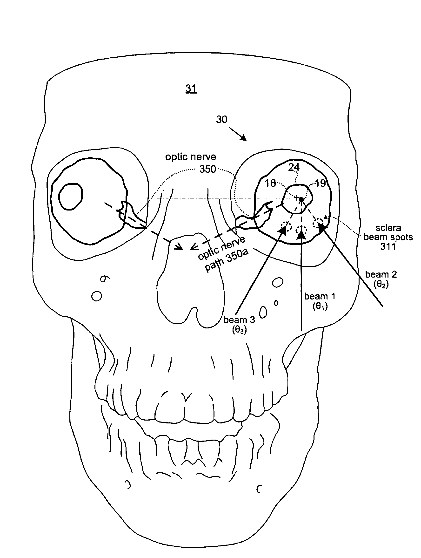
[0258]For example, a collimator embodiment 118 comprises aperture 1405 which provides crescent-shaped beam exit pattern configured to minimize dosage to the optic nerve adjacent a nearby retinal treatment target. The beam pattern created by aperture 1405 includes a maximal dose-intensity region which is shaped to match a retinal target lesion, and a corresponding minimal dose-intensity region of the pattern is shaped to align with the optic disk, thereby sparing that structure. The aperture 1405 may be rotated as mounted in the outer portion 118b, so as to align a minimal-intensity region of the pattern with the optic disk. Rotation of portion 118b may thus compensate for overall rotation of the collimator during repositioning of the X-ray source for successive stereotactic treatments.

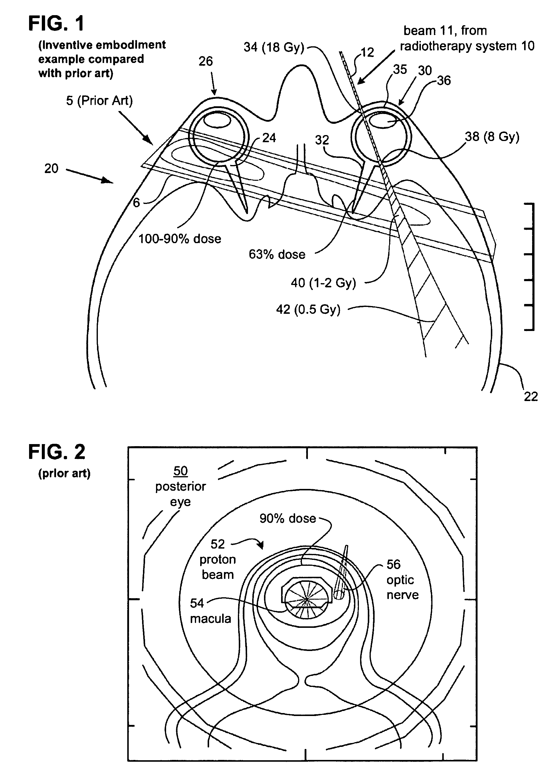
[0259]FIG. 29A is a plot showing the results of a Monte Carlo computational simulation for absorption of X-ray energy in a configuration generally similar to that shown in FIG. 21. See description abov...

exemplary embodiment 625

[0345]FIGS. 40A-B illustrate perspective views of an exemplary embodiment 625 having aspects of the invention of a contact device or eye-guide and eye alignment and stabilizing module configured for use with system 10 (it additionally may be usefully employed independent of system 10). This may be used together with head-chin restraint device 160, which includes a head support or support 170 for stabilizing the head of subject, and includes a chin rest 172.

[0346]FIGS. 40A-B and 41A-B depict one example embodiment of an method of aligning and / or stabilizing a patients eye 30 and engaged eye-guide 110 with the coordinates of radiotherapy system 10, using a laser beacon 150 a mechanism by which the contact device 110 can be used to align the eye with laser alignment system 800, including laser device 150 (alternative image-based alignment subsystems are described herein). Optionally, the alignment mechanism also directly aligns a treatment system, such as a radiotherapy system (not sho...


PUM


Abstract

A method, code and system for planning the treatment a lesion on or adjacent to the retina of an eye of a patient are disclosed. There is first established at least two beam paths along which x-radiation is to be directed at the retinal lesion. Based on the known spectral and intensity characteristics of the beam, a total treatment time for irradiation along each beam paths is determined. From the coordinates of the optic nerve in the aligned eye position, there is determined the extent and duration of eye movement away from the aligned patient-eye position in a direction that moves the patient's optic nerve toward the irradiation beam that will be allowed during treatment, while still maintaining the radiation dose at the patient optic nerve below a predetermined dose level.

Description

RELATED APPLICATIONS[0001]This application claims the benefit of priority of U.S. Application Nos. 61 / 101,013 filed Sep. 29, 2008, 61 / 093,092 filed Aug. 29, 2008, and 61 / 076,128 filed Jun. 26, 2008. This application is also a continuation-in-part of U.S. application Ser. No. 12 / 103,534 filed Apr. 15, 2008, now pending, which claims benefit of priority to U.S. Application Nos. 61 / 016,472 filed Dec. 23, 2007, and 61 / 020,655 filed Jan. 11, 2008. U.S. application Ser. No. 12 / 103,534 is also a continuation-in-part of U.S. application Ser. No. 12 / 100,398 filed Apr. 9, 2008, now pending. U.S. application Ser. No. 12 / 103,534 is also a continuation-in-part of U.S. application Ser. No. 12 / 027,083 filed Feb. 6, 2008, now pending. U.S. application Ser. No. 12 / 103,534 is also a continuation-in-part of U.S. application Ser. No. 12 / 027,094 filed Feb. 6, 2008, now pending. U.S. application Ser. No. 12 / 103,534 is also a continuation-in-part of U.S. application Ser. No. 12 / 027,069 filed Feb, 6, 2008,...

Claims


Application Information

Patent Timeline
Patent Type & Authority Patents(United States)
IPC IPC(8): A61N5/10
CPCA61N5/1017A61N5/103A61N5/1037A61N5/1064A61N2005/1091
Inventor GERTNER, MICHAELARNOLDUSSEN, MARKCHELL, ERIKHANSEN, STEVEN D.LIANG, JUNZHONG
Owner CARL ZEISS MEDITEC INC
Why Eureka
Try Eureka