

Method for in vitro production of three-dimensional vital cartilage tissue and use thereof as transplant material
a technology of vital cartilage and in vitro production, which is applied in the direction of skeletal/connective tissue cells, biocide, plant growth regulators, etc., can solve the problems of immune reaction or infection with animal or human pathogens
- Summary
- Abstract
- Description
- Claims
- Application Information
AI Technical Summary
Benefits of technology
Problems solved by technology
Method used
Image

Examples
example 1
In Vitro Production of Cartilage Tissue
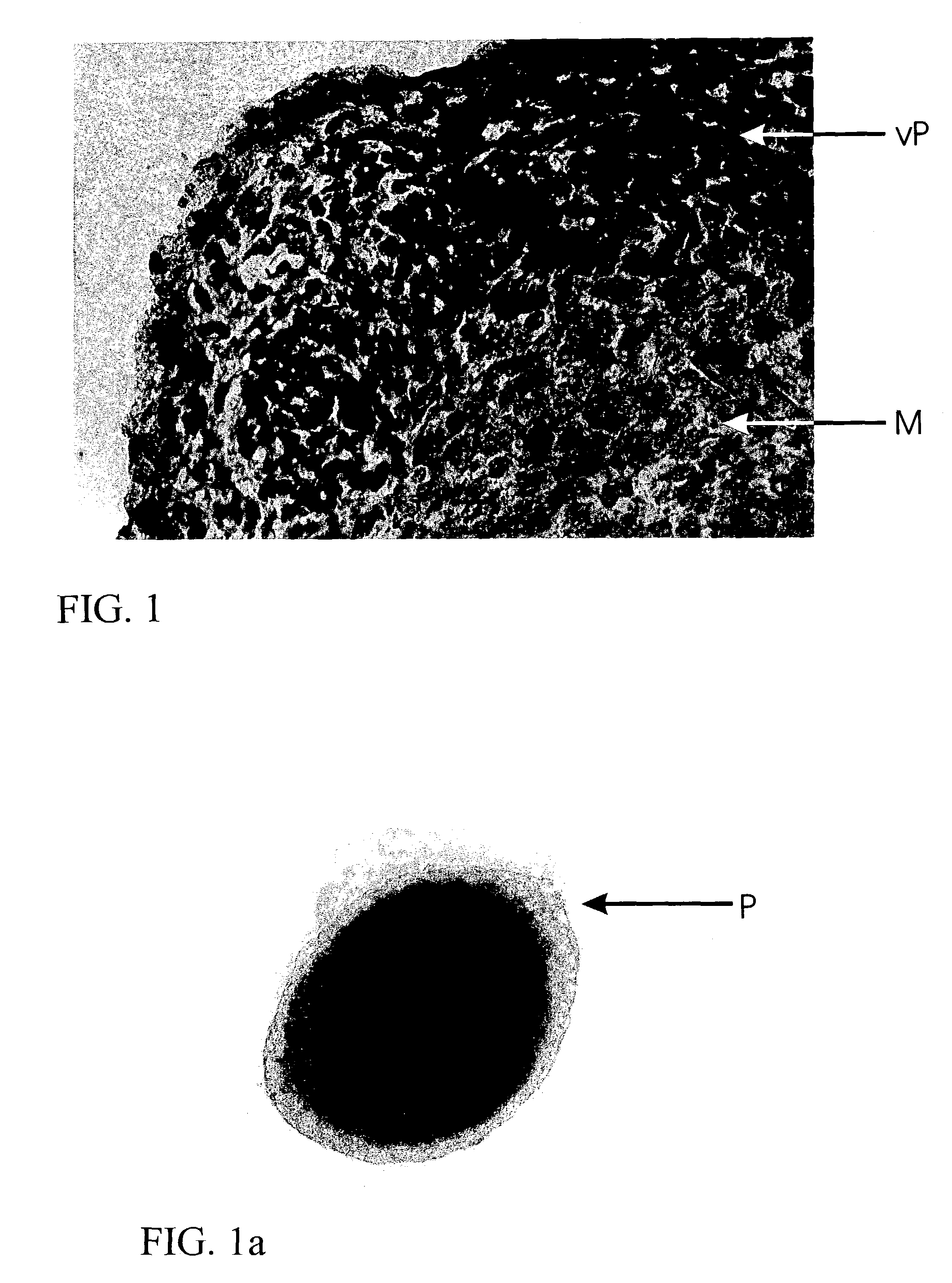
[0029]A biopsy is taken from a patient from a region of hyaline, healthy cartilage. Chondrocytes are isolated from this biopsy, using enzymatic digestion by incubation with collagenase solution. Following separation of the isolated cells from the undigested cartilage tissue, the cells are transferred in cell culture flasks and, following addition of DMEM / HAMS F12 culture medium (1 / 1) and 10% autologous serum from the patient, incubated at 37° C. and 5% CO2. The medium is exchanged twice a week. After reaching the confluent stage, the cell layer is washed with physiological saline solution and harvested from the cell culture surface using trypsin. Following another washing, 1×10⁵ cells each time are transferred in a cell culture vessel coated with agarose. After one day, the first cells arrange into aggregates. These aggregates are supplied with fresh medium every second day and cultured for at least 2 weeks.
[0030]After only one week, type II co...
example 2
Transplantation of Cartilage Tissue
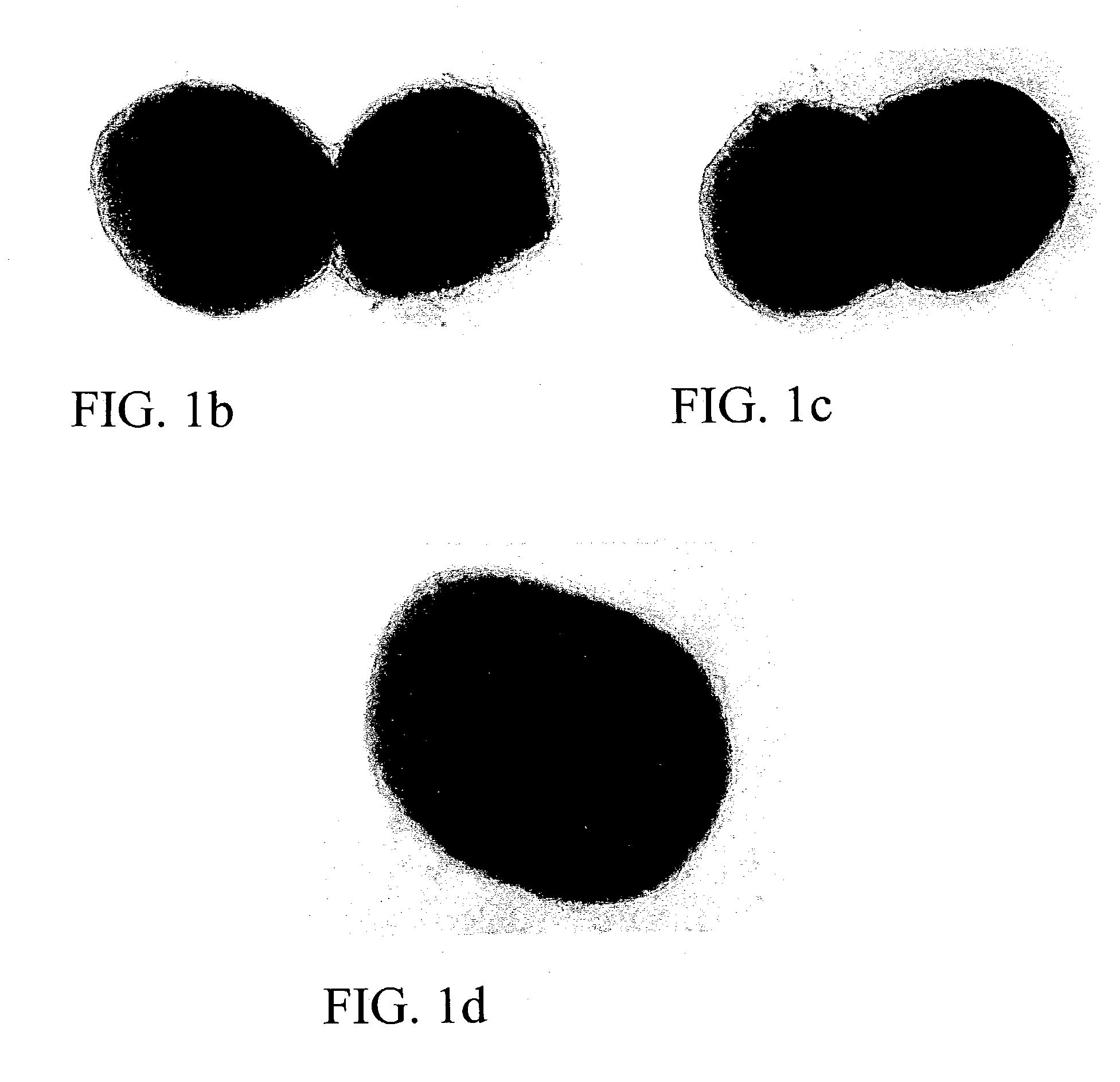
[0034]The tissue produced in Example 1 (about 200 spheroids, each one comprised of 1×10⁵ cells) was taken up in physiological saline solution and injected into a cartilage defect in the subject about 1 cm2 in size and covered with periosteum. Repletion of the cartilage defect with cartilage tissue was determined already within 1 to 2 months, while repletion of a defect of such a size can be detected not before 6-12 months when using the previous transplantation of juvenile cartilage cells merely grown in vitro. Thus, in addition to assuming the mechanical function of the tissue produced, the cartilage tissue of the invention produced in vitro ensures rapid integration of the produced tissue patches by virtue of the proliferative and migratory cells in the outer layer of the aggregates. Consequently, structure and function of the tissue patches also allow for rapid repair of defects and degenerate cartilage tissue.
example 3
In Vitro Production of Bone Tissue
[0035]A bone biopsy is taken from a patient from a spongiosa region. Osteoblasts are isolated from this biopsy, using enzymatic digestion by incubation with collagenase solution. Following separation of the isolated cells from the undigested bone tissue, the cells are transferred in cell culture flasks and, following addition of DMEM / HAMS F12 culture medium (1 / 1) and 10% autologous serum from the patient, incubated at 37° C. and 5% CO2. The medium is exchanged twice a week. After reaching the confluent stage, the cell layer is washed with physiological saline solution and harvested from the cell culture surface using trypsin. Following another washing, 1×10⁵ cells each time are transferred in a cell culture vessel coated with agarose. After one day, the first cells arrange into aggregates. These aggregates are supplied with fresh medium every second day and cultured for at least 2 weeks.
[0036]After only one week, type I collagen and proteoglycans we...
PUM
| Property | Measurement | Unit |
|---|---|---|
| diameter | aaaaa | aaaaa |
| hydrophobic | aaaaa | aaaaa |
| size | aaaaa | aaaaa |
Abstract
Description
Claims
Application Information