Devices and methods for interconnecting vessels

a technology of interconnected vessels and devices, applied in the field of anastomosis and anastomosis devices, can solve the problems of affecting the perfusion of the tissue served by the blood flow, restricting blood flow, and myocardial infarction, and achieves the effect of quick and efficient performing an anastomosis, minimizing tension and compression forces, and readily conforming to the inside wall

Active Publication Date: 2011-02-22
BIOCONNECT SYST
View PDF178 Cites 204 Cited by
  • Summary
  • Abstract
  • Description
  • Claims
  • Application Information

AI Technical Summary

Benefits of technology

[0025]Another object of the invention is to provide for a method of quickly and efficiently performing an anastomosis.
[0026]Another object of the present invention is to provide anastomotic devices and methods which minimize tension and compression forces at the site of the anastomosis.
[0027]Another feature of the invention is that the device is flexible and readily conforms to the inside wall of the native vessels to minimize flow resistance within the vessel and to reduce the risk of embolization.

Problems solved by technology

In the context of the peripheral vascular and / or the cardiovascular system, atherosclerosis, a common vascular disease, can cause partial blockage or complete occlusion of an arterial vessel, resulting in restricted blood flow and therefore compromised perfusion to the tissue served by the blood flow.
In the case of an occluded coronary vessel, for example, an area of the heart's myocardium would be compromised, which can lead to a myocardial infarction, or other ischemic heart syndrome such as congestive heart failure.
Patency may be compromised by technical, biomechanical or pathophysiological means.
Among the technical and biomechanical causes for compromised patency (also termed restenosis) is poorly achieved anastomoses, whether induced by poor placement, trauma at the anastomosis site or biological responses to the anastomosis itself.
Improperly anastomosed vessels may lead to leakage, create thrombus and / or lead to further stenosis at the communication site, possibly requiring re-operation and further increasing the risk of stroke.
Surgeons must delicately sew the vessels together being careful not to suture too tightly so as to tear the delicate tissue, thereby injuring the vessel which may then result in poor patency of the anastomosis.
On the other hand, surgeons sometimes inadvertently suture too loosely or do not properly place the sutures so as to provide continuous seal around the arteriotomy site, resulting in leakage of fluid from the anastomosis.
In addition to creating a surgical field in which it is difficult to see, leakage of fluid from the anastomosis can cause serious drops in blood pressure, acute or chronic.
The loss of blood may cause other deleterious effects on the patient's hemodynamics that may even endanger the patient's life.
In addition to the inherent inconsistencies in suture tightness, placement and stitch size and the lack of reproducibility, suturing an anastomosis can be very time consuming.
While stapling techniques have been found to be successful in gastrointestinal procedures, due to the large size and durability of the vessels, it is less adequate for use in vascular anastomosis where the vessels are much smaller.
The manufacturing of stapling instruments small enough to be useful for anastomosing smaller vessels, such as coronary arteries, is very difficult and expensive.
As stapling instruments are typically made of at least some rigid and fixed components, a stapler of one size will not necessarily work with multiple sizes of vessels.
This may significantly raise the cost of the equipment and ultimately the cost of the procedure.
Stapling instruments and staples which are adapted to conform to the smaller sized vessels are difficult to maneuver and, thus, a great deal of time, precision, and fine movement is necessary to successfully approximate the vessel tissue.
Everting may not always be practical especially for smaller arteries because of the likelihood of tearing when everted.
Another factor which may lead to damage or laceration of the vessel and / or leakage at the anastomosis site is the variability of the force that a surgeon may use to fire a stapling instrument causing the possible over- or under-stapling of a vessel.
Rectifying a poorly stapled anastomosis is itself a complicated, time-consuming process which can further damage a vessel.
The tension and / or compression forces exerted on the vessel walls as a result of suturing and stapling can result in damage to the vessel wall, even to the extent of causing tissue necrosis.
Damage to the intima of a vessel is particularly problematic as it may inhibit the natural bonding process that occurs between the anastomized vessels and which is necessary for sufficient patency.
Furthermore, damaged vessel walls are likely to have protuberances that when exposed to the bloodstream could obstruct blood flow or may produce turbulence which can lead to formation of thrombus, stenosis and possible occlusion of the artery.
These procedures are made more difficult due to the multiple characteristics that are unique to each anastomosis and to each patient.
For example, the arteries' internal diameter dimensions are difficult to predict and the inside walls are often covered with deposits of stenotic plaque which creates the risk of dislodging plaque into the patient's blood stream during the anastomosis procedure.
The resulting emboli in turn create a greater risk of stroke for the patient.
The vessel walls can also be friable and easy to tear, and are often covered with layers of fat and / or are deeply seated in the myocardium, adding to the difficulty of effectively and safely performing conventional anastomotic procedures.

Method used


Image

  • Devices and methods for interconnecting vessels
  • Devices and methods for interconnecting vessels
  • Devices and methods for interconnecting vessels

Examples


Embodiment Construction

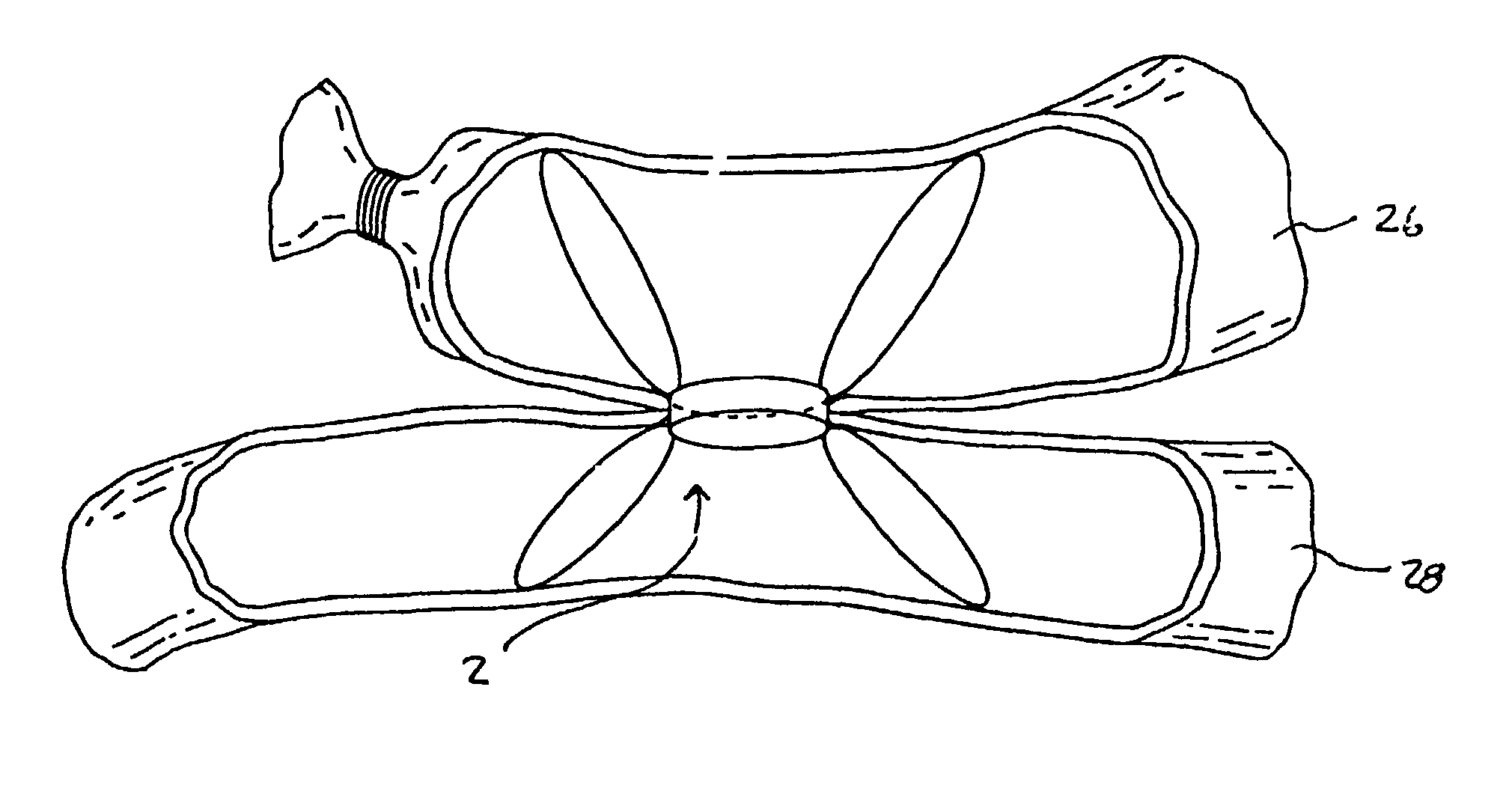
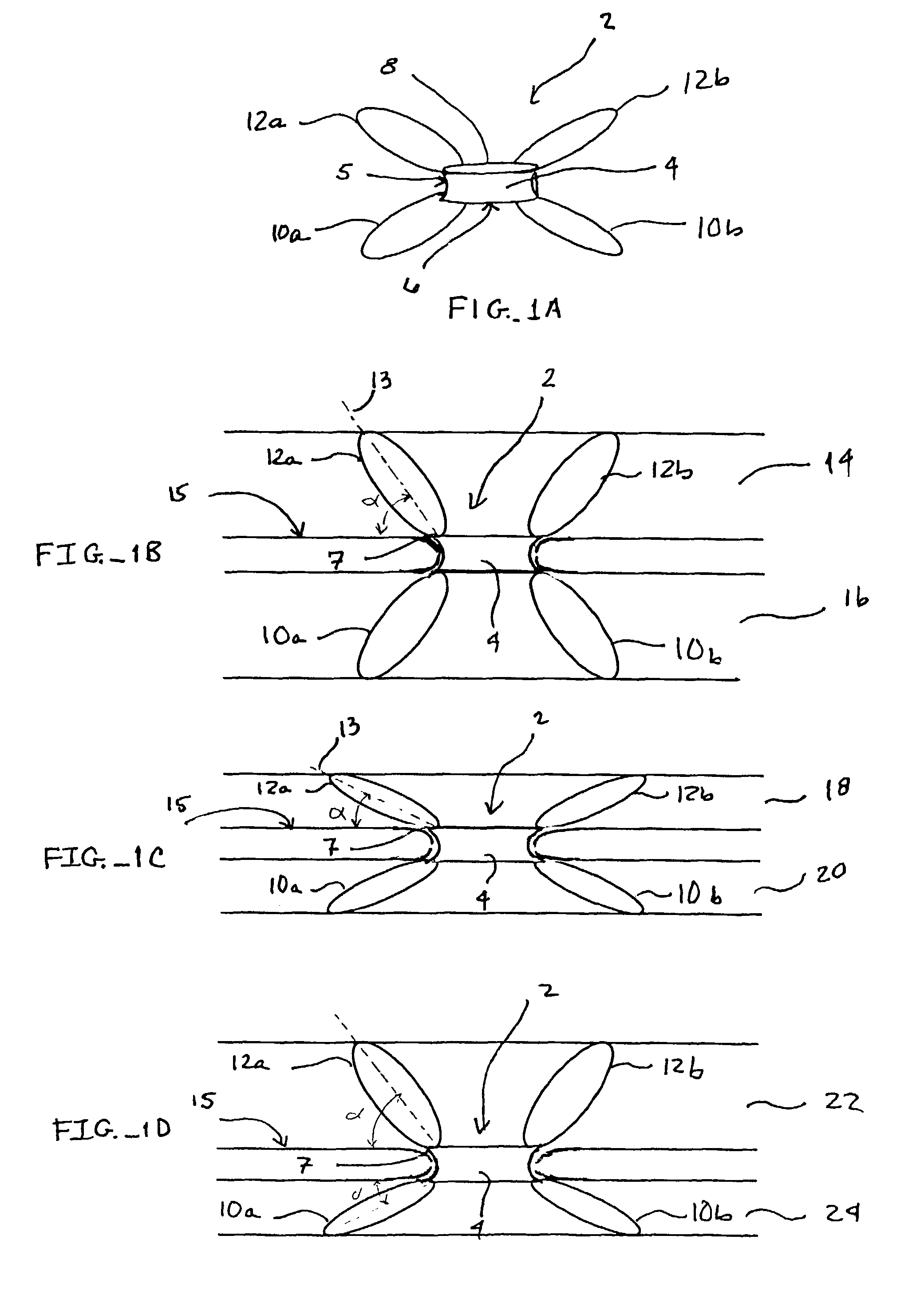
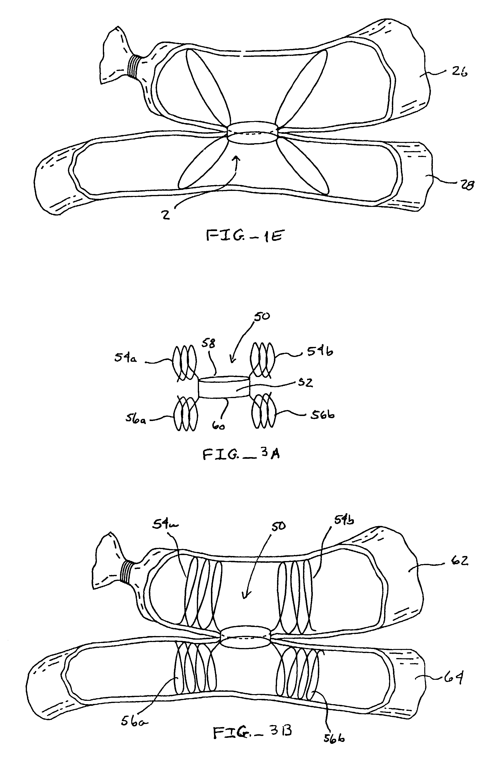
[0042]The present invention provides implantable devices and associated methods for interconnecting human vessels, lumens, ducts or other tubular organs rapidly, safely and in a minimally invasive manner. These device and methods are particularly helpful in surgical procedures involving the anastomosis of small vessels or the like within a limited surgical access field, such as coronary artery bypass graft procedures (CABG), and such devices and methods are exemplary described in detailed herein in the context of a CABG procedure. As such, a subject device is positioned within a target or native vessel, such as downstream of a diseased coronary artery, which allows the attachment of a second, graft vessel to form the anastomosis. The subject invention provides devices and methods for forming both side-to-side and end-to-side anastomosis.

[0043]Before the present invention, devices and methods used therein are disclosed and described, it is to be understood that this invention is not ...


PUM


Abstract

The present invention provides implantable devices and associated methods for interconnecting human vessels in a side-to-side or an end-to-side arrangement rapidly, safely and in a minimally invasive manner. The devices comprise a vessel connector and a intravascular support mechanism for establishing fluid communication between two vessels. Certain embodiments further include a sealing member for further sealing the openings in the vessels created for inserting the subject devices.

Description

CROSS-REFERENCE TO RELATED APPLICATIONS[0001]Pursuant to 35 U.S.C. §119 (e), this application claims priority to the filing date of the U.S. Provisional Patent Application Ser. No. 60 / 327,126 filed Oct. 3, 2001; the disclosure of which is herein incorporated by referenceFIELD OF THE INVENTION[0002]The field of this invention is anastomosis and anastomotic devices.BACKGROUND OF THE INVENTION[0003]The human body has numerous vessels carrying fluid to essential tissues and areas for circulation or excretion. When vessels become damaged, severed or wholly occluded due to physiological problems or diseases, certain sections must be bypassed to allow for the free and continuous flow of fluids. Anastomosis is performed for the purpose of connecting different conduits together to optimize or redirect flow around a damaged or occluded portion of a vessel.[0004]In the context of the peripheral vascular and / or the cardiovascular system, atherosclerosis, a common vascular disease, can cause pa...

Claims


Application Information

Patent Timeline
Patent Type & Authority Patents(United States)
IPC IPC(8): A61B17/08A61B17/11A61B19/02A61F2/06
CPCA61B17/11A61B50/362A61F2/064A61B2050/364
Inventor CONSTON, STANLEY R.YAMAMOTO, RONALD K.AKIN, JODI J.BARRY, GITANJALI V.
Owner BIOCONNECT SYST
Why Eureka
Try Eureka