Clean margin assessment tool

a technology of clean margins and assessment tools, which is applied in the field of medical tools, can solve the problems of inaccurate placement of guide wires, difficult to place guide wires at the correct depth, and radiation may not properly place guide wires through the lesion

Inactive Publication Date: 2011-03-08
DUNE MEDICAL DEVICES
View PDF119 Cites 12 Cited by
  • Summary
  • Abstract
  • Description
  • Claims
  • Application Information

AI Technical Summary

Benefits of technology

[0151]the system thereby enabling removal of additional tissues at locations adjacent to the periphery region of said tissue portion, upon detecting that there is no clean margin in the periphery region of the characterized tissue portion thus, confirming an existence of a clean margin of healthy tissue around an abnormal tissue.
[0159]According to yet another broad aspect of the invention, there is provided a medical tool for use in a clean margin assessment system for determining existence of a clean margin of healthy tissues in a periphery region of a tissue portion, the medical tool comprising: a tissue-type sensor unit comprising at least one tissue-type sensor configured and operable to determine a type of the tissue at various locations along the tissue portion surface and generating data indicative of existence of a clean margin of healthy tissue in a periphery region of the tissue portion, thereby enabling to decide about removal of additional tissues at locations adjacent to said periphery region of said tissue portion.
[0166]c) analyzing data generated by the medical tool and providing output data indicative of existence of a clean margin of a first tissue type in a periphery region of said tissue potion, thereby enabling to decide about removal of additional tissues at locations adjacent to said periphery region of the characterized tissue portion.
[0171]According to yet further broad aspect of the invention, there is provided a method for use in providing a clean-margin of healthy tissue around an abnormal tissue, the method comprising: characterizing a tissue segment of a tissue surface to determine and record the tissue segment margin status; analyzing the tissue segment margin status for identifying whether it corresponds to clean margin, thereby enabling removal of tissue segments which are adjacent or correspond to the tissue segments which were not identified as clean margin.

Problems solved by technology

There are several problems with conventional breast conserving therapy.1. There are technical challenges associated with placement of the guide wire tip, and the radiologist may not place the guide wire properly through the lesion.
It is particularly difficult to place the guide wire at the correct depth.
When the breast is decompressed for the surgical excision, the guide wire can move, resulting in inaccurate placement thereof.
Finally, the guide wire placement procedure is uncomfortable to the patient and logistically challenging; the procedure must be coordinated with the time of surgery.
Often, the easiest path for the radiologist to place the guide wire is different from the best surgical approach, so the surgeon cannot follow the guide wire down to the tumor.2. It is difficult to estimate correctly the full extent of the disease and the exact volume of the cancerous portion of the tumor, especially with non-palpable lesions.
Non-palpable lesions are similar in their properties to normal tissue, hence harder to detect by ultrasound and mammography.
Thus, the guide wire may be inaccurately placed.
This leads to a negative impact aesthetically and emotionally on the patient.
The drawback of such a procedure is that it requires exact knowledge as to the location of the malignant tissue and its boundaries while creating the cage.
Therefore liver scans, for instance, did not show possible carcinomas or other pathological states.
As such, it is unsuitable as a handheld probe or an endoscopic probe, because the tissue to be imaged has to be in the internal region of the imager,

Method used


Image

  • Clean margin assessment tool
  • Clean margin assessment tool
  • Clean margin assessment tool

Examples


Embodiment Construction

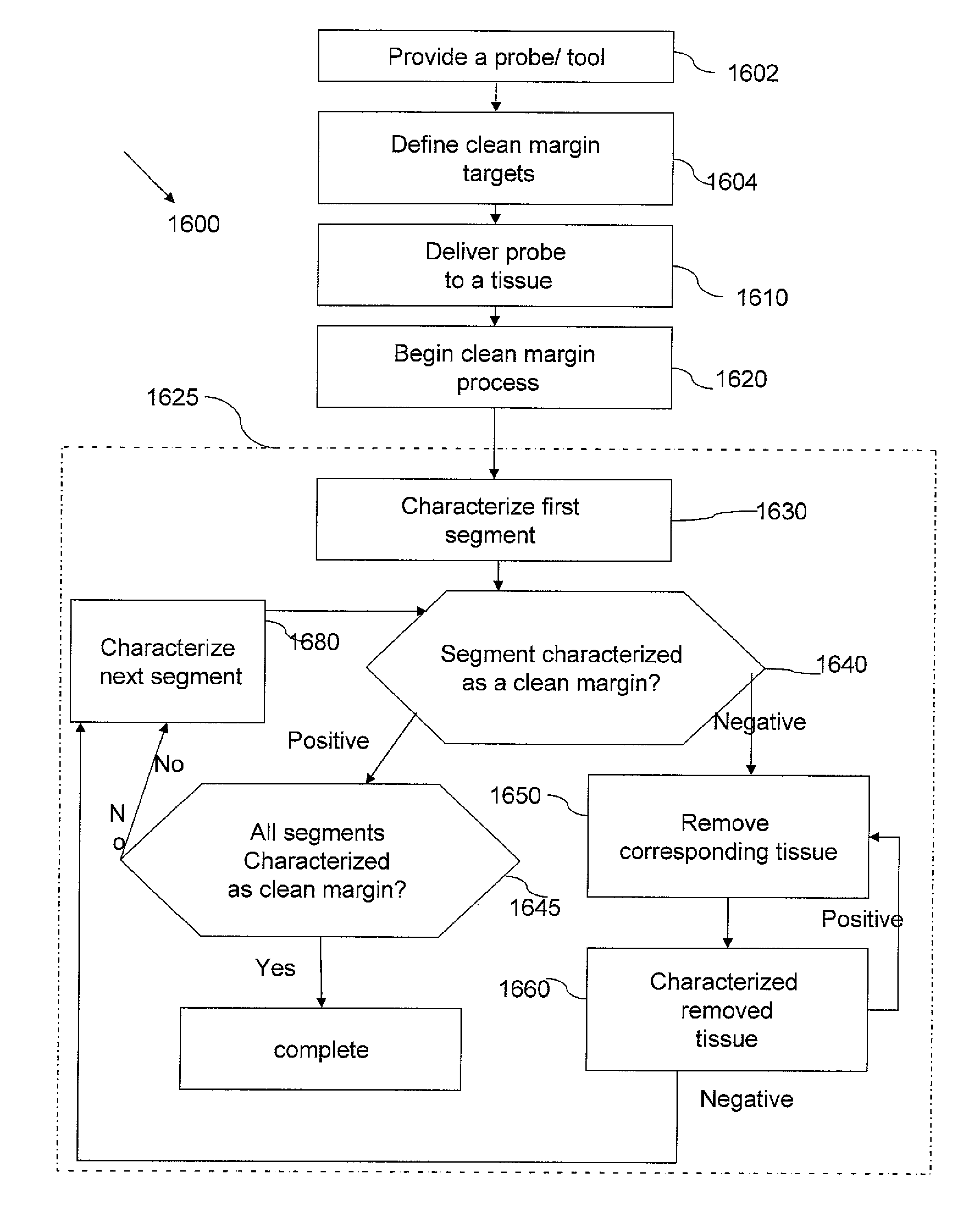
[0194]The present invention is of an integrated tool having a tissue-type sensor, for determining the tissue type at a near zone volume of a tissue surface, and a distance-measuring sensor, for determining the distance to an interface with another tissue type. The tool is operable for (i) confirming an existence of a clean margin of healthy tissue around a malignant tumor, which is being removed, and (ii) determining the width of the clean margin, wherein both are performed in real time, while the malignant tumor is being removed. The tissue-type sensor may be selected from the following: a sensor for tissue electromagnetic properties, a dielectric sensor, an impedance sensor, a sensor for optical fluorescence spectroscopy, a sensor for optical reflectance spectroscopy, an MRI sensor, an RF sensor, an MW sensor, a temperature sensor, and infrared thermography sensor, or another tissue-characterization sensor, as known. The distance-measuring sensor may include at least one of the fo...


PUM


Abstract

An integrated tool is provided, having a tissue-type sensor, for determining the tissue type at a near zone volume of a tissue surface, and a distance-measuring sensor, for determining the distance to an interface with another tissue type, for (i) confirming an existence of a clean margin of healthy tissue around a malignant tumor, which is being removed, and (ii) determining the depth of the clean margin. The integrated tool may further include a position tracking device and an incision instrument. The soft tissue may be held within a fixed frame, while the tumor is being removed. Additionally a method for malignant tumor removal is provided, comprising, fixing the soft tissue within a frame, performing imaging with the hand-held, integrated tool, from a plurality of locations and orientations around the soft tissue, reconstructing a three-dimensional image of the soft tissue and the tumor within, defining a desired clean margin on the reconstructed image, calculating a recommended incision path, displaying the recommended path on the reconstructed image, and cutting the tissue while determining its type, at the near zone volume of the incision surface. The method may further include continuously imaging with the cutting, continuously correcting the reconstructed image and the recommended incision path, and continuously determining the tissue type, at the near zone volume of the incision surface.

Description

[0001]This Application is a continuation-in-part of U.S. application Ser. No. 11 / 743,028 filed May 1, 2007, the entire contents of which being hereby incorporated by reference.FIELD AND BACKGROUND OF THE INVENTION[0002]The present invention relates generally to a medical tool and method for use in removal of tissues from a subject's body.[0003]When a malignant tumor is found in a breast, the patient currently has two primary treatment options, 1) mastectomy or 2) breast conserving therapy, which means, lumpectomy, followed by radiation therapy. Generally, breast conserving therapy is indicated for patients with Stage T1 or T2 cancers, of between less then about 0.5 and about 5 cm in greatest dimension.[0004]To localize the tumor within the breast, a radiologist may place a guide wire under x-ray or ultrasound guidance, so that the proximal tip of the guide wire, with respect to the tissue, is in the tumor. Alternatively, an imaging modality alone, for example, mammography, CT, ultra...

Claims


Application Information

Patent Timeline
Patent Type & Authority Patents(United States)
IPC IPC(8): A61B5/05A61B5/04
CPCA61B5/0071A61B5/0073A61B5/0075A61B5/0084A61B5/0091A61B5/01A61B5/0507A61B5/415A61B5/4312A61B8/0858A61B19/5244A61B5/0086A61B5/015A61B5/053A61B5/055A61B5/06A61B8/12A61B8/4472A61B19/2203A61B2017/00026A61B2017/00057A61B2017/00061A61B2017/00106A61B2019/461A61B2019/507A61B2019/5251A61B2019/5295A61B8/4245Y10S607/901A61B2090/367A61B34/20A61B34/30A61B2090/061A61B2034/107A61B2034/2051A61B5/068A61B5/708
Inventor HASHIMSHONY, DANCOHEN, GILGELTNER, IDDO
Owner DUNE MEDICAL DEVICES
Why Eureka
Try Eureka