Method for analyzing blood for the presence of cancer cells
a cancer cell and blood technology, applied in the direction of microbiological testing/measurement, sugar derivatives, biochemistry apparatus and processes, etc., can solve the problems of complex antioxidative protection, death of cells, and lack of comprehensiveness
- Summary
- Abstract
- Description
- Claims
- Application Information
AI Technical Summary
Problems solved by technology
Method used
Image
Examples
example 1
Healthy Donors
[0185]The amounts of MNSOD, TXNRD1 and GPX1 mRNA determined in the isolated cells (fraction C) for healthy donors (number N) are indicated in table 1, specifically as ratio to the amounts of corresponding mRNAs in the comparative cell fraction (fraction A).
[0186]
TABLE 1MNSOD, TXNRD1 and GPX1 mRNA expression in theblood of healthy donorsNAverageStandard deviationLimitMNSOD90.93000.28911.2TXNRD190.91330.41661.3GPX183.68881.55335.2
[0187]The cells isolated from blood show a slightly elevated GPX1 expression. The expression of MNSOD and TXNRD1 is unchanged relative to the comparative cell fraction, that is the lymphocytes.
[0188]For the subsequent assessment of the levels of expression measured in tumor patients, the levels regarded as positive are those which exceed the average level plus standard deviation. These levels are indicated as limit in table 1.
example 2
Tumor Patients
[0189]A number of tumor patients with different tumors are included in the investigation. The tumors had been diagnosed by various other medical methods.
1. Sensitivity
[0190]The number of cases among tumor patients (number N) in which the expression of MNSOD, TXNRD1 and GPX1 mRNA is elevated (positive) in the isolated cells (fraction C) in relation to the cells of the complete cell fraction (fraction A′) is indicated below, grouped according to type of tumor.
[0191]
MNSODInvestigationsN = 93PatientsN = 90of whichbreast41 (40 positive)colon 8 (8 positive)prostate 8 (7 positive)ovary 8 (5 positive)lung 5 (2 positive)bladder 4 (3 positive)liver 2 (1 positive)thyroid 2 (2 positive)others12 (10 positive)→ 78 / 90 = 87% POSITIVETXNRD1InvestigationsN = 93PatientsN = 90of whichbreast41 (31 positive)colon 9 (6 positive)prostate 8 (6 positive)ovary 7 (3 positive)lung 5 (1 positive)bladder 4 (3 positive)liver 2 (1 positive)thyroid 2 (1 positive)others12 (8 positive)→ 60 / 90 = 67% POSIT...
example 3
Tumor Patients
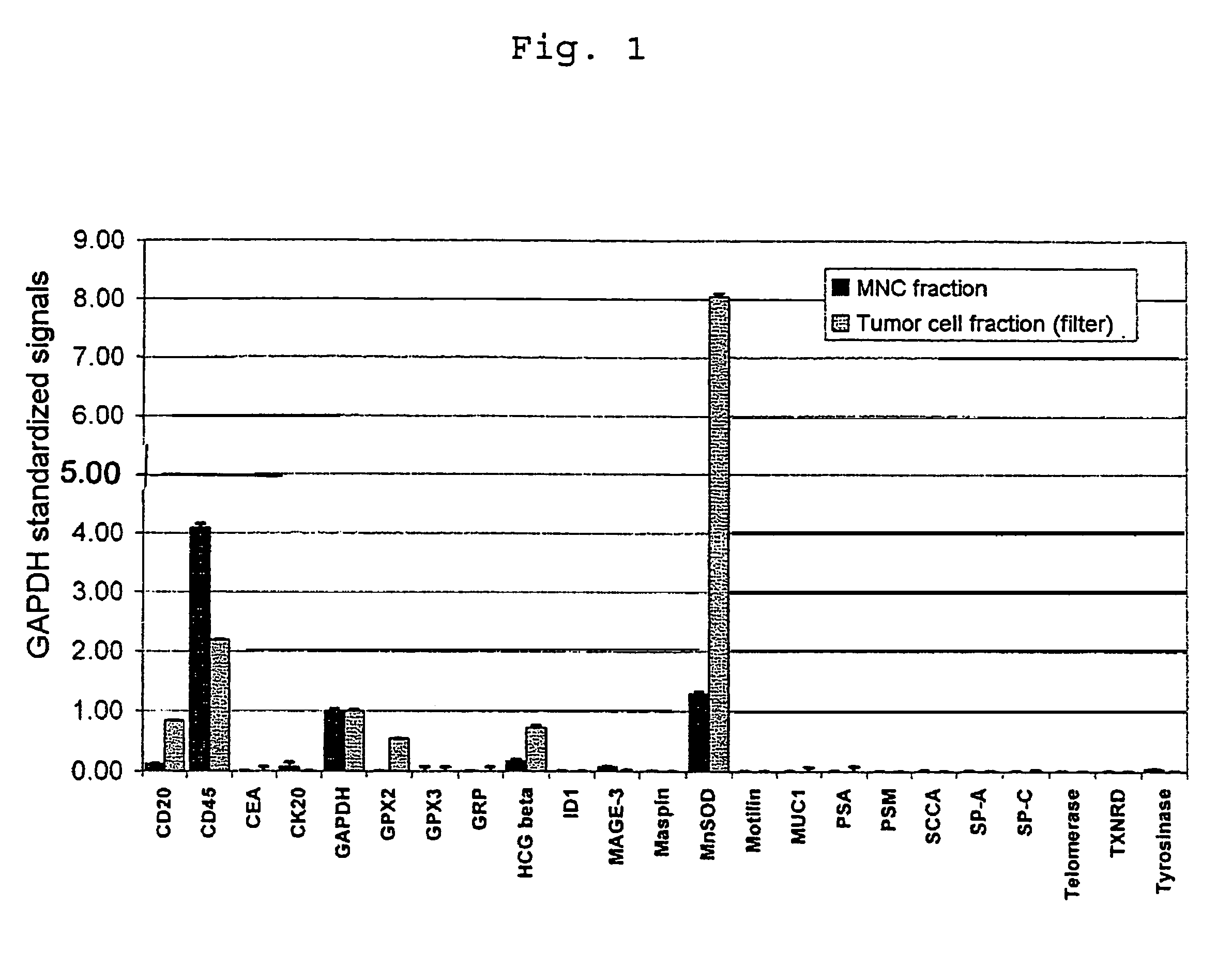
[0236]FIG. 1 shows a CCD contact exposure image of the fluorescence radiation emitted from the array after hybridization with cDNA single-stranded fragments which were generated in the manner described above by means of mRNA total amplification from the tumor cell fraction C and the comparative cell fraction A′.
[0237]Overexpression of MNSOD and GPX2 in the tumor cell fraction is clearly evident. The measurement underlines the increased sensitivity and specificity of the detection method of the invention compared with other tumor cell-detecting genes.
PUM
| Property | Measurement | Unit |
|---|---|---|
| Length | aaaaa | aaaaa |
| Width | aaaaa | aaaaa |
| Fraction | aaaaa | aaaaa |
Abstract
Description
Claims
Application Information
 - R&D
- Intellectual Property
- Life Sciences
- Materials
- Tech Scout
- Unparalleled Data Quality
- Higher Quality Content
- 60% Fewer Hallucinations
Browse by: Latest US Patents, China's latest patents, Technical Efficacy Thesaurus, Application Domain, Technology Topic, Popular Technical Reports.
© 2025 PatSnap. All rights reserved.Legal|Privacy policy|Modern Slavery Act Transparency Statement|Sitemap|About US| Contact US: help@patsnap.com

