
Method and apparatus for automated vascular function testing

a vascular function and automated technology, applied in the field of methods and systems for testing the vascular function of patients, can solve the problems of poor reproducibility of ultrasound measurements, the use of ultrasound probes poses problems for the assessment of the vascular endothelial function, etc., and achieve the effect of improving the performance of the combined system

Active Publication Date: 2011-10-25
GENERAL ELECTRIC CO
View PDF15 Cites 5 Cited by
  • Summary
  • Abstract
  • Description
  • Claims
  • Application Information

AI Technical Summary

Benefits of technology

[0009]In one embodiment, the method and system further includes a heartbeat sensing system that is operable to detect each heartbeat cycle for the patient. Information related to the heartbeat cycle can be relayed to the ultrasound system such that the ultrasound system can obtain measurements at specific points during the heartbeat cycle to enhance the performance of the combined system. In one embodiment, the NIBP monitor, ultrasound monitor and heartbeat sensing system communicate with each other either directly or through a patient monitor such that the determination of the endothelial dysfunction of a patient can be determined automatically without intervention by a technician.

Problems solved by technology

However, the use of ultrasound probes poses problems for assessment of the vascular endothelial functions.
Specifically, in presently used methods, the ultrasound measurements may be poorly reproducible since the technique is highly operator-dependent with regard to the positioning of the probe on the patient.
Further, since the prior methods and systems require a trained technician to manually locate the blood vessel within the patient limb and simultaneously integrate the functions of the blood pressure cuff and the ultrasound probe, testing typically takes place in a research environment and is not widely used as a clinical tool.

Method used


Image

  • Method and apparatus for automated vascular function testing
  • Method and apparatus for automated vascular function testing
  • Method and apparatus for automated vascular function testing

Examples


Embodiment Construction

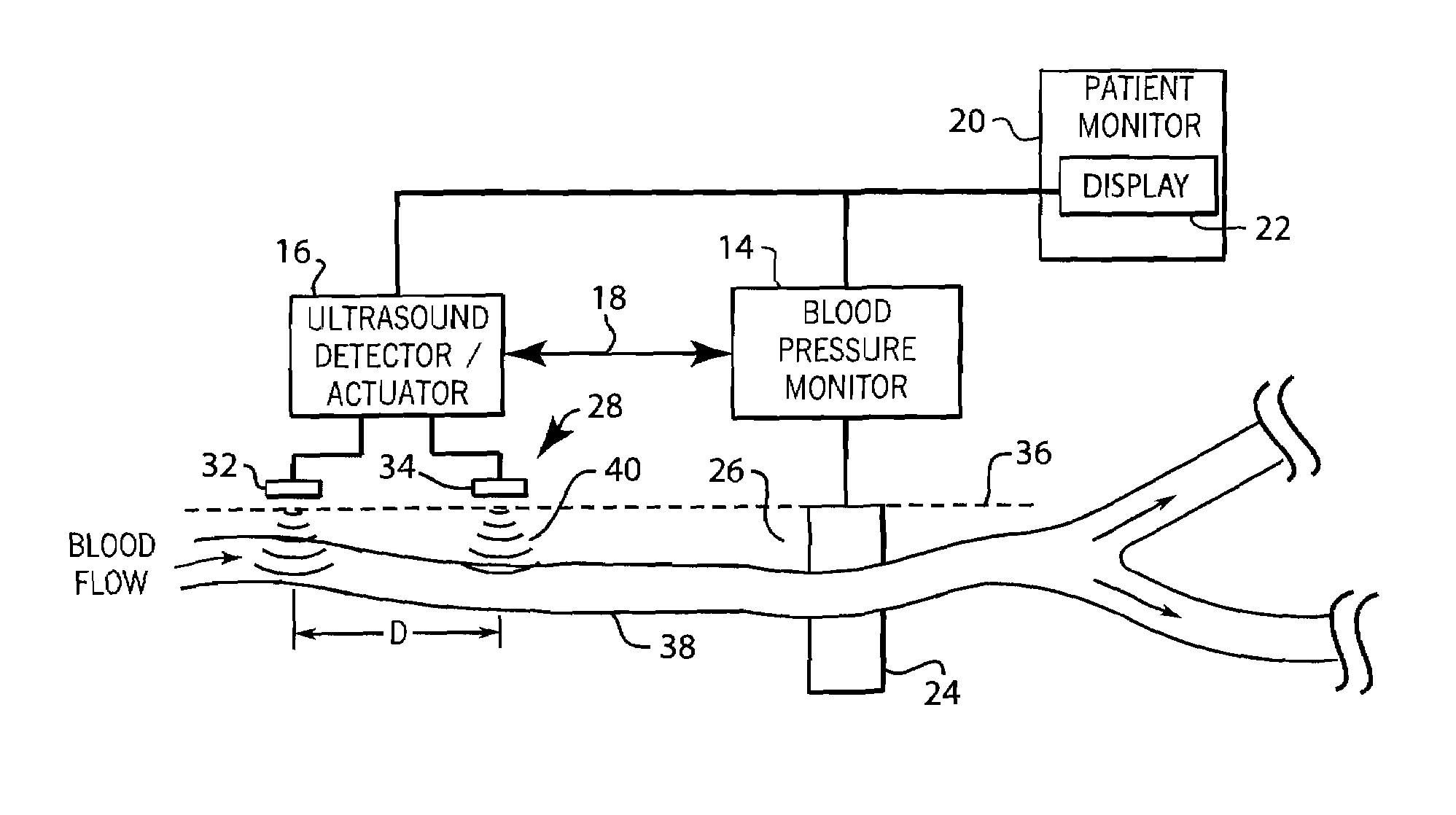
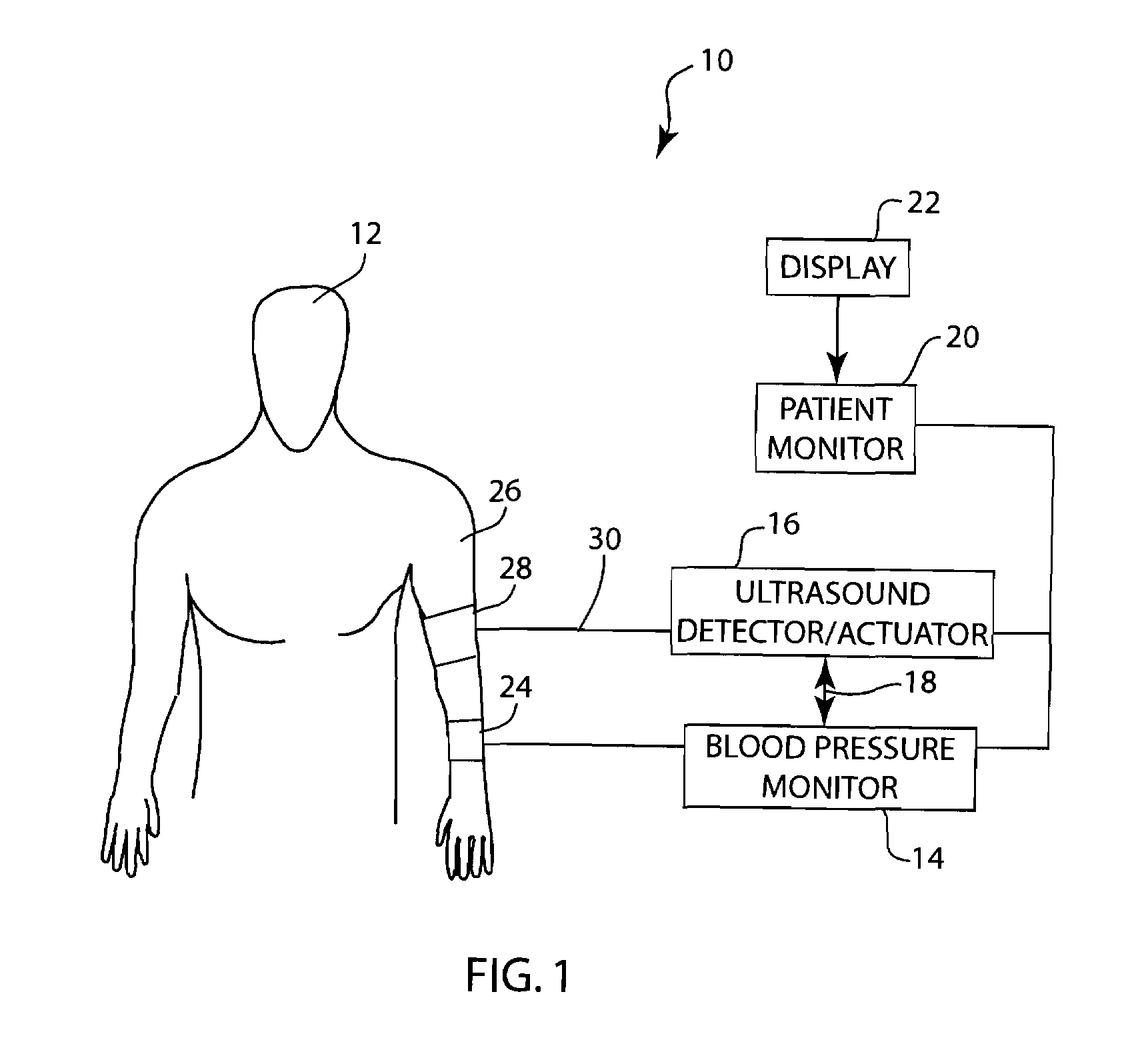
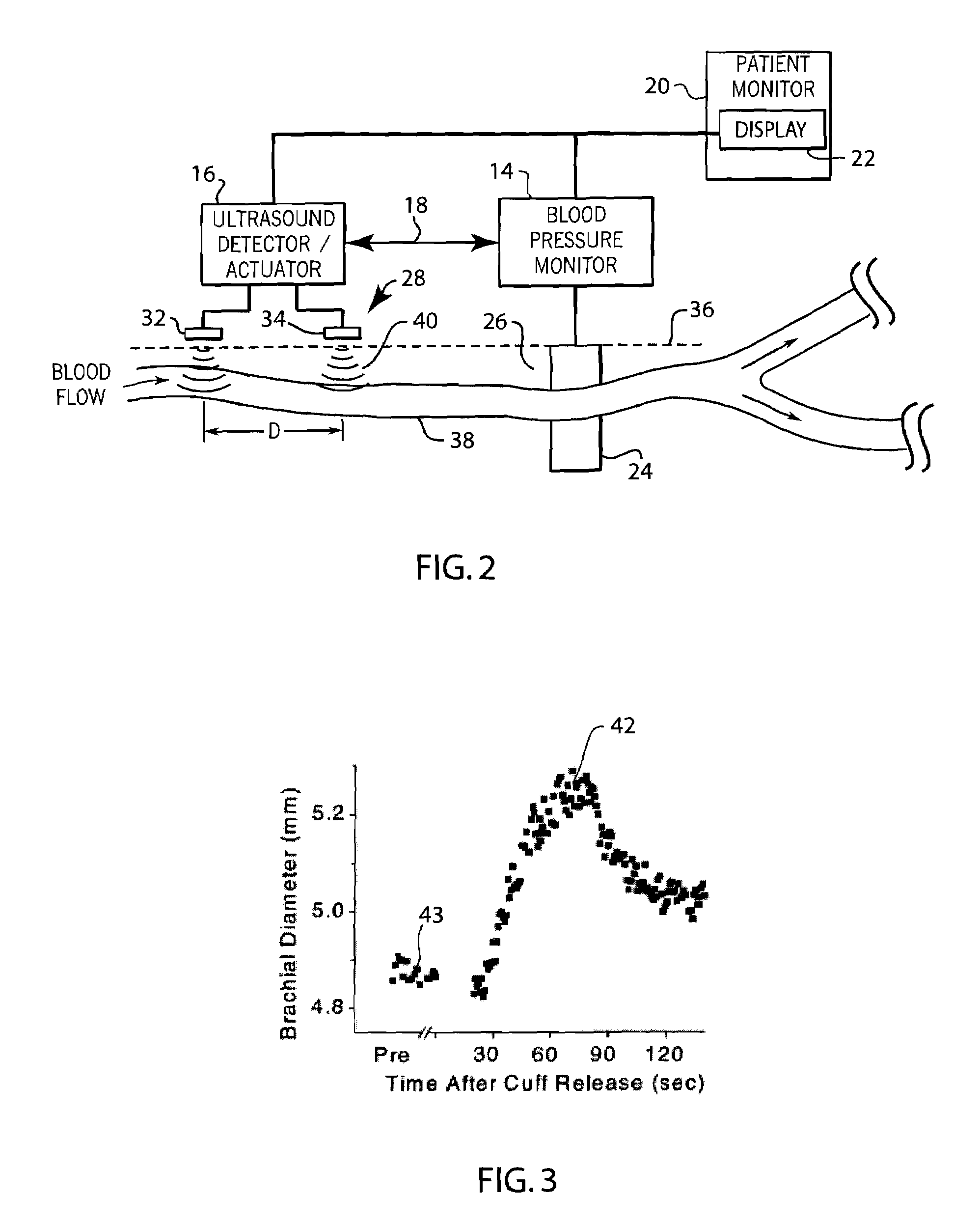
[0021]FIG. 1 generally illustrates a system 10 for the automated determination of the endothelial dysfunction of a patient 12 using flow mediated dilation (FMD). The system 10 shown in FIG. 1 preferably includes a non-invasive blood pressure (NIBP) monitor 14 and an ultrasound system 16 that are preferably in direct communication with each other through the communication line 18. In the embodiment shown in FIG. 1, both the ultrasound system 16 and the blood pressure monitor 14 communicate to a patient monitor 20 having a display 22. However, it is contemplated that the patient monitor 20 and display 22 could be eliminated and the blood pressure monitor 14 could communicate directly to the ultrasound system 16 and may include its own internal display to display the measurement of the endothelial dysfunction calculated by the system 10.

[0022]In the embodiment shown, the NIBP monitor 10 includes a blood pressure cuff 24 placed on the arm 26 of the patient 12. The blood pressure cuff 24...


PUM


Abstract

A method and system for use in measuring the endothelial dysfunction utilizing flow mediated dilation and determining arterial health of a patient. The system includes a non-invasive blood pressure monitor, an ultrasound system and a pulse oximeter monitor that all communicate with each other to perform the flow mediated dilation. Initially, the ultrasound transducer, blood pressure cuff and pulse oximeter sensor are positioned on an arm of the patient. The blood pressure cuff is inflated to occlude an artery for an occlusion period. Following the occlusion period, the ultrasound system is automatically signaled to begin determining a parameter of the artery, such as diameter, and the flow rate of blood through the artery without any operator intervention. At the same time, the pulse wave velocity PWV is calculated between the ultrasound transducer and the finger probe of the pulse oximeter following the occlusion period. Based upon the detected characteristics of the artery before and after occlusion, as well as the PWV, the system can determine the endothelial dysfunction and arterial health of the patient.

Description

FIELD OF THE INVENTION[0001]The subject matter described herein generally relates to a method and system for testing the vascular function of a patient using multiple distinct measurements, including flow mediated dilation (FMD). More specifically, the subject matter described herein relates to an automated system for integrating the use of an ultrasound system, a optical plethysmographic monitor (such as a pulse oximeter monitor) and an automated NIBP monitor to determine endothelial dysfunction and either pulse wave velocity or pulse wave transit time.BACKGROUND OF THE INVENTION[0002]Endothelial dysfunction is a disruption of the normal biochemical processes carried out by the endothelium, which is a cell group that lines the inner surface of all blood vessels, including arteries and veins. The normal function of the endothelial cells is to produce or release substances that control the biological characteristics of the blood vessels, including the dimension, elasticity, permeabil...

Claims


Application Information

Patent Timeline
Patent Type & Authority Patents(United States)
IPC IPC(8): A61B5/02
CPCA61B5/02007A61B5/02125A61B5/022A61B8/4227A61B5/14551A61B5/02416A61B8/06
Inventor FRIEDMAN, BRUCE A.THOMENIUS, KAI E.DENTINGER, AARON M.
Owner GENERAL ELECTRIC CO
Learn More