Nanoparticulate probe for in vivo monitoring of tissue oxygenation
a tissue oxygenation and nanoparticulate technology, applied in the direction of porphines/azaporphines, diagnostic recording/measuring, organic chemistry, etc., can solve the problems of complex measurement of intracellular po/sub>2 and tissue damag
- Summary
- Abstract
- Description
- Claims
- Application Information
AI Technical Summary
Benefits of technology
Problems solved by technology
Method used
Image
Examples
example 1
Protocols for Isolation of Cells
[0194]Human-derived stem cells were isolated from human umbilical cord blood collected following healthy fetus delivery at the Ohio State University Medical Center. All blood collection procedures were based on IRB protocol and in all cases a written consent approval was obtained from participants in this study.
[0195]In contrast to adult bone marrow derived HSCs, cord blood progenitors have distinctive proliferation potential, including the capacity to form a greater number of colonies, a higher cell-cycle rate, and a longer telomere. All of these properties favor the growth of the cord blood progenitors compared with adult peripheral blood or bone marrow progenitors. In addition, cord blood could be obtained noninvasively, in contrast to invasive bone marrow or G-CSF stimulated adult peripheral blood progenitor cells isolation.
[0196]Umbilical cord blood in the amount of 50-150 ml was obtained from healthy donors following normal fetus delivery into s...
example 2
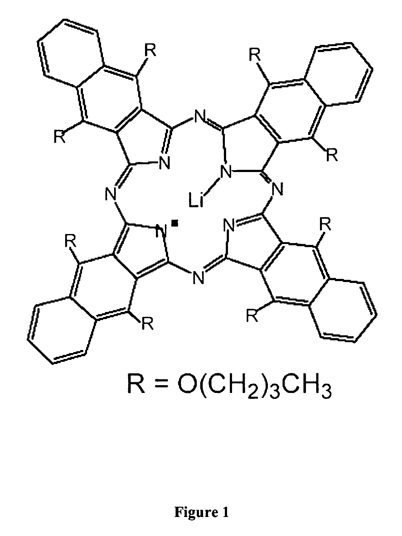
Internalization of Cultured Cells with Oximetry Spin Probes LiNc-BuO
[0199]Cultured cells (either human umbilical cord blood cells or animal derived skeletal myoblasts) were transferred to sterile Petri dish containing cell culture media. Sonicated oximetry spin probes were added concomitantly to cultured media cells. Cells and spin probes were co-cultured for a period ranging from 24-96 hours in order to determine dynamics of spin probes incorporation into cultured cells. Co-cultured cells-spin probes were frequently (every 30-45 min) gently shaken. Subsequently, cells were intensively (3-6 times) washed and collected for oximetry.
example 3
Internalization of EPR Particles (LiNc-BuO) in Skeletal Myoblasts
[0200]Isolated myoblasts were cultured in petridishes to obtain 60-70% confluence. Freshly sonicated LiNc-BuO particles were added. The petridishes were than periodically shaken every 15 minutes for 4-5 hours to achieve maximum internalization of the particles into the cells. After 24-48 hours, the cocultutred cells with the LiNc-BuO were washed thoroughly 10 times to remove the free particles. The cells were trypsinized, and counted to obtain approximately 500,000 cells for EPR spectrum analysis. Similarly cultured cells without particles treatment served as control. FIG. 23 shows sheep cultured skeletal myoblasts internalized with LiNc-BuO spin probes. The arrows show places of spin probes concentration inside the cells. FIG. 24 shows cellular uptake of LiNc-Bu) by sheep skeletal myoblast cells measured using EPR spectroscopy. Values are expressed as mean+ / −SD (n=3).
PUM
 Abstract
Description
Claims
Application Information
 - R&D
- Intellectual Property
- Life Sciences
- Materials
- Tech Scout
- Unparalleled Data Quality
- Higher Quality Content
- 60% Fewer Hallucinations
Browse by: Latest US Patents, China's latest patents, Technical Efficacy Thesaurus, Application Domain, Technology Topic, Popular Technical Reports.
© 2025 PatSnap. All rights reserved.Legal|Privacy policy|Modern Slavery Act Transparency Statement|Sitemap|About US| Contact US: help@patsnap.com


