Cell death-inducing agent

a cell death and inducing agent technology, applied in the field of cell death inducing agents, can solve the problems of difficult clinical application of 2d7 antibody to tumors or autoimmune diseases, diabody hardly acted on normal peripheral blood-derived lymphocytes and adherent cells, etc., and achieve the effect of large effect on activated t cells

Inactive Publication Date: 2012-04-17
CHUGAI PHARMA CO LTD
View PDF179 Cites 11 Cited by
  • Summary
  • Abstract
  • Description
  • Claims
  • Application Information

AI Technical Summary

Benefits of technology

[0019]As described, because cross-linking with an anti-mouse IgG antibody is necessary for 2D7 antibody to induce cell death, it is difficult to clinically apply the 2D7 antibody to tumors or autoimmune diseases. Therefore, the present inventors examined the effect of reducing the molecular weight of the 2D7 antibody on cell death induction More specifically, genes encoding the variable regions of the 2D7 antibody were cloned from hybridomas. The 2D7 antibody was then made into diabodies using genetic engineering techniques and the effects on cell death-inducing activity was examined. Surprisingly, the 2D7 antibody converted to diabodies showed strong cell death-inducing activity within a very short time and at low doses, even without cross-linking with an anti-mouse IgG antibody. Furthermore, the diabody hardly acted on normal peripheral blood-derived lymphocytes and adherent cells, and specifically induced cell death in various myeloma cells, T cell leukemia cell lines, and activated lymphocytes. The above-mentioned results show that the minibodies of antibodies recognizing HLA can be utilized as cell death-inducing agents.
[0050]In the present invention, administration of the minibodies that recognize HLA can treat or prevent diseases such as tumors including blood tumors (hematopoietic tumors) (specific examples include leukemia, myelodysplastic syndrome, malignant lymphoma, chronic myelogenic leukemia, plasmacytic disorder (myeloma, multiple myeloma, macroglobulinemia), and myeloproliferative disease (polycythemia vera, essential thrombocythemia, idiopathic myelofibrosis)), and autoimmune diseases (specific examples include rheumatism, autoimmune hepatitis, autoimmune thyroiditis, autoimmune bullosis, autoimmune adrenocortical disease, autoimmune hemolytic anemia, autoimmune thrombycytopenic purpura, autoimmune atrophic gastritis, autoimmune neutropenia, autoimmune orchitis, autoimmune encephalomyelitis, autoimmune receptor disease, autoimmune infertility, Crohn's disease, systemic lupus erythematosus, multiple sclerosis, Basedow's disease, juvenile diabetes, Addison's disease, myasthenia gravis, lens-induced uveitis, psoriasis, and Behchet's disease).
[0061]Furthermore, a diabody comprising the amino acid sequence of SEQ ID NO: 6, or a diabody comprising the sequence of a CDR (or a variable region) of SEQ ID NO: 2 and a CDR (or a variable region) of SEQ ID NO: 4 may be humanized or chimerized to reduce heterologous antigenicity against humans.
[0065]Antibodies that recognize HLA are not particularly limited, so long as they bind to HLA; mouse antibodies, rat antibodies, rabbit antibodies, sheep antibodies, human antibodies, and such may be used as necessary. Alternatively, artificially modified, genetically recombinant antibodies, such as chimeric and humanized antibodies, may be used to reduce heterologous antigenicity against humans. These modified antibodies can be produced using known methods. A chimeric antibody is an antibody comprising the variable regions of the heavy and light chains of an antibody from a non-human mammal such as a mouse, and the constant regions of the heavy and light chains of a human antibody. The chimeric antibody can be produced by linking a DNA encoding the variable regions of the mouse antibody with a DNA encoding the constant regions of the human antibody, incorporating this into an expression vector, and then introducing the vector to a host.

Problems solved by technology

As described, because cross-linking with an anti-mouse IgG antibody is necessary for 2D7 antibody to induce cell death, it is difficult to clinically apply the 2D7 antibody to tumors or autoimmune diseases.
Furthermore, the diabody hardly acted on normal peripheral blood-derived lymphocytes and adherent cells, and specifically induced cell death in various myeloma cells, T cell leukemia cell lines, and activated lymphocytes.

Method used


Image

  • Cell death-inducing agent
  • Cell death-inducing agent
  • Cell death-inducing agent

Examples


example 1

Expression Analysis of 2D7 Antigen in Each Type of Cell Line

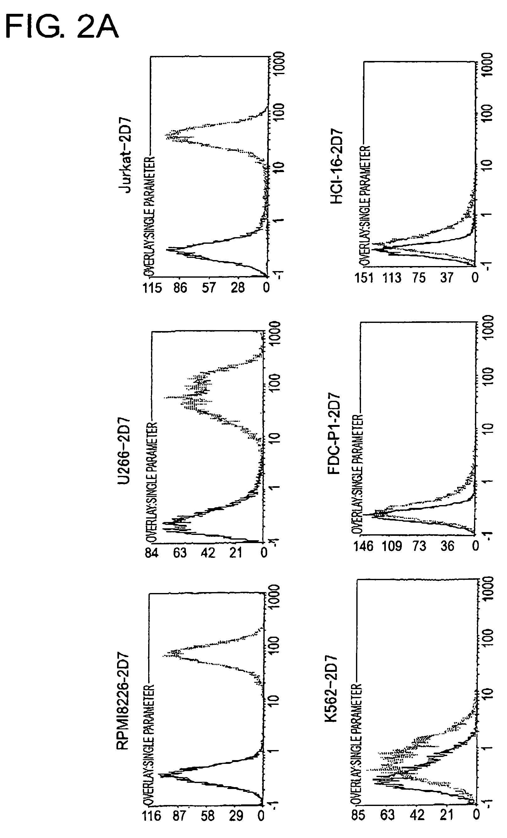
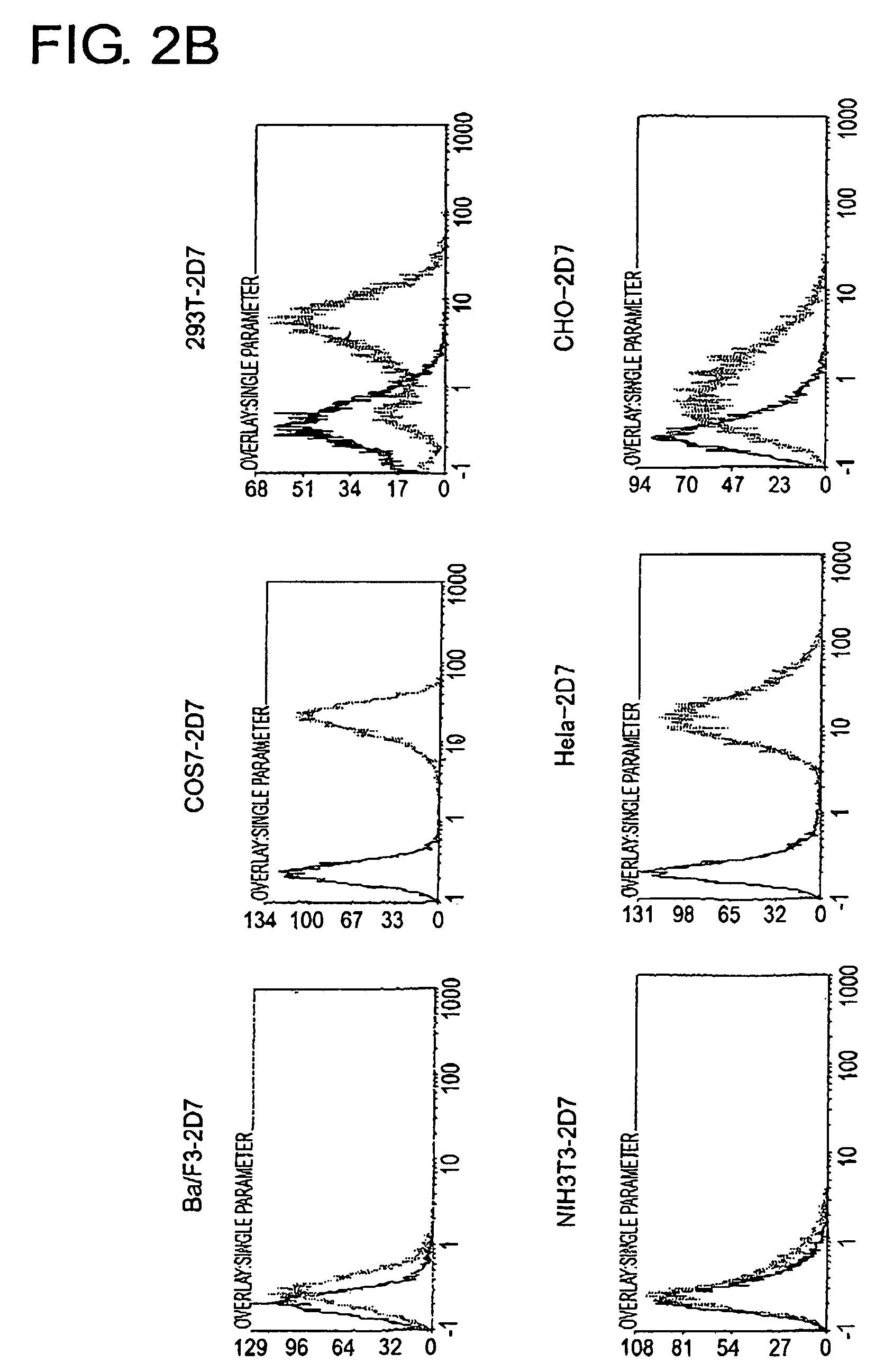
[0162]To determine the cell line that should become the source to produce a cDNA expression library and the cell line that should become the host, 2D7 antigen expression in each type of animal cell was analyzed using FACS (FIG. 2A and FIG. 2B). As a result, among human-derived white blood cells, extremely strong expression of the 2D7 antigen was observed in lymphocytic tumor cell lines, RPMI8226, U266, and in Jurkat, but expression was found to be weak in K562. In Ba / F3, FDC-P1, and HCI-16, which are white blood cells derived from mice, expression was very weak, perhaps due to differences between the species. Of the adherent cells, expression was observed in COS7, 293T, and HeLa Expression was hardly observed in mouse NIH3T3 cells.

[0163]From the expression patterns mentioned above, RPMI8226 cells were judged to be appropriate as a source of a cDNA library to be used for expression cloning, and NIH3T3 cells were determined t...

example 2

Cloning of 2D7 Antigen

[1] Cloning from a Protein

[0164]Cell lysates were prepared from RPMI8226 cells and U266 cells, which express the 2D7 antigen, and NIH3T3 cells, which do not express the 2D7 antigen, and immunoprecipitation was performed using the 2D7 antibody. As a result, a molecule (approximately 12 kD) that precipitates specifically in RPMI8226 and U266 cells was observed (FIG. 3). This molecule was not detected by Western blotting using the 2D7 antibody, but since it is at least reproducibly precipitated by the 2D7 antibody, it was strongly predicted to be the 2D7 antigen itself, or a molecule that co-precipitates with the 2D7 antigen.

[0165]Coomassie staining was performed on this band; it was then cut out and the peptides were sequenced. As a result, this 12 kD molecule was identified as β2 microglobulin (β2M). Since β2M is one of the class I MHC protein complexes that associate with HLA class I through non-covalent bonds, the 2D7 antibody is considered to have co-precipit...

example 3

Examination of Growth Inhibitory Effect

[0172]Several types of leukemia cell lines (K562, Jurkat, and RPMI8226) were used to investigate whether the 2D7 antibody has a cytocidal effect. The expression level of the 2D7 antigen in the three cell lines is: K562, weakly positive; Jurkat and RPMI8226, strongly positive.

[0173]K562 and Jurkat cells were plated in the presence or absence of PHA and PMA, and 10 μg / ml of the 2D7 antibody was added thereto. On observing the cells 24 hours later, weakly 2D7-positive K562 cells did not show obvious differences in their morphology due to the presence or absence of the 2D7 antibody, however, addition of 2D7 antibody resulted in significant cell aggregation in Jurkat cells strongly expressing 2D7 (FIG. 7B and FIG. 7C). However, growth inhibition due to addition of the 2D7 antibody was not observed (FIG. 7A). Growth inhibition due to 2D7 in Jurkat cells activated by PHA and PMA stimulation was also not observed.

[0174]Unexpectedly, addition of 2D7 ant...


PUM

PropertyMeasurementUnit
temperatureaaaaaaaaaa
body weightaaaaaaaaaa

Abstract

To identify antigens of the 2D7 antibody, the present inventors cloned the 2D7 antigen. The results suggested that the 2D7 antigen is an HLA class I molecule. Based on this finding, the present inventors examined whether the 2D7 antibody has cell death-inducing activity. Nuclei fragmentation was observed when the 2D7 antibody was cross-linked with another antibody, indicating that cell-death was induced. Further, diabodies of the 2D7 antibody were found to have very strong cell death-inducing activities, even without the addition of another antibody. These results indicate that minibodies of an HLA-recognizing antibody can be used as cell death-inducing agents.

Description

[0001]The present invention relates to minibodies of antibodies that recognize HLA.BACKGROUND ART[0002]The HLA class I antigen is formed by a heterodimer of a 45-KD α chain comprising three domains (α1, α2, α3), and a 12-KD β2 microglobulin. The main role of the HLA molecule is to present CD8+T cells with antigenic peptides, formed from about eight to ten amino acids produced inside cells. As such, it plays a very important role in the immune response and immune tolerance induced by this peptide presentation.[0003]By ligating HLA class IA antigens with antibodies, cell growth-suppressing and cell death-inducing effects have been observed in lymphocytes, suggesting that HLA molecules may also be signal transduction molecules.[0004]More specifically, for example, there are reports showing cell growth suppression of activated lymphocytes by the B9.12.1 antibody against the α1 domain of human HLA class IA, the W6 / 32 antibody against the α2 domain, and the TP25.99 and A1.4 antibodies aga...

Claims


Application Information

Patent Timeline
Patent Type & Authority Patents(United States)
IPC IPC(8): C12P21/04C12P21/08A61P35/00A61P37/02A61P43/00C07K14/74C07K16/28
CPCC07K14/70539C07K16/2833A61K2039/505C07K2317/565C07K2317/626C07K2317/73C07K2317/34A61P35/00A61P37/02A61P43/00A61P7/00
Inventor OZAKIABETSUCHIYA, MASAYUKIKIMURA, NAOKIKAWAI, SHIGETO
Owner CHUGAI PHARMA CO LTD
Why Eureka
Try Eureka