Method for determination of protein, peptide or peptoid aggregation, stability, and viability and system using the same

Active Publication Date: 2012-09-18
UNIVERSITY OF PUERTO RICO +1
View PDF0 Cites 29 Cited by
  • Summary
  • Abstract
  • Description
  • Claims
  • Application Information

AI Technical Summary

Benefits of technology

[0019]According to one aspect of the invention, the method involves transmission Fourier transform infrared (FT-IR) and/or attenuated total reflectance (ATR) FT-IR spectroscopy and two-dimensional correlation spectroscopy (2DCOS) for the analysis of these proteins, peptides or peptoids. The graphical treatment provides greater resolution

Problems solved by technology

Aggregation is considered a primary mode of protein degradation, often leading to immunogenicity of the protein and a loss of bioactivity.
The formation of protein aggregates is critical in industrial applications, because it can highly affect the production of protein-based drugs or commercial enzymes, greatly lowering the production yields.
These prior art methods are either de

Method used


Image

  • Method for determination of protein, peptide or peptoid aggregation, stability, and viability and system using the same
  • Method for determination of protein, peptide or peptoid aggregation, stability, and viability and system using the same
  • Method for determination of protein, peptide or peptoid aggregation, stability, and viability and system using the same

Examples


example 1

Epidermal Growth Factor (EGF)

[0043]The first example chosen is a human recombinant mitogenic polypeptide, known as epidermal growth factor (EGF) comprised of amino acids and three disulfide bridges. Its secondary structure is highly conserved primarily beta sheet, turns and random coil. The cellular pathways associated to the binding of these growth-factor polypeptides to growth factor receptors lead to cell proliferation and act in an atypical manner in tumor cells.

[0044]Results of the thermal dependence studies for EGF within the spectral region of 1720-1500 cm−1 are shown in FIG. 3. The decrease in intensity of the amide I band along with a shift to lower wavenumbers from 1645 to 1638 cm−1 and the appearance of a shoulder at 1620 cm−1 is indicative of thermal denaturation and aggregation, decrease of the amide II band associated with side chain modes. Therefore the method serves to determine the melting temperature (Tm) of the polypeptide as well. A plot of the FWHH of the amide ...

example 2

Methionine Adenosyltransferase (MAT)

[0046]In the thermal denaturation of the MAT (Methionine adenosyltransferase) protein (1,584 amino acids in its homotetramer form, for a molecular weight of 198 kDa), apparently a cooperative two-state denaturation process is assumed, as seen at the 3D plot of the deconvolved spectra for MAT in FIG. 7. Further studies using 2DCOS show that it's not a fully two state process, with a more complex denaturation pattern. The aggregation is characterized by the appearance of two bands at 1620 cm−1 and 1680 cm−1.

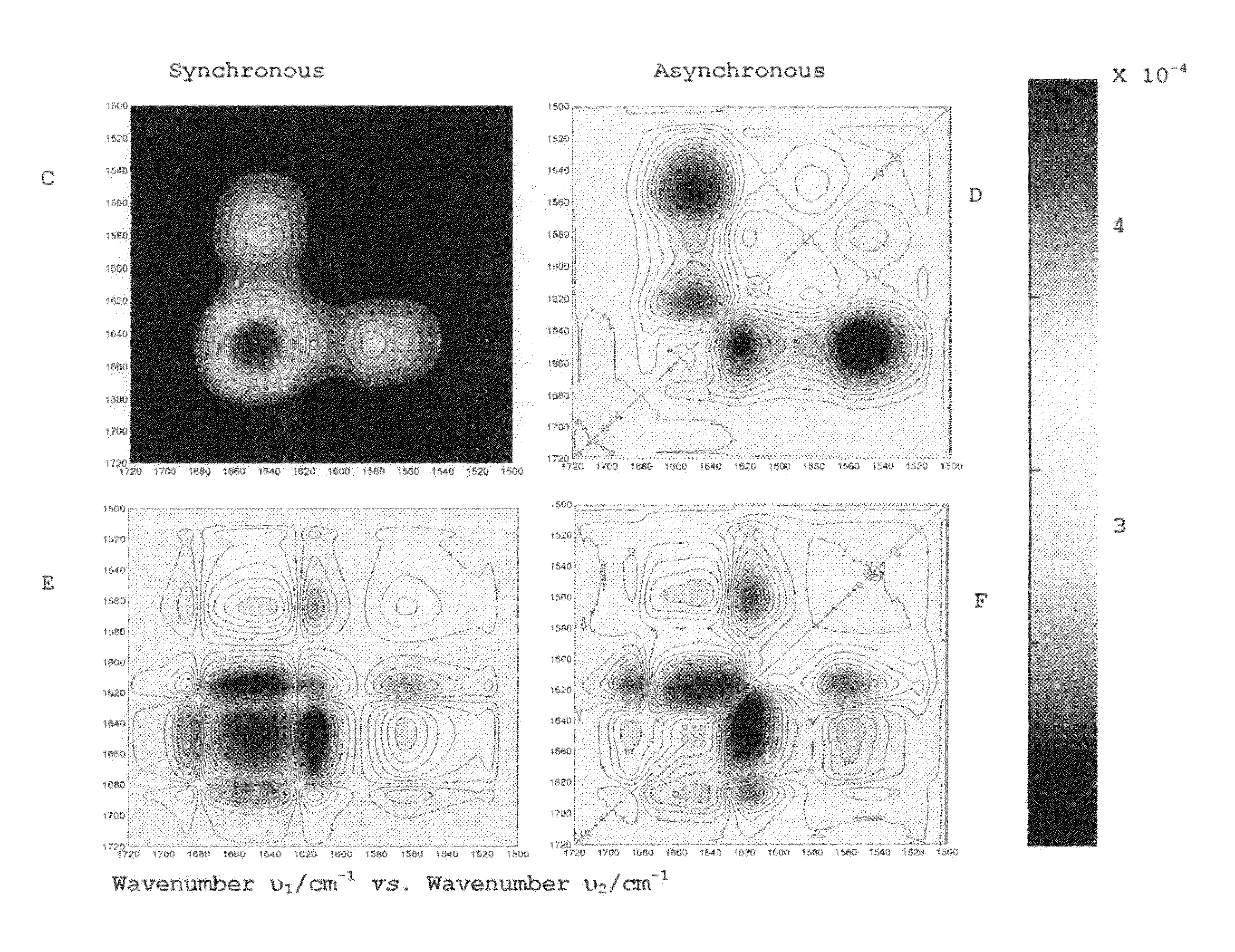
[0047]In the temperature range of 37-53° C., the activity of the protein is being gradually lost. 2DCOS plots as shown in FIG. 8 have been used to show that aggregation has occurred at the expense of the α-helix and β-sheet structural motifs and also of the residual β-turns. The synchronous plot has autopeaks at 1652 cm−1 and 1635 cm−1, with a positive cross-peak between them, showing that both structures (α-helix and β-turn, respectively) are de...

example 3

Insulin

[0048]Insulin is an α-helical peptide with a total of 51 amino acids (about 6.5 kDa). Heating this peptide up to 70° C. produces amyloid fibers, formed by β-sheet structure. FT-IR spectroscopy can be used to study this process as the appearance of a large band at 1620 cm−1 as shown in FIG. 9, associated to fiber formation. Thus, this method is comparably easy when compare to other methods, which require preparing several samples, the use of external probes and does not analyze the secondary structure, so the mechanism of the aggregation can't be studied.

[0049]FIG. 10 shows the asynchronous and synchronous plots summarizing the events related to fiber formation. An autopeak at 1620 cm−1 on the Synchronous plot is associated to the fiber formation with the concomitant loss of the α-helix native structure (1650 cm1). The asynchronous plot shows this process involving a new structure at 1670 cm−1 (β-turns).


PUM


Abstract

The invention describes a method for determining aggregation in protein, peptide or peptoid formulation, without the use of probes or additives. The method uses FTIR spectroscopy combined with the two-dimensional correlation analysis (2DCOS) which allows for the determination of the presence of aggregates, the determination of the mechanism of aggregation, allowing for correction in the pipeline manufacturing process of the protein to once again generate viable protein. In addition, the thermal transition of the protein can also be determined and a 2DCOS plot generated to compare with the established viable protein, allowing for quality control, stability and viability of the desired protein product. The ease of sample preparation and data analysis allows for the automation of this method.

Description

STATEMENT REGARDING FEDERALLY SPONSORED RESEARCH OR DEVELOPMENT[0001]This invention was made with government support under grant numbers NIH-COBRE P20 RR16439-01 (BPR), NIH-SCORE 5-S06GM08103 (BPR) awarded by the National Institute of Health (NIH). The government has certain rights in the invention.BACKGROUND OF THE INVENTION[0002]Proteins are large organic compounds made of amino acids arranged in a linear chain and joined together by peptide bonds between the carboxyl and amino groups of adjacent amino acid residues. The sequence of amino acids in a protein is defined by a gene and encoded in the genetic code. Although this genetic code specifies 20 “standard” amino acids plus selenocysteine and—in certain archaea—pyrrolysine, the residues in a protein are sometimes chemically altered by post-translational modification: either before the protein can function in the cell, or as part of control mechanisms. Proteins can also work together to achieve a particular function, and they of...

Claims


Application Information

Patent Timeline
IPC IPC(8): G01N33/00
CPCG01N33/1826G01N33/18G01N21/3577G01N21/552G01N21/82G01N2021/3595
Inventor PASTRANA-RIOS, BELINDALLORO-MANZANO, IBON
Owner UNIVERSITY OF PUERTO RICO
Why Eureka
Try Eureka