Antibody complexes and methods for immunolabeling

an antibody complex and immunolabeling technology, applied in the field of immunolabeling compositions, can solve the problems of false positives, inability to remove, and inability to direct label antibodies in ascites fluid or other media containing interfering compounds, etc., to achieve the effect of removing excess labeling protein, reducing false positives, and reducing false positives

Inactive Publication Date: 2012-12-04
LIFE TECH CORP
View PDF184 Cites 8 Cited by
  • Summary
  • Abstract
  • Description
  • Claims
  • Application Information

AI Technical Summary

Benefits of technology

[0081]When preparing the immunolabeling complex, stock solutions of both the labeling protein and the target-binding antibody are typically near 1 mg / mL in an appropriate buffer, although more or less concentrated solutions are also suitable. Generally, the labeling protein is mixed in a molar ratio of at least one to 50 moles of labeling protein to one mole of the region of the target-binding antibody to be complexed. More commonly a ratio of at least one to as many as 10 moles of labeling protein per mole of target-binding antibody is combined. With an Fab fragment of an antibody to the Fc region of a target-binding antibody, a molar ratio of approximately 2 to 10 is typical, more typically 3 to 5 (particularly for complexes in which the labeling protein has been labeled while immobilized on an affinity matrix). The ease of formation of the complex permits rapid optimization of the complex and assessment of the effect of variation in experimental parameters. A particularly unique advantage of the invention is that the stoichiometry of the complex is easily adjusted to provide complexes with different ratios of labeling protein to target-binding antibody, and thus there is some control over the ultimate detectability of the target in the sample. This feature is impractical with primary antibodies that have been directly chemically labeled with dyes. Complexes that have been labeled with the same dye but at different molar ratios can be separately detected by the differences in their intensities.
[0082]Complex formation appears to occur almost within the mixing time of the solutions (<1 minute) but the reaction typically is allowed to proceed for at least 5 minutes and can be longer before combining the antibody complex with the sample. Although complex formation can be reversed by addition of an unlabeled antibody that contains the same binding region, reversibility is very slow; furthermore, following staining of the sample with the complex, the sample can be “fixed” using aldehyde-based fixatives by methods that are commonly practiced by those skilled in the art of immunolabeling.
[0083]A unique and important advantage of the method of the invention is the ability to form the immunolabeling complex from exceedingly small quantities of the target-binding antibody and labeling protein, which is impractical when using chemically reactive dyes. Complexes of the invention have been prepared from sub-micrograms of a target-binding antibody with sub-micrograms of a labeling protein. The process of forming a labeling complex can easily be automated. An important use of this characteristic is that of screening hybridoma supernatants in the generation and optimization of new antibodies, where the very small quantity of antibody present in a single well makes it impractical to perform chemical labeling.
[0084]In one aspect of the invention, a capture component is added to the labeling mixture, prior to combining the labeling mixture with the sample, to remove excess labeling protein. For applications in which immunolabeling complexes of multiple primary antibodies from the same species (e.g. mouse monoclonal antibodies) or cross-reacting species (e.g. mouse and human antibodies) are to be used simultaneously or sequentially, it is necessary to quench or otherwise remove any excess labeling protein by use of a capture component or by other means to avoid inappropriate labeling of the sample. The most effective capturing components to capture excess labeling protein are those that contain the binding site of the labeling protein but are themselves not labeled, preferably an antibody or antibody fragment. Capture components may be free in solution or immobilized on a solid phase, such as agarose, cellulose, or a natural or synthetic polymer, to facilitate separation of the excess capture component from the labeled protein. The capture component is optionally attached to a microsphere or magnetic particle. Any of the isolated labeling mixtures optionally include a capture component to remove excess labeling protein. However, separation of excess labeling protein is not essential for successful utilization of the invention, particularly when using a single target-binding antibody.
[0085]Included in the invention are isolated labeling mixtures comprising one or more immunolabeling complexes, wherein at least one immunolabeling complex is comprised of a target-binding antibody, is selective for a target in a sample, and a labeling protein, as described above, that binds selectively and with high affinity to a selected region of the target-binding antibody. More preferably the labeling mixture contains at least three such complexes. The potential number of immunolabeling complexes that can be used simultaneously is principally limited by the number of targets for which selective antibodies are available and the ability to spatially or spectrally resolve the detectable signals, such as on a Western blot, a protein array, a chromosome “spread”, or a microsphere or “chip” array (e.g., U.S. Pat. Nos. 5,981,180 and 5,736,330).
[0086]These labeling mixtures optionally include additional detection reagents such as those known to those skilled in the art, including reagents that localize the same or additional targets in the sample. Preferred additional detection reagents include stains for cell organelles, reagents to indicate the state of cell viability or proliferation, and enzyme substrates. Other preferred additional detection reagents are additional antibodies that can be detected in conjunction with direct or indirect labeling methods, wherein the targets of such additional antibodies and of the target-binding antibody are optionally the same or different.

Problems solved by technology
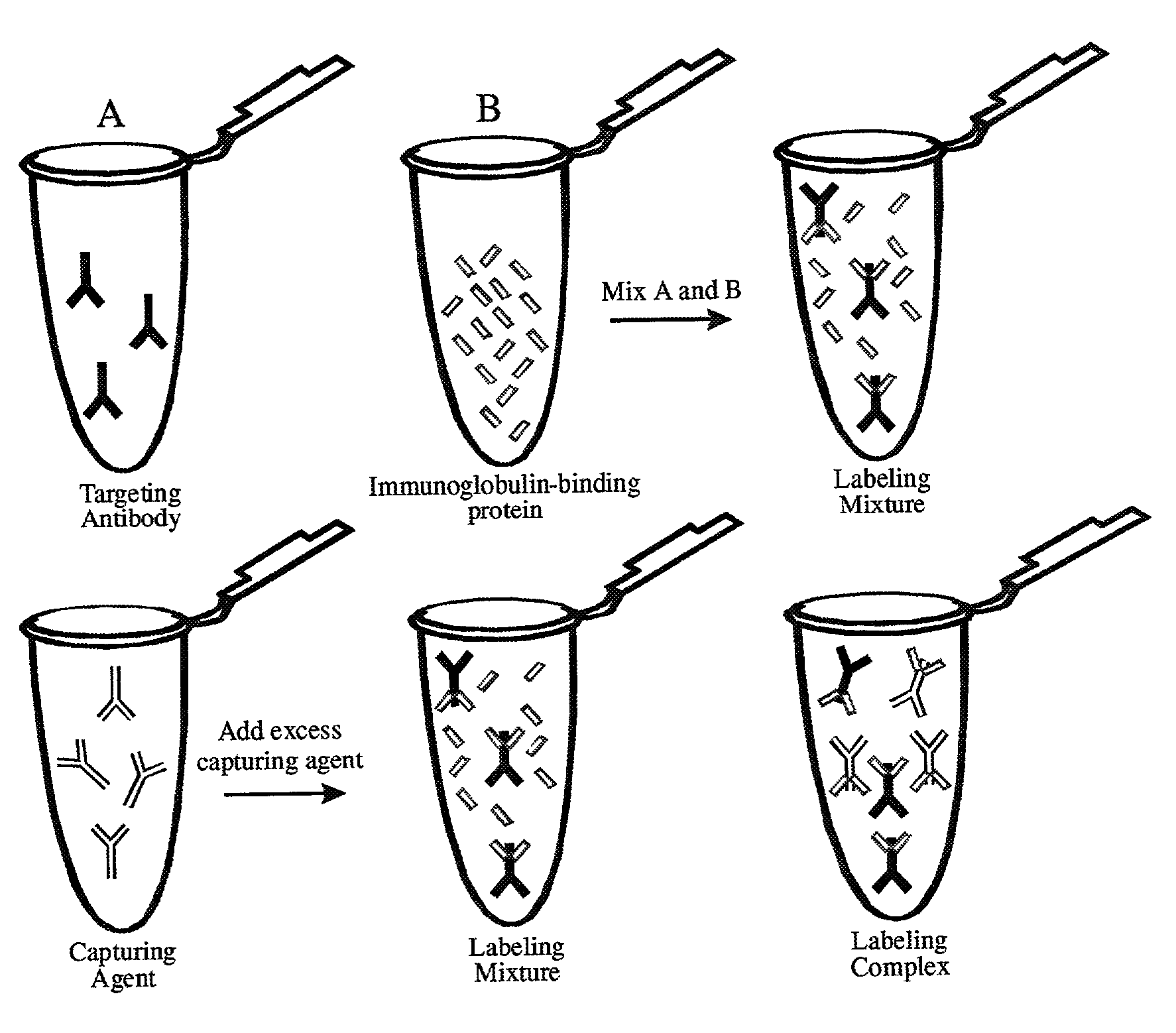
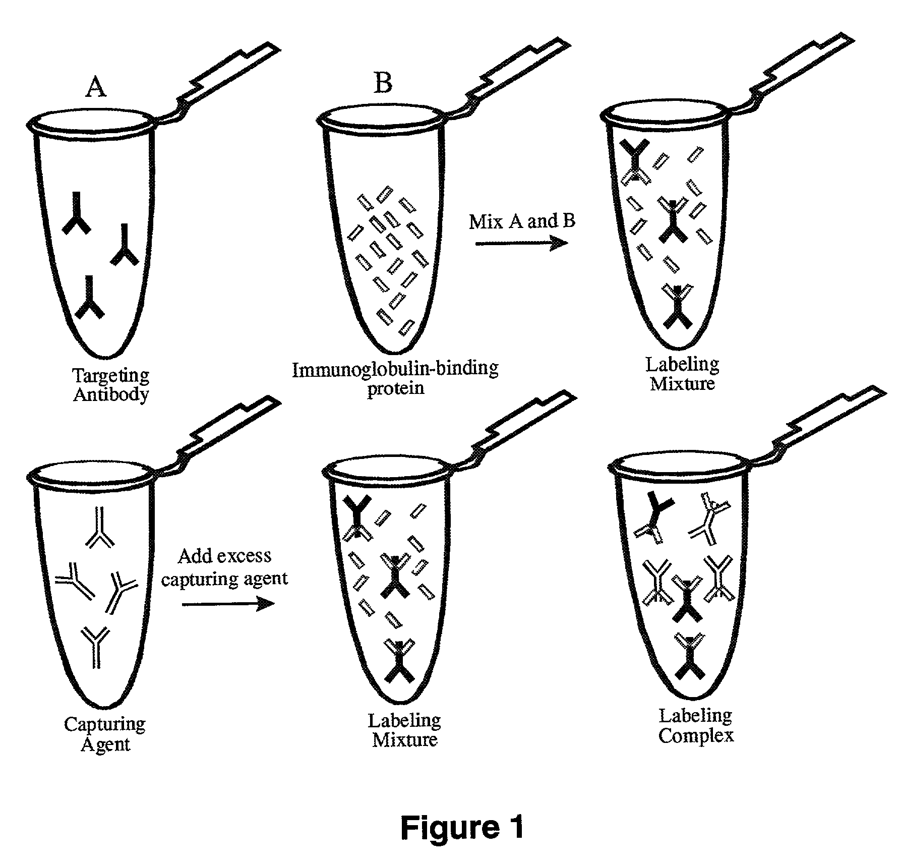
These materials are, however, common components in antibody buffers and purification methods, and it may not be possible or feasible to remove them prior to the coupling reaction.
Thus, direct labeling of antibodies in ascites fluid or other medias containing interfering compounds is not attainable.
Indirect immunolabeling often results in false positives and high background.
This causes a significant background problem, especially in diseased tissues, which reduces the usefulness and sensitivity of the assay.
This method is limited by the necessity to utilize an appropriate ratio of primary-secondary complex.
Too low a ratio of primary-secondary complex will cause a decrease in specific staining and increased background levels due to the uncomplexed secondary anti-mouse antibody binding to endogenous mouse antibodies.
However, the ability of a whole IgG antibody (as was used in the referenced method) to simultaneously bind and crosslink two antigens results in too high a ratio, causing the complex to precipitate or form complexes that are too large to penetrate into the cell or tissue.
This approach requires large quantities of expensive Fab′ fragments and gives mixed results and adds at least two steps (block and wash) to the overall staining procedure.
The addition of a cross-linking reagent has resulted in improved reduction of background levels (Tsao, et al., U.S. Pat. No. 5,869,274 (1997)) but this is problematic when used with fluorophore-labeled antibodies.
This process is complex in that it requires multiple incubation steps and washing steps and it still cannot be used with mouse antibodies to probe mouse tissue.

Method used


Image

  • Antibody complexes and methods for immunolabeling
  • Antibody complexes and methods for immunolabeling
  • Antibody complexes and methods for immunolabeling

Examples


example 1

Preparation of Mouse Fc Antigen

[0118]Purified mouse IgG was fragmented with the proteolytic enzyme papain (CURRENT PROTOCOLS IN CELL BIOLOGY, 16.4.1-16.4.10 (2000)). A 12 mL solution of mouse IgG was prepared at ˜2 mg / mL in phosphate-buffered saline (PBS). A solution containing 0.1 mg of papain in digestion buffer (PBS, 0.02 M EDTA, 0.02 M cysteine) was added to the antibody and allowed to react at 37° C. for 16 hours. The digestion was terminated by the addition 20 μL of 0.3 M iodoacetamide in PBS. The fragments were dialyzed against 2 L of PBS for 16 hours at 4° C. The Fc fragment was purified on a protein G-Sepharose CL-4B column. The bound fraction containing the Fc fragment was eluted from the column using 50-100 mM glycine / HCl buffer, pH 2.5-2.8. The eluate was collected in 1 mL fractions. The pH of the protein fractions was immediately raised to neutral by addition of 100 μL of either 500 mM phosphate or Tris buffer, pH 7.6, to each 1 mL fraction. The solution was then loaded...

example 2

Goat Anti-(Mouse Fc) Production

[0119]To produce polyclonal antibodies, the immunogen used to immunize the animal was the purified mouse or rabbit IgG Fc domain (see Example 1). Methods of immunizing animals are well known and conventional, and suitable immunization protocols and immunogen concentrations can be readily determined by those skilled in the art (Current Protocols in Immunology 2.4.1-9 (1995); ILAR Journal 37, 93 (1995)). Briefly, individual goats were immunized with purified mouse Fc or purified rabbit Fc. The initial immunization in 50% Freund's complete adjuvant (1000 μg conjugate (half subcutaneous, half intramuscularly)) was followed by 500 μg conjugate per goat in Freund's incomplete adjuvant two and four weeks later and at monthly intervals thereafter. Antibodies were harvested from serum using protein A-Sepharose chromatography. Antibodies against mouse Fc isotypes can be prepared by starting with isotype-selected mouse Fc antigens. Rabbits have a single Fc isotyp...

example 3

Preparation of Fab Fragments

[0120]Fragmentation of the goat anti-(mouse Fc) antibody to the monovalent Fab fragment was carried out using the proteolytic enzyme, papain, as described in Example 1. Following dialysis against PBS, the Fab fragment was purified on a protein A-Sepharose CL-4B column. The unbound fraction containing the Fab fragment and the papain was collected. This solution was then loaded onto a Sephacryl S-200 Superfine size-exclusion column and fractions corresponding to a molecular weight of ˜50 kDa were collected and analyzed by SDS-PAGE. The Fab fragments of goat anti-(rabbit Fc) can be prepared similarly.


PUM

PropertyMeasurementUnit
molar ratioaaaaaaaaaa

Abstract

The present invention provides novel immunolabeling complexes and certain components of such complexes, as well as methods of preparing and using such complexes, and kits for use in preparing labeling proteins and for immunolabeling. The pre-formed immunolabeling complexes of the invention comprise both a target-binding antibody and a labeling protein that contains covalently attached labels, where the labeling protein binds selectively and with high affinity to a selected region of the target-binding antibody. Novel labeling proteins of the invention include non-antibody peptides and proteins, such as a complex of protein G and a labeled albumin, and monovalent antibody fragments, such as labeled Fab fragments of an anti-Fc antibody. In methods of the invention, the preformed immunolabeling complexes are added to the sample alone or in combination, for purposes of labeling and optionally detecting the target of interest.

Description

CROSS-REFERENCE TO RELATED APPLICATIONS[0001]This application claims priority to provisional applications, Ser. No. 60 / 329,068, filed Oct. 12, 2001 and provisional application No. 60 / 369,418 filed Apr. 1, 2002, which disclosures are herein incorporated by reference.FIELD OF INVENTION[0002]The present invention relates to novel immunolabeling compositions and methods for use in the detection and measurement of one or more targets in a biological sample. The invention has applications in the fields of molecular biology, cell biology screening, immunohistochemistry, diagnostics, and therapeutics.BACKGROUND OF THE INVENTION[0003]Immunolabeling is a method for qualitative or quantitative determination of the presence of a target in a sample, wherein antibodies are utilized for their specific binding capacity. The antibodies form a complex with the target (antigen), wherein a detectable label is present on the antibody or on a secondary antibody. The detectable label is a key feature of i...

Claims


Application Information

Patent Timeline
Patent Type & Authority Patents(United States)
IPC IPC(8): G01N33/53G01N33/58G01N33/68
CPCB82Y5/00B82Y10/00B82Y30/00G01N33/53G01N33/58G01N33/6857
Inventor ARCHER, ROBERT A.BEECHEM, JOSEPH M.HAGEN, DAVID C.HAUGLAND, RICHARD P.HAUGLAND, ROSARIA P.
Owner LIFE TECH CORP
Why Eureka
Try Eureka