
Methods of detection GPVI

a technology of platelet activation and detection method, which is applied in the direction of instruments, extracellular fluid disorder, peptide/protein ingredients, etc., can solve the problems of inconsistentness, difficult to obtain consistent, and inability to meet the clinical efficacy of the drug, so as to assess the susceptibility to such a disease and evaluate the risk of developing

Inactive Publication Date: 2012-12-04
MOCHIDA PHARM CO LTD
View PDF22 Cites 2 Cited by
  • Summary
  • Abstract
  • Description
  • Claims
  • Application Information

AI Technical Summary

Benefits of technology

This approach provides a reliable, sensitive, and consistent method for detecting platelet activation and vascular endothelial injury, aiding in the diagnosis of thrombotic diseases and selecting appropriate antiplatelet drug responders, with improved detection sensitivity down to 1.0 pg / mL or less.

Problems solved by technology

However, the clinical efficacy have by no means come up to satisfactory levels.
However, these require consideration from the influence of the blood sampling technique and the fact that they assume high values when renal function declines, and thus they are not always categorized as an appropriate markers.
However, sample handling can cause a pseudo-positive result for the expression of P-selectin in platelets, making it difficult to obtain consistent results.
However, because P-selectin originates in platelets and vascular endothelial cells, the problem of its origin arises when the soluble P-selectin in plasma is measured.
However, a number of problems have been noted here, such as (1) differences in sensitivity between instruments, (2) how the microparticle region is gated, (3) the fact that detection limit is near the size of the microparticles, and (4) differences between facilities based on the antibody used, and standardization of the measurement method has not been achieved.
However, this phenomenon was artificially induced ex vivo under conditions that can almost never occur physiologically, and it was completely unclear as to whether the shedding of platelet GPVI is actually occurred in vivo, under physiological or pathological conditions.
Moreover, the detection of platelet GPVI and sGPVI by western blotting is itself encumbered by a number of problems with regard to its generalization into the clinical setting, such as preparation of the sample for testing and detection sensitivity.

Method used


Image

  • Methods of detection GPVI
  • Methods of detection GPVI
  • Methods of detection GPVI

Examples


example 1

Cloning of the Rat GPVI Gene

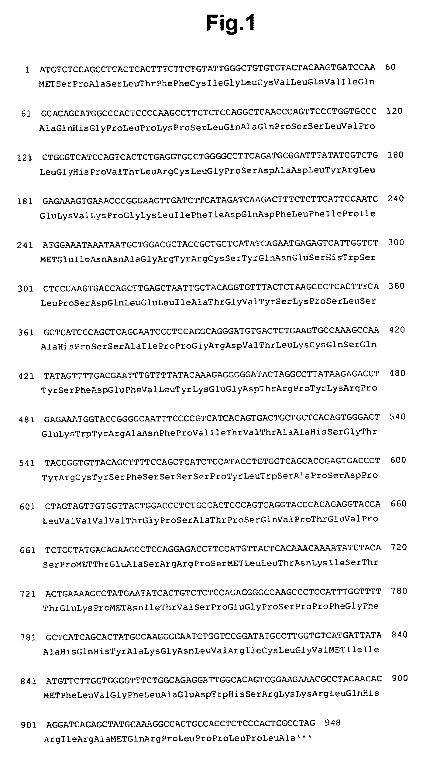
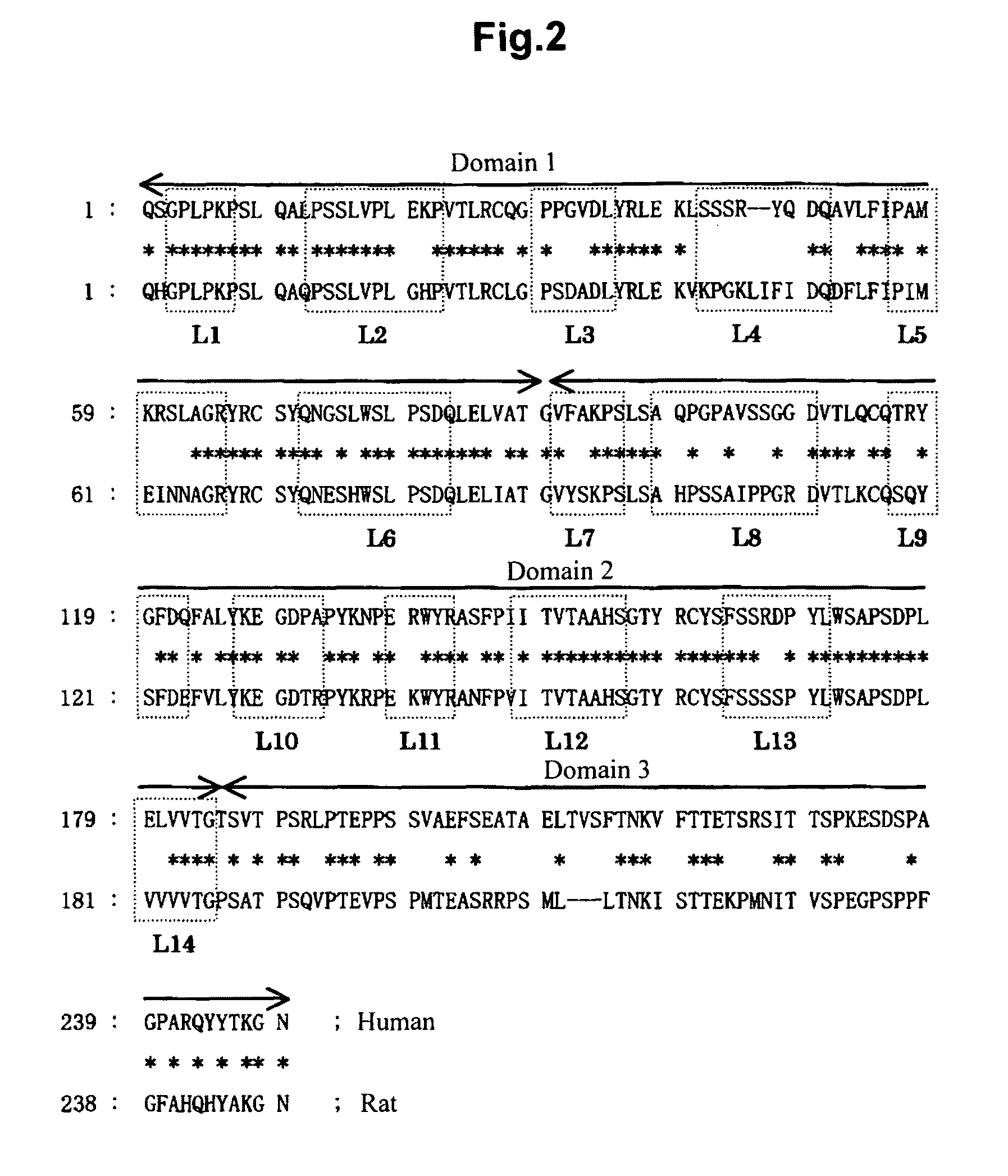
[0199]First of all, primers (6 pairs) capable of amplifying each of the exons of the mouse GPVI gene were designed based on the known data of the mouse GPVI gene. With these primers, PCRs were carried out using rat genomic DNA as the template; the gene fragments that were specifically amplified were sequenced; and the nucleotide sequence of rat GPVI gene was estimated by connecting them. Next, based on this nucleotide sequence data, primers for rat GPVI (ratGPVI-#a, mGPVI-d) were redesigned, and the full-length rat GPVI gene was amplified by PCR using rat bone marrow cDNA (reverse transcripted from rat bone marrow RNA with oligo dT primer) as the template. The amplificated product was extracted from the gel and was cloned into TA-cloning vector pT7-Blue(T) (TAKARA BIO INC.), and its nucleotide sequence was determined. It was confirmed to be the same as the estimated sequence; this plasmid was named pTK-2478. The nucleotide sequence of the rat GPVI gene is...

example 2

Construction of a Rat GPVI (D1D2) Mouse GPVI (D3)-Mouse Fc Fusion Protein (rGPVI-mFc) Expression Plasmid

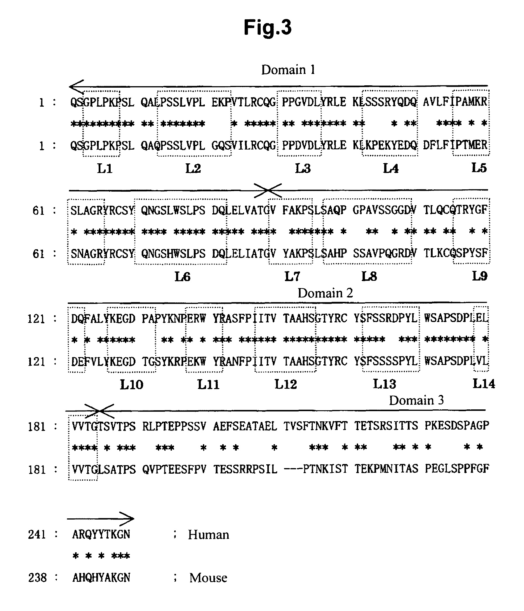
[0201]Using pTK-2478 as the template, a fragment ‘A’ encoding D1 and D2 of rat GPVI (extracellular domain 1 and domain 2 of GPVI) was amplified by PCR with the primer pair (rat GPVI-#a and rat GPVI-#t). Similarly, using pTK-2440 (described in WO 2006 / 117910 and WO 2006 / 118350) as the template, a fragment ‘B’ encoding D3 of mouse GPVI (region of extracellular domain of GPVI except D1 and D2) was amplified by PCR using the primer pair (rat GPVI-#s and IgG1-i). Then, using a mixture of ‘A’ and ‘B’ as the template, a fragment ‘C’, which was ligated rat GPVI (D1 and D2) and mouse GPVI (D3), was obtained by re-PCR using the primer pair (rat GPVI-#a and IgG1-i). After digestion of the 5′ end of this fragment ‘C’ with Eco RI and the 3′ end with Bam HI, the fragment ‘C’ was inserted into Eco RI and Bam HI sites of the plasmid (pTK-2299: described in WO 2006 / 117910 and WO 2006 / 118350) which...

example 3

Expression and Purification of rGPVI-mFc Fusion Protein

[0204]COS-1 cells were maintained in Dulbecco's MEM culture medium containing 10% fetal bovine serum. After mixing of the transfection reagent (FuGENE6, Roche Diagnostics) and serum-free Dulbecco's MEM culture medium, suitable amount of pTK-2483 was added and mixed, and this mixture was added to COS-1 cells that had been medium-exchanged to Hybridoma-SFM culture medium (Gibco). The supernatant was collected after cultivation for 3 days at 37° C. / 5% CO2; cultivation was then carried out for an additional 3 days in fresh culture medium; and these supernatants were purified with a protein A column (Prosep-vA, Millipore) to yield antigen for use in the preparation of anti-rat GPVI antibody. The obtained rGPVI-mFc fusion protein was subjected to SDS-PAGE analysis by an established method. In brief, the sample and molecular weight markers (Bio-Rad, Precision Plus Protein Unstained Standard) were applied to a 5 to 20% gradient gel foll...


PUM

PropertyMeasurementUnit
concentrationaaaaaaaaaa

Abstract

The present invention provides a convenient and highly sensitive method of determining sGPVI present in plasma; this is accomplished by establishing a plurality of mouse hybridomas that produce antibody against GPVI and combining the antibodies produced therefrom. Provided thereby are a novel platelet activation marker, a reagent and method for determining this novel platelet activation marker, and novel applications of this marker in, for example, the diagnosis of diseases associated with platelet activation / vascular endothelial injury.

Description

[0001]This application is a National Stage application of PCT / JP2007 / 056530, filed Mar. 20, 2007, which claims priority from Japanese application JP 2006-100241, filed Mar. 31, 2006.TECHNICAL FIELD[0002]The present invention relates to a novel platelet activation marker, a reagent and method for determining this novel platelet activation marker, and novel applications of the marker to the diagnosis of diseases associated with platelet activation / vascular endothelial injury.BACKGROUND OF THE INVENTION[0003]The leading cause of death in Japan is malignant neoplasm, while the second leading cause is heart disease and the third leading cause is cerebrovascular disease. The majority of heart disease and cerebrovascular disease is accounted for by thrombosis, such as myocardial infarction and cerebral infarction, and together the two of these rival the number of deaths from cancer. The prevention and treatment of thrombosis continues to be an important issue. Controlling platelet activati...

Claims


Application Information

Patent Timeline
Patent Type & Authority Patents(United States)
IPC IPC(8): G01N33/53
CPCC07K16/28C07K2317/34G01N33/86G01N2333/70503G01N2800/32
Inventor HOSAKA, YOSHITAKANAITOH, KATSUKIHONDA, MOTOYASU
Owner MOCHIDA PHARM CO LTD
Learn More