

Engineered human broncho-epithelial tissue-like assemblies
a technology of bronchoepithelial tissue and assembly, which is applied in the field of engineering human bronchoepithelial tissuelike assemblies, can solve the problems of inability to maintain consistent cultures, limited in vivo lung epithelium models, and inability to achieve the functional and structural characteristics of airway epithelium in situ, etc., to achieve rapid vaccine development and improve the efficiency of clinical response and treatment of diseases
- Summary
- Abstract
- Description
- Claims
- Application Information
AI Technical Summary
Benefits of technology
Problems solved by technology
Method used
Image

Examples
example 1
Materials & Methods
[0043]Viruses and strains used in certain embodiments of the invention are set forth in Table 3.
[0044]
TABLE 3VIRUSES & STRAINSVirusGenotypeRefwtRSVA2wild-type human respiratory ATCC VR-1540 ™syncytial viruswtPIV3 JSwild-type parainfluenza ATCC VR-94 ™virus type 3StrainDescriptionRefhBTCprimary human mesenchymal CAMBREXbronchial-tracheal cellsBEAS-2Bhuman bronchial epithelial ATCC CRL-9609 ™immortalized cell lineHEp-2HeLa contaminantATCC CCL-23 ™LLC-MK2KidneyATCC CCL-7 ™
Cell Cultures and Media
[0045]Mesenchymal cells (hBTC) from human bronchi and tracheae were obtained from the lung mucosa of multiple tissue donors through CAMBREX BIOSCIENCES® (Walkersville, Md.). BEAS-2B epithelial cells were obtained from ATCC® (Manassas, Va.). All were harvested and banked at the NASA Johnson Space Center's Laboratory for Disease Modeling and shown to be free of viral contamination by survey of a panel of standard adventitious viruses (e.g. HIV, hepatitis, herpes) conducted by th...
example 2
Growth Kinetics of TLAs
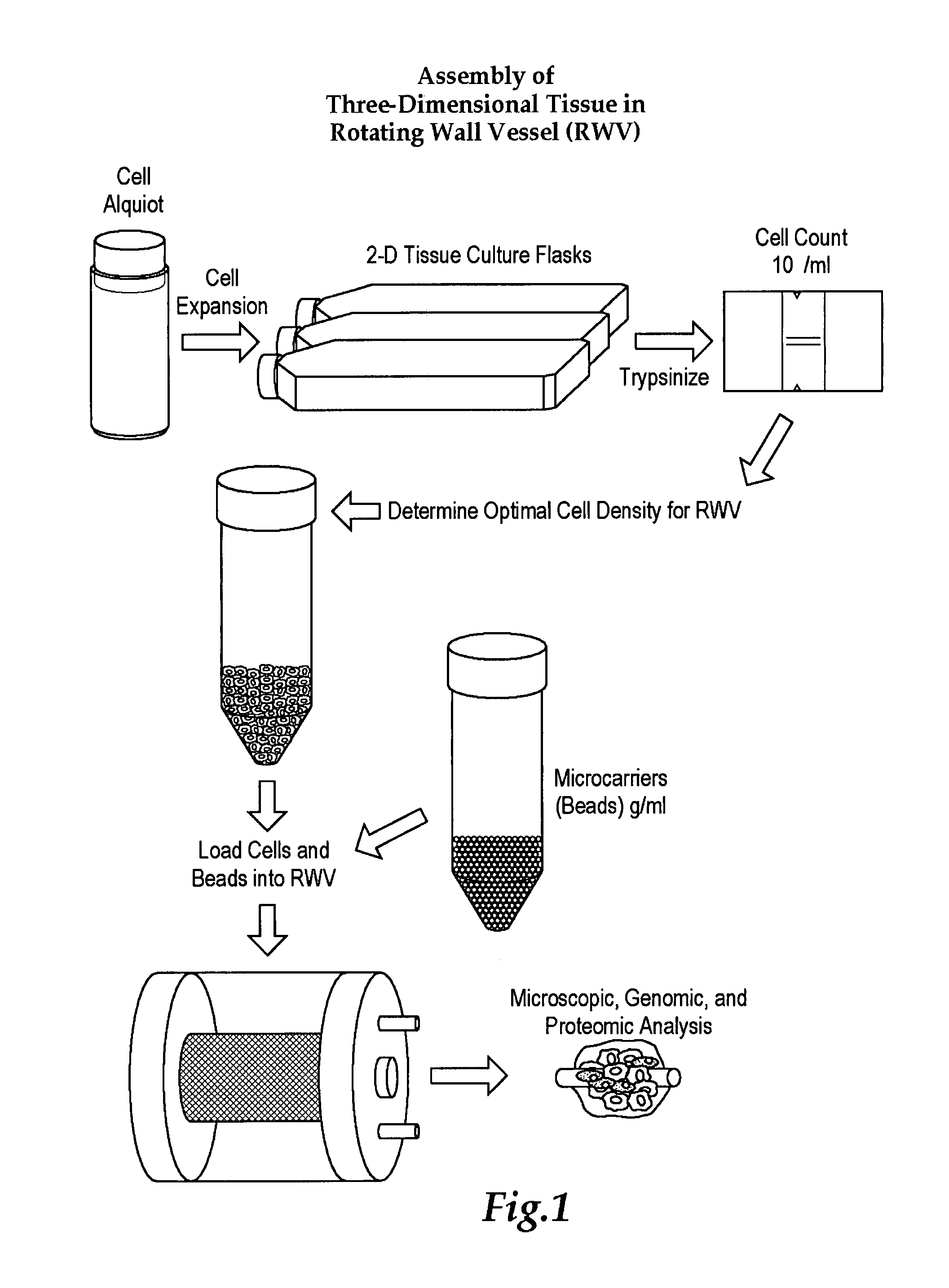
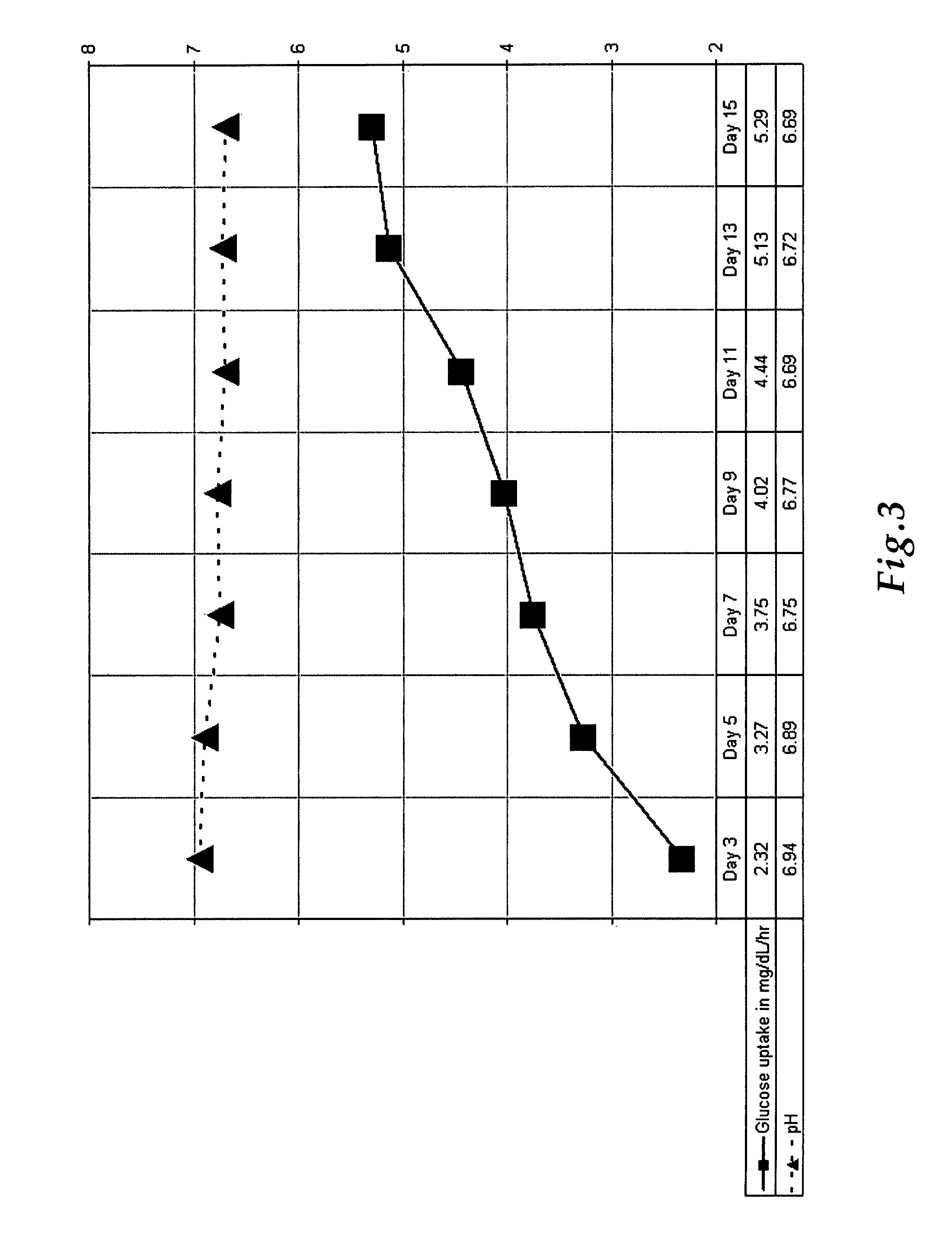
[0054]TLAs were produced by inoculating a RWV using GTSF-2 media with mesenchymal and epithelial lung cells in the presence of microcarriers. In one embodiment an aliquot of lung cells is expanded using 2D tissue culture flasks, trypsinized to dislodge the cells from the conical tube, bring the cells to a known cell density in an RWV with GTSF-2 media, and incubated under microgravity conditions allowing the formation of TLAs. The cultures were monitored at 24-hour time points for glucose utilization and pH. FIG. 3 reflects a typical metabolic profile for these cultures. These data clearly demonstrate rapid uptake of glucose by TLAs with a slight decrease in pH over the initial growth period. Together these factors indicate an increase in cellular metabolism commensurate with an increase in the size of the aggregates.
[0055]In one example, hBTC and BEAS-2B cells were initiated as monolayers in human fibronectin coated flasks and propagated in GTSF-2 media suppl...
example 3
TLAs Express Markers of In Vivo Respiratory Epithelium (IHC)
[0056]To compare the cellular composition and differentiation state of TLAs to normal human respiratory epithelium, fixed TLAs and normal human lung sections were immunostained for epithelial specific cell markers (FIG. 4, Table 4). The cytokeratins (Ke, 1988; Sutherland, 1988) (FIG. 4G, FIG. 4H, FIG. 4O, FIG. 4P) and Factor VIII (FIG. 4I, FIG. 4J) antibodies detect epithelial, mesenchymal, and endothelial cells, respectively (Tsao, et al, 1992, Moyer, 1990, Woodcock-Mitchell, et al, 1982, Vogel, et al, 1984, Shima, et al 1988). Tubulin (FIG. 4E, FIG. 4F), is a cytoskeletal protein found in epithelial cells. Endothelial markers, PECAM-1 (FIG. 4A, FIG. 4B) and Factor VIII (FIG. 4I, FIG. 4J), are present in subsets of precursor endothelial cells, particularly dividing cells. Basement membrane and extracellular matrix components (e.g., collagen IV; FIG. 4Y, FIG. 4Z) were also assayed to determine their expression in the TLAs. ...
PUM
 Abstract
Description
Claims
Application Information