Dielectric spectroscopy assays for screening of ion channel ligands

a technology of ion channel ligand and dielectric spectroscopy, which is applied in the direction of resistance/reactance/impedence, instruments, library screening, etc., can solve the problems of limiting the clamping technique, not easily automated, and limiting the optical detection system

Active Publication Date: 2013-04-16
PURDUE PHARMA LP +1
View PDF3 Cites 15 Cited by
  • Summary
  • Abstract
  • Description
  • Claims
  • Application Information

AI Technical Summary

Problems solved by technology

The patch clamp technique is significantly limited by its low throughput.
Further, it is not easily amenable to automation.
Similarly, optical detection systems are limiting in that they require the use of one or more dyes and do not provide the ability to regulate, or clamp, the transmembrane potential of a cell during the measurement.
However, most either become non-reliable in the low frequency range or involve extremely large electric fields.
However, dielectric spectroscopy has not been shown to be useful for measuring membrane potential of mammalian cells, particularly in a screening or high throughput assay.
However, the theory has not been sufficient to accurately and quickly calculate the membrane potential until now.

Method used


Image

  • Dielectric spectroscopy assays for screening of ion channel ligands
  • Dielectric spectroscopy assays for screening of ion channel ligands
  • Dielectric spectroscopy assays for screening of ion channel ligands

Examples


example 1

[0183]hERG cells used in the experiments are treated following the standard protocol: incubated at 37 degrees at 5% CO2 in DMEM media (with 10% fetal calf serum, penicillin streptomycin and 2 mM L-glutamine). When 80% confluent they are split and re-incubated. When the cells are needed for the experiment the flasks is treated with trypsin, incubated for 10 minutes. Trypsin is an enzyme that removes the cells from the flasks by cleaving. The cells are then gently diluted into DMEM media with 20% fetal calf serum (to annihilate the effects of trypsin) then span at 1000 g for 8 minutes. The pellet is gently and slowly resuspended in Tyrode's buffer for dielectric measurements.

example 2

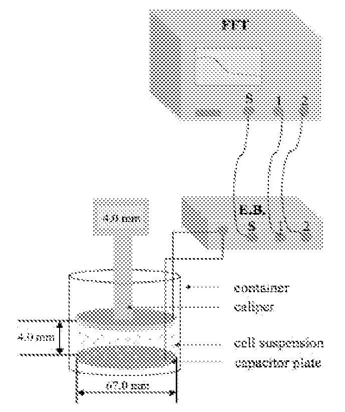
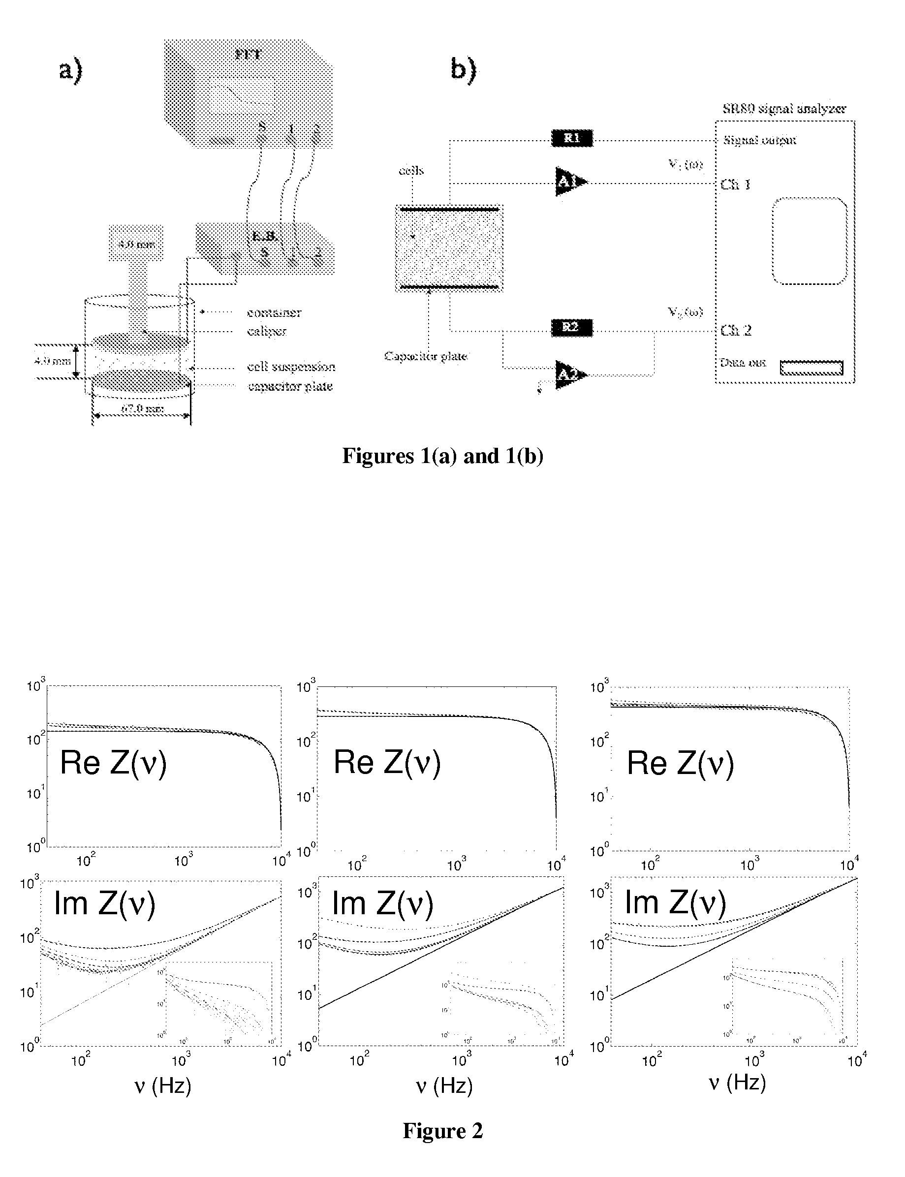
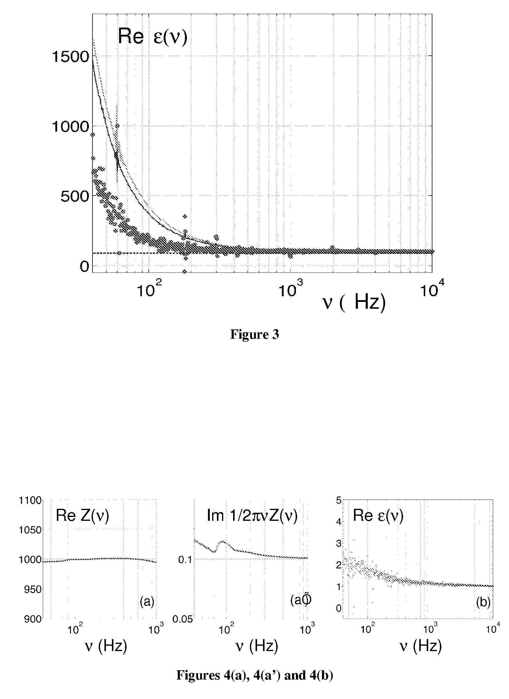
[0184]The solution to be measured is placed in a small, 30 ml glass beaker with 2 gold-plated flat circular plate electrodes as shown in FIG. 1. A Signal Analyzer SR80A provides a sinusoidal voltage at its signal output. It also digitizes the voltage at Channels 1 and 2 and takes the ratio of these two as function of frequency. The real and imaginary of this ratio is stored on a computer. The bottom electrode plate is held at ground potential through the negative input of the Amplifier A2. The output voltage from the Signal Analyzer is applied to the upper electrode, through resistor R1. As a result, the current I that flows through the cell suspension produces a voltage V1, equal to the product of I and impedance Z of the probe. The voltage V2 is equal to minus the product of I and resistance R2. Thus the transfer function, T, is directly related to the cell suspension impedance by: T=V1 / V2=Z / R2. The complex dielectric function is then extracted from Z. All the electronic component...

example 3

Activity of hERG Ion Channels in the Presence of Antiarrhythmic Agents and Antihistamines with Carditoxicity Effects

[0185]The hERG ion channel activity is studied on HEK-293 cell line with overexpresses hERG ion channels. Antiarrhythmic agents tested include quinidine, a class 1 agent, and propranolol, a class 2 agent. An antihistamine agent, astemizole, was also tested. For these compounds, the membrane potential variation was determined by dielectric spectroscopy and the IC50 curve is compiled. The results from the dielectric spectroscopy are then closely compared with patch clamping experiments done in parallel. The compounds analyzed herein were chosen to test a large range of potency. For example, 2 μLM quinidine is needed to block 50% of the hERG channels while only 1.3 nM astemizole is required to produce the same effect.

HEK-293 cells: Determining Drug Potency Using DS. Comparison with Traditional Methods

[0186]The cells used in the experiments were treated following the stand...


PUM


Abstract

A method for measuring membrane potential using dielectric spectroscopy is described. A new theoretical model allows for the determination of membrane potential from low-frequency impedance measurements to provide a non-evasive method which is both rapid and inexpensive.

Description

[0001]This application claims priority under 35 U.S.C. §119(e) to U.S. Provisional Patent Application Ser. No. 60 / 953,179 filed Jul. 31, 2007. This application is incorporated herein by reference in its entirety.FIELD OF THE INVENTION[0002]The present invention relates generally to methods for measuring membrane potentials of living mammalian cells using dielectric spectroscopy.BACKGROUND OF THE INVENTION[0003]Finding new drugs which have specific modulatory effects on ion channels requires methods for measuring and manipulating the membrane potential of cells with the ion channels present in the membrane. A number of methods exist today that can be used to measure cell transmembrane potentials and to measure the activities of specific ion channels. Probably the best known approach is the patch clamp, originally developed by Neher, Sakmann, and Steinback. (The Extracellular Patch Clamp, A Method For Resolving Currents Through Individual Open Channels In Biological Membranes”, Pflueg...

Claims


Application Information

Patent Timeline
Patent Type & Authority Patents(United States)
IPC IPC(8): G01R27/08
CPCG01N33/5008G01N33/6872
Inventor PRODAN, CAMELIAILYIN, VICTOR I.KYLE, DONALD J.CARLIN, KEVINWU, GANG
Owner PURDUE PHARMA LP
Why Eureka
Try Eureka