Ophthalmic microsurgical system
a microsurgical and ophthalmology technology, applied in the field of microsurgical systems, can solve the problems of surgical treatment, marked increase in intraocular pressure, and low success ra
- Summary
- Abstract
- Description
- Claims
- Application Information
AI Technical Summary
Benefits of technology
Problems solved by technology
Method used
Image
Examples
example 1
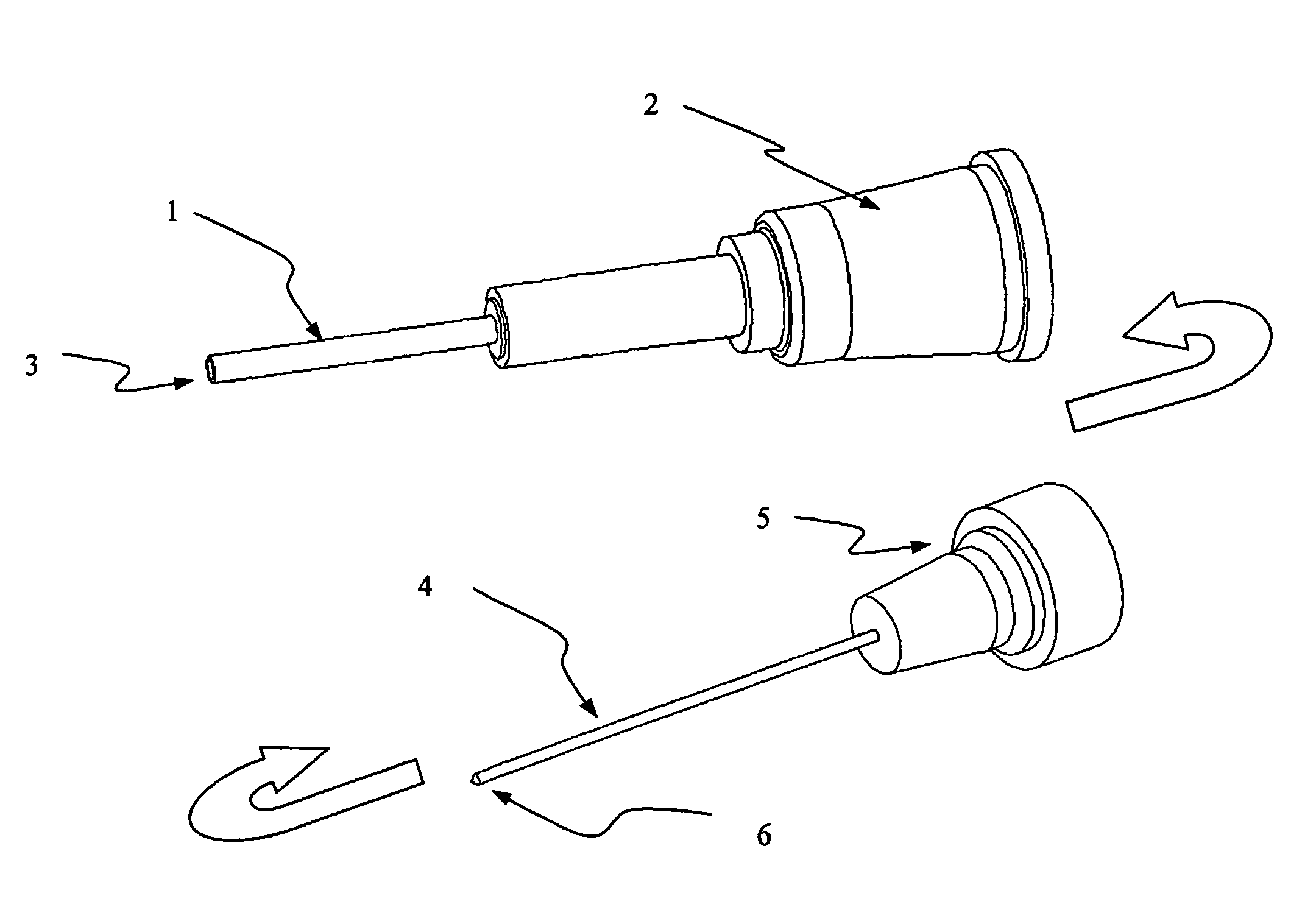
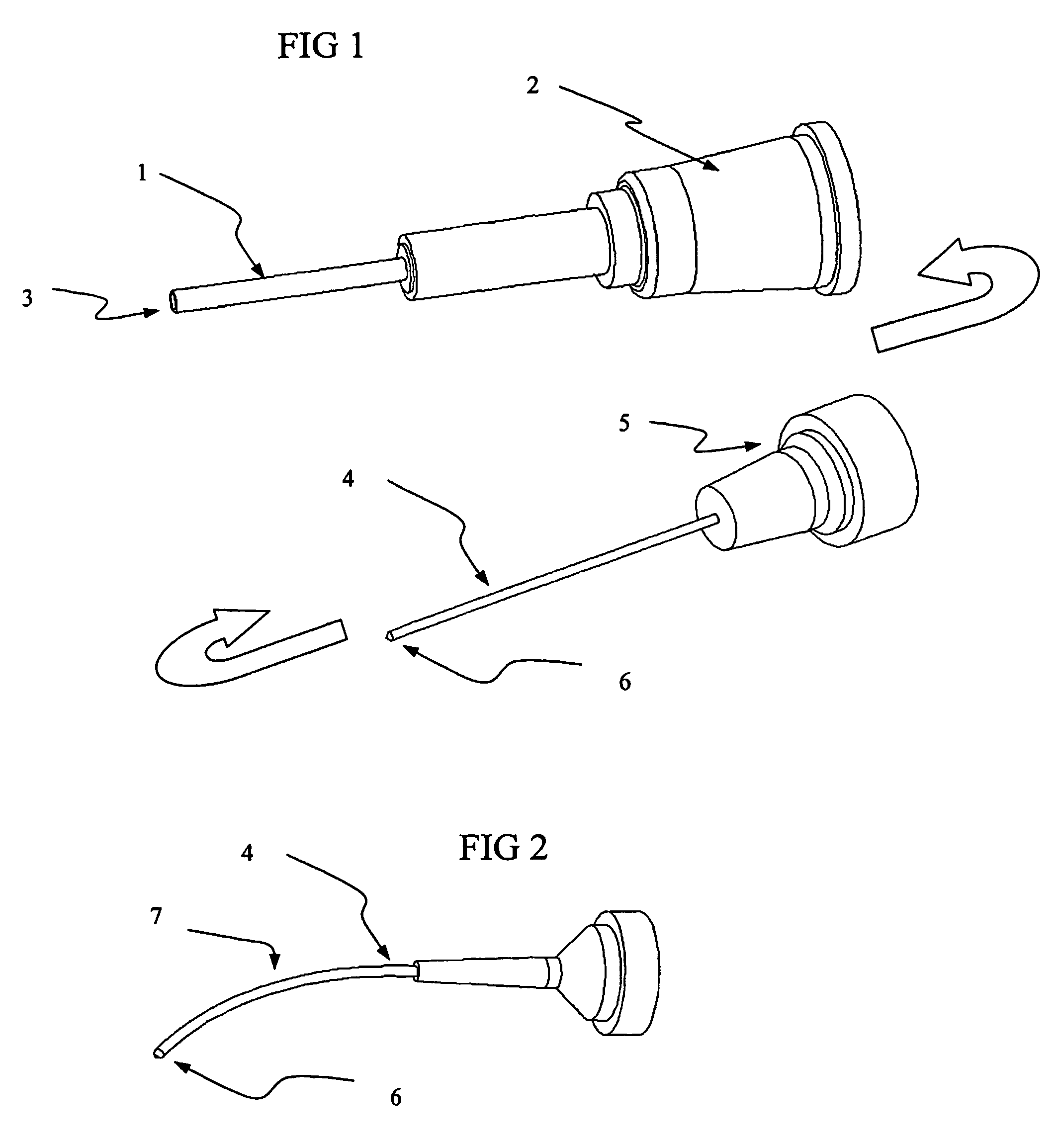
[0045]A microcannula system was fabricated for experimentation on ex-vivo human eyes obtained from an eye bank. The microcannula consisted of a 30 gauge tubing adapter (Small Parts, Inc., Miami Lakes, Fla.) with a distal tip comprised of polyimide tubing bonded into the lumen of the tube adapter. The tube adapter is a standard hypodermic needle, cut to ½″ (12.5 mm) length with a perpendicular (straight) cut distal end and a female Luer at the proximal end. The tube adapter has an inner diameter of 150 microns and an outer diameter of 300 microns. A section of polyimide tubing (MicroLumen, Tampa, Fla.) with inner diameter of 110 microns and a wall thickness of 14 microns was bonded into the distal tip of the tube adapter with cyanoacrylate adhesive and allowed to cure overnight. Assemblies were fabricated with 1.0 and 1.5 cm of polyimide tubing extending from the tube adapter. A 2 cm section of stainless steel wire (Fort Wayne Metals, Fort Wayne, Ind.) 100 microns diameter was mounte...
example 2
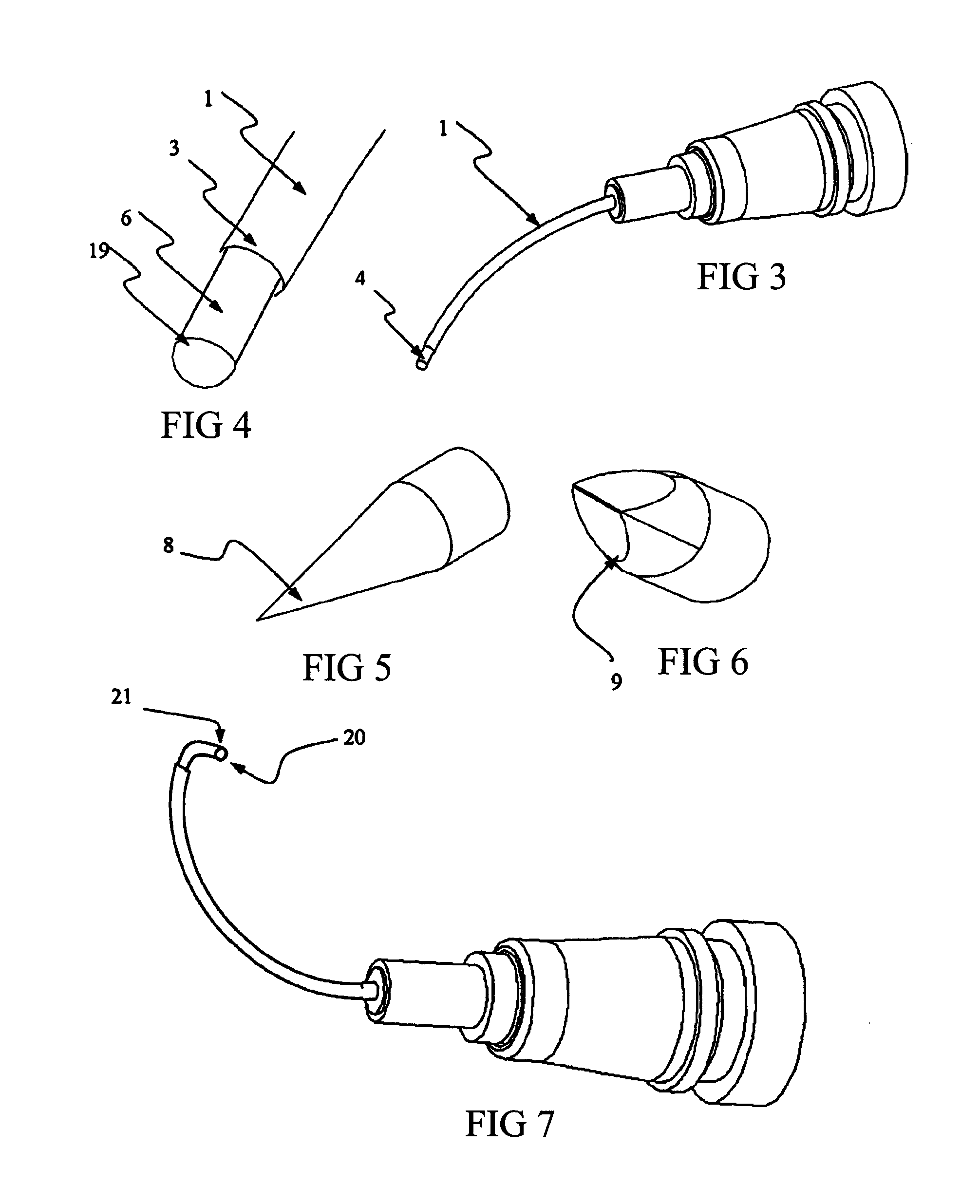
[0049]In another example, a surgical tool to provide for controlled punctures in the trabecular meshwork was created using Nitinol (nickel titanium alloy) wire, 0.004″ (100 microns) diameter (Ft. Wayne Metals, Ft. Wayne, Ind.). The wire was formed with a 10 mm diameter curve for the distal 3 cm. The distal 2 mm of the tip was further formed with a small radius bend at approximately 90 degrees from the axis of the wire, directed toward the inside and remaining in the plane of the curve.
[0050]A microcannula was fabricated comprised of a 3 cm long polyimide tube (Microlumen, Tampa, Fla.), with an inner diameter of 140 microns and an outer diameter of 200 microns, adhesively bonded to a section of 26 gauge hypodermic tubing (Small Parts, Inc, Miami Lakes, Fla.). The hypodermic tubing was mounted in a short plastic sleeve for ease of manipulation. The polyimide tubing was heat set with a curvature of approximately 2.5 cm. A stainless steel guiding sheath was fabricated from sections of h...
example 3
[0052]In another example, a signaling means for determining the location of the microcannula distal tip was fabricated. A small battery powered laser diode light source illuminator was constructed, with the diode operating in the visible red light range. A single plastic optical fiber (POF) (South Coast Fiber Optics, Achua, Fla.) of approximately 100 microns in diameter and 20 cm in length was mounted to an adapter which provides adjustable alignment capabilities to bring the fiber tip into the focus of the laser illuminator. The POF distal tip was cut flat, and hence the illumination was directed toward all radial angles from the tip. A cylindrical handpiece mount was fabricated to hold a microcannula. The microcannula was constructed of nylon with dimensions of approximately 120 microns inner diameter and 180 microns outer diameter. The operative end of the microcannula was 15 mm in length and the proximal end was flared for mounting on the handpiece. The fiber is disposed through...
PUM
| Property | Measurement | Unit |
|---|---|---|
| outer diameter | aaaaa | aaaaa |
| diameter | aaaaa | aaaaa |
| angle | aaaaa | aaaaa |
Abstract
Description
Claims
Application Information
 - R&D
- Intellectual Property
- Life Sciences
- Materials
- Tech Scout
- Unparalleled Data Quality
- Higher Quality Content
- 60% Fewer Hallucinations
Browse by: Latest US Patents, China's latest patents, Technical Efficacy Thesaurus, Application Domain, Technology Topic, Popular Technical Reports.
© 2025 PatSnap. All rights reserved.Legal|Privacy policy|Modern Slavery Act Transparency Statement|Sitemap|About US| Contact US: help@patsnap.com


