Treating inflammatory diseases with antibodies that inhibit fractalkine-CXCR1 interaction

a technology of fractalkine and cxcr1 is applied in the field of inflammatory disease treatment agents, which can solve the problems of inability to inhibit the function of these cells, no specific report on whether the production of tnf and inos by these cells is inhibited, and no promising drug that inhibits the inos activity has been launched. to achieve the effect of improving pathological conditions and inhibiting inos production

Inactive Publication Date: 2013-08-27
EISIA R&D MANAGEMENT CO LTD
View PDF32 Cites 1 Cited by
  • Summary
  • Abstract
  • Description
  • Claims
  • Application Information

AI Technical Summary

Benefits of technology

[0042]That is, investigation was performed by using antibodies inhibiting the interaction of fractalkine and CX3CR1 and the function of CX3CR1 in various animal models described in the examples mentioned later, and as a result, it was found for the first time that they inhibited iNOS production and improved pathological conditions. Further, although it had been reported that the antibodies inhibiting the interaction of fractalkine and CX3CR1 and the function of CX3CR1 are effective for ConA-induced hepatitis, it was found for the first time that they inhibited expression of iNOS mRNA in the liver in ConA-induced hepatitis and iNOS production in inflammatory cells. On the basis of these findings, they showed that an antibody inhibiting the interaction of fractalkine and CX3CR1 and the function of CX3CR1 or a compound inhibiting the interaction of fractalkine and CX3CR1 and the function of CX3CR1 (henceforth also referred to as “CX3CR1 antagonist”) was useful for the treatment of inflammatory diseases associated with the fractalkine-CX3CR1 cell infiltration system and caused by excessive NO production by the iNOS activity or inflammatory cells (inflammatory bowel diseases [in particular, ulcerative colitis and Crohn's disease], psoriasis, atopic dermatitis, asthma, arteriosclerosis, tissue damage caused by ischemic reperfusion, acute respiratory distress syndrome etc.), and thus accomplished the present invention.

Problems solved by technology

However, functions and significance of the CX3CR1highCCR2− monocytes upon inflammation have not been mentioned, and it is rather said that they are necessary for constitutive supply of tissue macrophages when there is no inflammation, and it is undesirable to inhibit functions of these cells.
However, there has been no specific report about whether production of TNFα and iNOS by these cells is inhibited by inhibition of the functions of CX3CR1.
However, since excessive NO production leads to tissue damage, it is considered that excessive iNOS production in lesions may be a major factor for progressing pathological conditions.
However, no promising drug that inhibits the iNOS activity has been launched.
Further, while inhibition of excessive NO production by inhibitors of enzymatic activity of iNOS improves hypotension commonly observed in septic shock, tissue destruction such as that in vasoparalysis and organ disorders, a risk of blocking the fundamental physiological functions of NO such as regulation of blood pressure and blood flow has been suggested (Non-patent document 26).

Method used


Image

  • Treating inflammatory diseases with antibodies that inhibit fractalkine-CXCR1 interaction
  • Treating inflammatory diseases with antibodies that inhibit fractalkine-CXCR1 interaction
  • Treating inflammatory diseases with antibodies that inhibit fractalkine-CXCR1 interaction

Examples


example 1

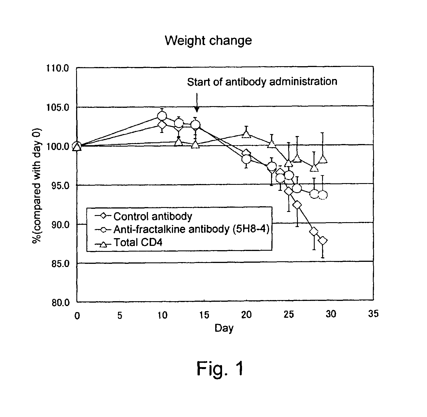
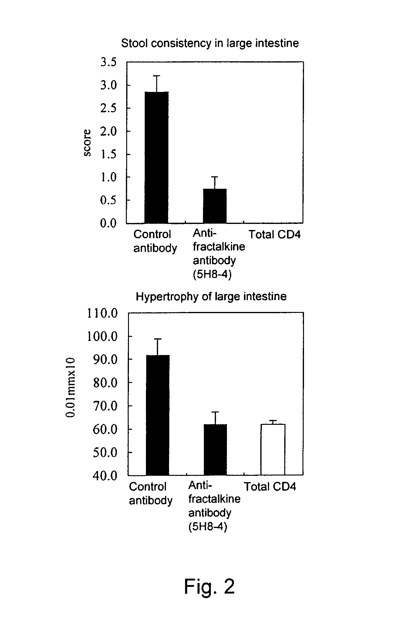
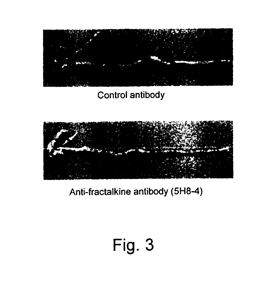
Effect of Anti-Fractalkine Antibody (5H8-4) in Mouse Inflammatory Bowel Disease Model Transfused with CD4-Positive and CD45RB Strongly-Positive (CD4+CD45RBhigh) T Lymphocytes

(1) Preparation of Anti-Fractalkine Antibody (5H8-4)

[0103]The anti-fractalkine antibody (5H8-4) was prepared by the following method (Japanese Patent Laid-open No. 2002-345454). Mouse fractalkine (R&D) was used as an antigen. The antigen was mixed with TiterMax adjuvant, Armenian hamsters were immunized with the mixture, and booster immunizations were performed thereafter with the antigen alone. The antibody titer in the serum was measured by ELISA. Lymphocytes were isolated from the Armenian hamsters in which the antibody titer increased, the lymphocytes and P3 myeloma cells were mixed at a ratio of 5:1, and the cells were fused by using PEG (Boehringer). The hybridomas were cultured on a 96-well plate for one week by using RPMI-1640 / 10% FCS / HAT / 10% Origen HCF (ISGN). Then, ELISA was performed for the culture s...

example 2

Effect of Anti-Fractalkine Antibody (5H8-4) in Mouse Oxazolone-Induced Inflammatory Bowel Disease Model

(1) Method

[0110]As for the mouse oxazolone-induced inflammatory bowel disease model, reference was made to Iijima et al., J. Exp. Med., 199, 471-482, 2004. The abdomens of 8 to 10-week old male Balb / c mice (Charles River Laboratories Japan, Inc.) were shaved in an about 2-cm square. To each mouse, 150 μl each of 100% ethanol solution containing 3% 4-ethoxymethylene-2-phenyl-2-oxazolin-5-one (hereinafter referred to as oxazolone, Sigma) was applied. The animals were starved on the fourth day after the oxazolone sensitization, and on the fifth day, given an intestinal injection of 100 μl each of 50% ethanol / physiological saline containing 0.5% oxazolone at a site about 3 cm from the anus under diethyl ether anesthesia. As a negative control group, five normal Balb / c mice were given an intestinal injection of 100 μl each of 50% ethanol / physiological saline. To seven mice in each group...

example 3

Effect of Anti-Fractalkine Antibody (#126) in Mouse Oxazolone-Induced Inflammatory Bowel Disease Model

(1) Preparation of Anti-Fractalkine Antibody (#126)

[0114]Mouse fractalkine (Genzyme) and Titer Max™ Gold adjuvant were mixed, and then used to immunize Armenian hamsters two or more times, and the final immunization was further performed with the mouse fractalkine alone. The antibody titer in the serum was measured by ELISA using solid-phased fractalkine, and lymphocytes were isolated from Armenian hamsters in which the antibody titer increased. The lymphocytes and P3 myeloma cells were mixed at a ratio of 5:1, and cell fusion was performed by using PEG (Rosh). The hybridomas were cultured on a plate for one week by using RPMI-1640 / 10% FCS / HAT / 10% Origen HCF (ISGN). Then, the culture supernatant was assayed by ELISA using solid-phased fractalkine to identify positive wells. The hybridomas producing the anti-fractalkine antibodies were cloned by performing limiting dilution twice. By...


PUM

PropertyMeasurementUnit
body weightaaaaaaaaaa

Abstract

A therapeutic agent for an inflammatory disease comprising an antibody or a CX3CR1 antagonist that inhibits an interaction of fractalkine and CX3CR1 is provided.

Description

TECHNICAL FIELD[0001]The present invention relates to a therapeutic agent for an inflammatory disease.BACKGROUND ART[0002]Chemokines are major cell migration factors in the living bodies and regulate lymphocyte infiltration into tissues via increase of cell motility and activation of cell adhesion molecules. Chemokines are classified into four subfamilies of CC, CXC, C and CXXXC on the basis of the sequence types of the first two cysteine residues. The members of the CC, CXC and C chemokine subfamilies are secretory proteins consisting of about 70 amino acids, and although they do not have activity as adhesion molecules themselves, they can induce cell adhesion. A secreted chemokine binds to a seven-transmembrane receptor on the surface of a target cell and activates integrin via the trimer G protein to induce cell adhesion or migration.[0003]Recently, a novel simple lymphocyte infiltration mechanism was identified in addition to the known cell migration mechanism. This mechanism is...

Claims


Application Information

Patent Timeline
Patent Type & Authority Patents(United States)
IPC IPC(8): A61K39/395
CPCA61K31/00C07K16/24A61K2039/505C07K2316/96A61P1/00A61P1/04A61P9/00A61P9/10A61P11/00A61P11/06A61P17/00A61P17/04A61P17/06A61P29/00A61P37/08C07K2317/76
Inventor KUBOI, YOSHIKAZUIMAI, TOSHIONISHIMURA, MIYUKIMIZUNO, KEIKO
Owner EISIA R&D MANAGEMENT CO LTD
Why Eureka
Try Eureka