Assemblies, systems, and methods for vacuum assisted internal drainage during wound healing

a vacuum assisted and internal drainage technology, applied in the field of assemblies, systems and methods for vacuum assisted internal drainage during wound healing, can solve the problems of insufficient clearing of wound voids, fluid still in the body, and current wound drain technology is not effective at adequately clearing wound voids, etc., to achieve enhanced wound healing benefits, promote tissue adherence, and increase the nature and extent of wound drainag

Active Publication Date: 2013-10-08
IC SURGICAL INC
View PDF258 Cites 30 Cited by
  • Summary
  • Abstract
  • Description
  • Claims
  • Application Information

AI Technical Summary

Benefits of technology

[0026]The invention provides a solution to the persistent, unsolved clinical problem of seromas, and, beyond that, makes it possible to actually close down a dead space or surgical wound, to approximate tissues so that a seroma cannot form, thereby accomplishing what current wound drains fail to do.
[0027]The invention provides assemblies, systems, and methods for draining a wound that is created by surgery or trauma. The wound is defined by an interior dead space having a volume enclosed between interior tissue surfaces consisting of muscle, connective, or skin tissue containing blood vessels that have been separated by surgery or trauma within a body beneath substantially intact skin. As part of the natural wound healing process, extracellular exudates comprising blood, serous fluid, and byproducts of wound healing including blood clots can accumulate during wound healing. The invention provides assemblies, systems, and methods that do not only manage blood and fluid collection of the extracellular exudates in the interior dead space, but also serve to close and eliminate the dead interior space itself, by drawing the separated interior tissue surfaces together to promote adherence of the tissue surfaces and a normal wound healing process.
[0031]In one embodiment, the housing, the perforations, and open cell component are mutually sized and configured, while the extracellular exudates taken in by the open cell material are conveyed in response to the negative pressure from the wound, to draw together the separated interior tissue surfaces, thereby promoting adherence of the tissue surfaces and a normal wound healing process, effectively closing the operative dead space.
[0038]The assemblies, systems, and / or methods that embody the technical features of the invention apply a vacuum of significant negative pressure internally and directly in a wound void or body cavity for enhanced wound healing benefits. By applying a vacuum of significant consistent negative pressure internally and directly in the wound void or body cavity, the assemblies, systems, and / or methods reduce the “dead-space” or open area inside the wound or cavity. The assemblies, systems, and / or methods increase the nature and extent of wound drainage, promote tissue adherence, facilitate closure of wounds, and thus decrease seroma formation and promote primary wound healing. The assemblies, systems, and / or methods thereby decrease the costly and increased patient morbidity caused by seroma formation and the resultant delay in primary wound healing or need for additional surgical procedures or drainage.

Problems solved by technology

However, the body still has fluid that collects in the open space.
Wound healing also needs to be accompanied by an absence of continued irritation, so that there is not a continuous initiation of new wound healing.
There is a recognized problem with conventional wound drain technology, which is directly related to the nature of the wound healing process itself.
As a result, current wound drain technology is not effective at adequately clearing wound voids.
Concurrently, current drains are ineffective at formally closing down larger dead spaces, and can only manage small amounts of fluid directly around the drain itself.
Finally, conventional wound drains provide non-uniform blood and fluid removal with low inconsistent suction pressure, often with long drain duration and the potential of infection.
The problem with these devices is that they may become plugged by blood clots carried by the wound fluid, or may be overwhelmed by the fluid generated in the space, or may have such low continuous pressure that they are ineffective at closing the internal space down.
Further, although they may, when not plugged, drain fluid, fluid drainage is limited to fluid directly around the drain itself.
As a result, current drains again, at best, do not effectively clear all of the fluid in the space and, more importantly, they do not clear enough fluid to effectively seal down and close off the dead space.
Conventional VAC devices, however, are only approved and used for external wounds.
Conventional VAC devices are not approved or used for internal wounds or operative sites, and may create bleeding upon withdraw and leave particulate matter from the foam inside the wound base.
Current wound drain devices assemblies at times do not remove a substantial amount of fluid from within a wound and have other performance issues.
For example, external VAC devices clear fluid directly around external wounds (as FIG. 3 shows), and they are limited to the application to external wounds only.
Furthermore, the clinical use of external VAC devices may not make wound drainage more cost-effective, clinician-friendly, and patient-friendly.
Removal of the foam structures in the presence of granulating tissue and the force of pressure on the wound bed that this removal can cause pain or discomfort.
Furthermore, the multiple steps of the conventional external VAC procedure—removing the adhesive membrane, then removing the old foam structures, then inserting the new foam structures, and then reapplying the adhesive member along the entire periphery of the wound—are exacting, tedious and time consuming.
They only prolong pain or discomfort, and cause further disruption to the patient, and also demand dedicated nursing time and resources.
Obtaining such a seal can be difficult, particularly in body regions where the surrounding skin is tortuous, and / or mucosal and / or moist.
Furthermore, prolonged wearing of wet dressings can cause further breakdown and maceration of the surrounding skin thereby increasing the wound size.
This can cause further discomfort to the patient, and the exudate can often be offensive in odor and color causing further morbidity to the patient.
This may, in turn, require more numerous dressing changes and re-padding throughout the day, which is disruptive to the patient and costly both in terms of nursing time and resources.
Such complications result in a significant amount of lost income to patients, as well as expenses to insurers and physicians who have to care for these patients that require serial drainage.
Such complications also delay wound healing, may entail additional surgical procedures, and ultimately delay the patient's return to work and routine functional activity.
Seroma management can also be costly and, further, can place health care workers to additional needle exposure risks and related outcomes such as hepatitis, etc.
The inability to prevent or treatment seromas that form in closed interior wounds is a problem that has persisted in the field of elective surgery since the beginning of surgery, and has been documented in the surgical literature for all specialties over the last fifty years.
Seromas and abnormal fluid collection are so common, that physicians and surgeons will acknowledge that seromas are, unfortunately, an expected part of wound healing following surgery.

Method used


Image

  • Assemblies, systems, and methods for vacuum assisted internal drainage during wound healing
  • Assemblies, systems, and methods for vacuum assisted internal drainage during wound healing
  • Assemblies, systems, and methods for vacuum assisted internal drainage during wound healing

Examples


example

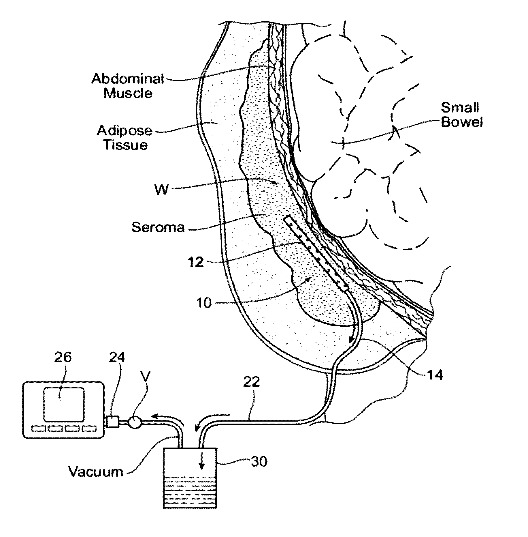
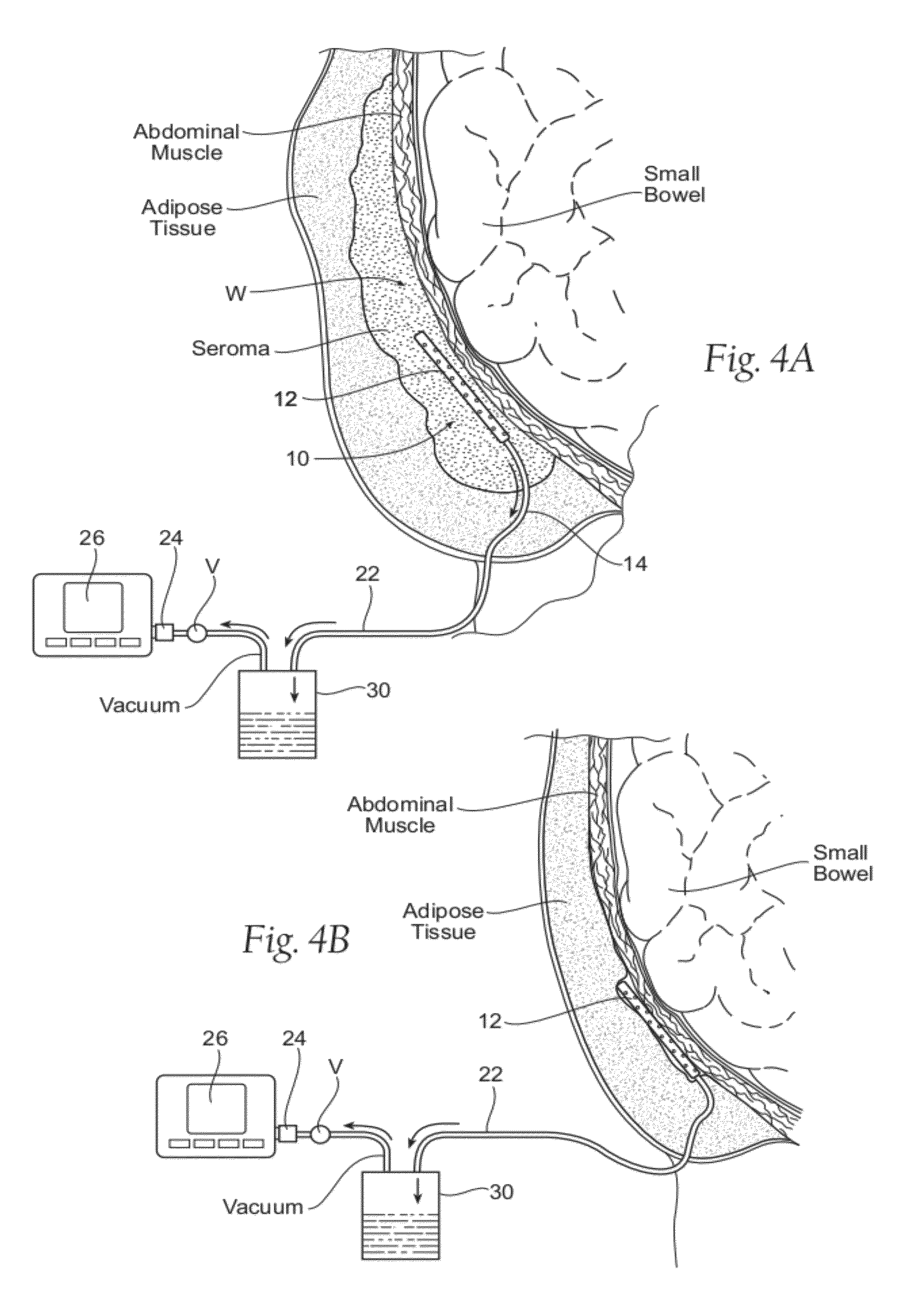
[0107]Wound drain assemblies having the technical features described above were placed into internal wound voids surgically created in a porcine model. Also concurrently placed into surgical created wound voids in the same porcine model were conventional wound drains. The performance of efficacy of the wound drain assemblies were compared to the performance and efficacy of the conventional drains over a period of eight days.

[0108]More particularly, following induction of general anesthesia, prefascial pockets were elevated with scissor dissection through ten (10) cm incisions on left and right lateral sides of a pig over the latissimus dorsi muscles and external oblique muscles, just posterior to the front legs. The left and right side pockets were placed six (6) cm off the midline to assure the pockets were kept separate. Bovie cautery was used for hemostatsis and pockets were irrigated with a triple antibiotic solution used in implant surgery, comprising 1 gm of Ancef, 80 mg of Ge...


PUM


Abstract

Assemblies, systems, and methods convey fluid from an internal wound site or body cavity by applying negative pressure from a source outside the internal wound site or body cavity through a wound drain assembly that is placed directly inside the internal wound site or body cavity.

Description

RELATED APPLICATION[0001]This application is a continuation-in-part of co-pending U.S. patent application Ser. No. 12 / 661,293, filed Mar. 15, 2010, which is a continuation of U.S. patent application Ser. No. 11 / 810,027, filed Jun. 4, 2007 (now U.S. Pat. No. 7,699,831), which is a continuation-in-part of U.S. patent application Ser. No. 11 / 646,918, filed Dec. 28, 2006, which claims the benefit of U.S. Provisional Patent Application Ser. No. 60 / 810,733, filed Jun. 2, 2006, which are each incorporated herein by reference.FIELD OF THE INVENTION[0002]This application relates generally to the drainage of fluid from the body during the wound healing process, e.g., following surgery, trauma, or placement of implants or surgical devices.BACKGROUND OF THE INVENTION[0003]During surgery, or as a result of trauma, subcutaneous tissue can be removed or altered, and an open void or dead space or cavity is created within the tissue that was previously attached to other tissue. This may also occur i...

Claims


Application Information

Patent Timeline
Patent Type & Authority Patents(United States)
IPC IPC(8): A61M27/00
CPCA61M1/008A61M1/0039A61M2209/088A61M1/84A61M1/90A61M1/982A61M1/76
Inventor BENGTSON, BRADLEY
Owner IC SURGICAL INC
Why Eureka
Try Eureka