Viral polyhedra complexes and methods of use

a polyhedra complex and polyhedra technology, applied in the field of viral polyhedra complexes and methods of use, can solve the problems of hampered polyhedra protein structure determination, and achieve the effects of preserving, prolonging or protecting the functionality of a target molecule, facilitating the identification of regions, and enhancing the functionality of the target molecul

Active Publication Date: 2013-10-08
NAT UNIV KYOTO INST OF TECH +1
View PDF0 Cites 6 Cited by
  • Summary
  • Abstract
  • Description
  • Claims
  • Application Information

AI Technical Summary

Benefits of technology

[0014]It was found that polyhedra are made of trimers of the viral polyhedrin protein and contain nucleotides. Although the shape of these building blocks is reminiscent of some capsid trimers, polyhedrin has a new fold and has evolved to assemble in vivo into 3-D cubic crystals rather than icosahedral shells. The polyhedrin trimers are extensively cross-linked in polyhedra by non-covalent interactions and pack with an exquisite molecular complementarity similar to that of antigen-antibody complexes. The resulting ultra-stable and sealed crystals shield the virus particles from environmental damage. The structure suggests that polyhedra can serve as the basis for a large number of applications including the development of robust and versatile nanoparticles for biotechnological applications (Douglas and Young 2006) such as microarrays (Ikeda et al 2006) and bio-pesticides (Chang et al 2003).
[0015]Specifically, it has been found that the crystal structure of the cypovirus polyhedra facilitates the identification of regions of the polyhedrin protein that play central roles in the architecture of the crystal structure and that attaching at least a portion of these regions to a target molecule allows the target molecule to be sequestered by the polyhedron crystal—either by incorporation into the lattice structure or by attachment to the surface of the crystal.

Problems solved by technology

Protein structure determination of polyhedra has been hampered by the small size of the crystals and a number of other factors.

Method used


Image

  • Viral polyhedra complexes and methods of use
  • Viral polyhedra complexes and methods of use
  • Viral polyhedra complexes and methods of use

Examples


example 1

Polyhedra Expression and Characterization

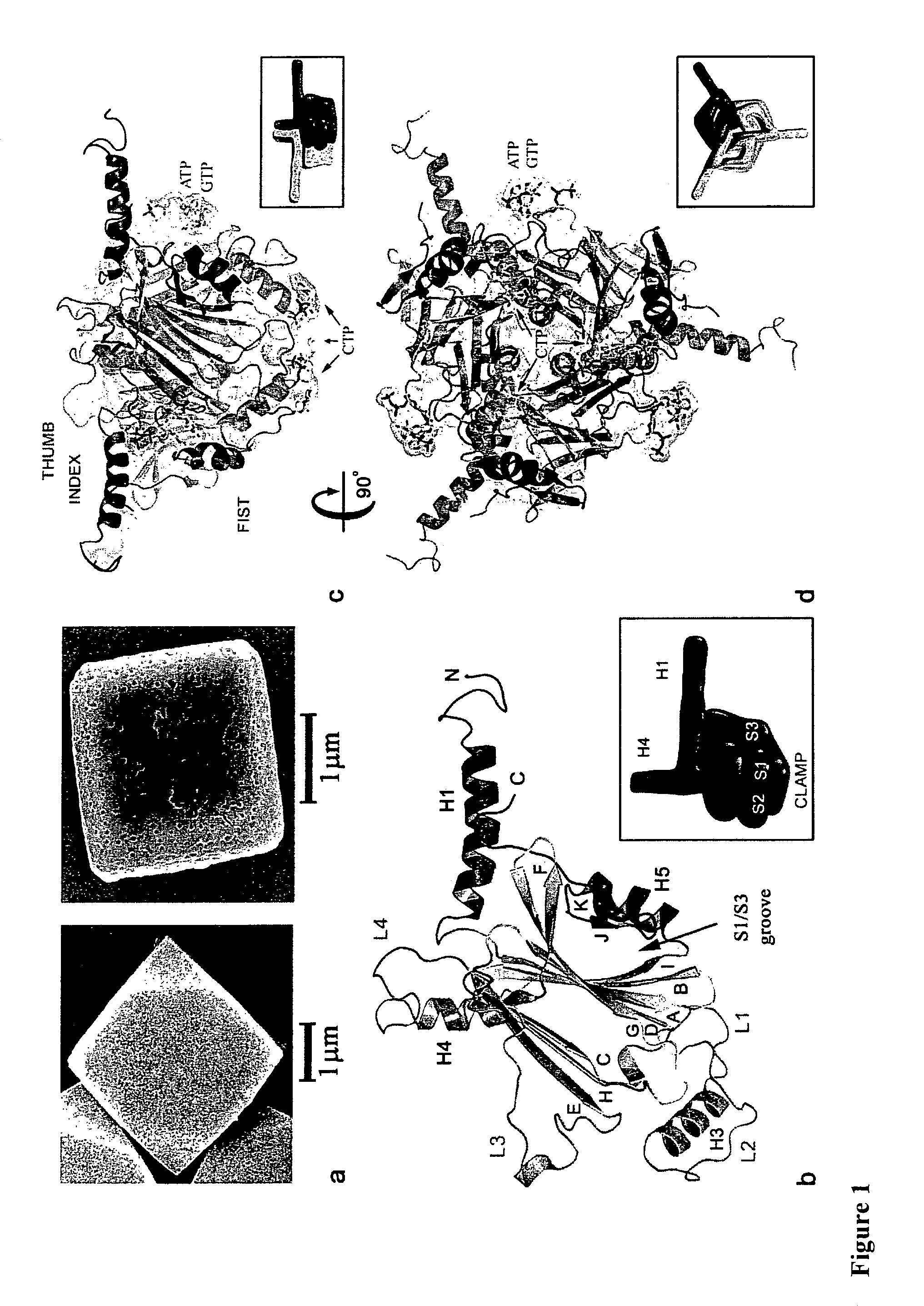
[0105]Expression and purification of recombinant and wild-type Bombyx mori CPV polyhedra in insect cells were carried as described elsewhere (Mori et al 1993). Polyhedra containing the human ZIP-kinase (Accession BAA24955) fused to an N-terminal fragment of the CPV turret protein were produced as previously described (Ikeda et al 2006). Scanning electron microscopy was performed on Pt coated samples. Mass spectrometry analyses confirmed the presence of ribonucleotide tri-phosphates in purified polyhedra and showed that acetylation of the N-terminal alanine is the only polyhedrin post-translational modification.

example 2

Structure Determination

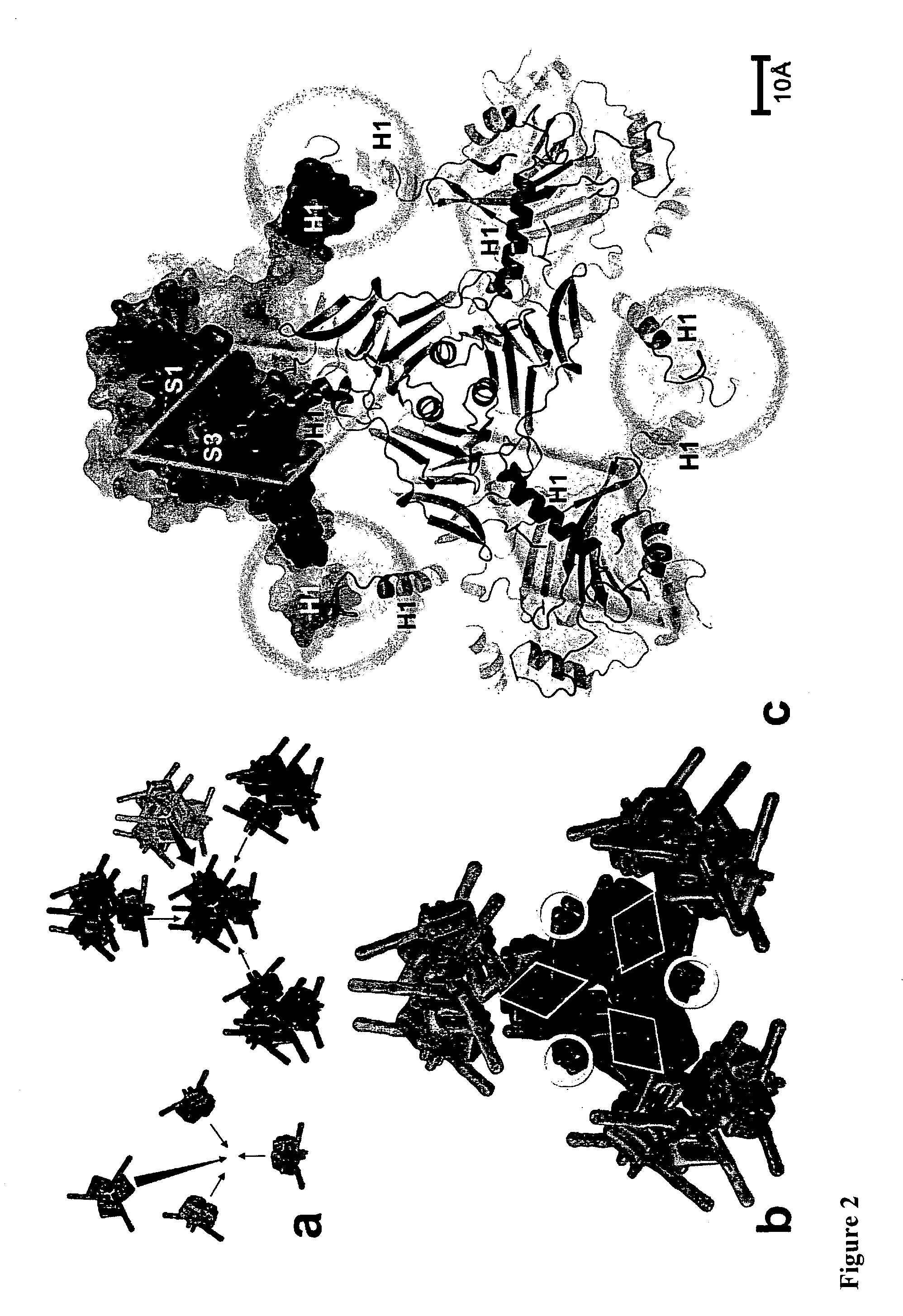
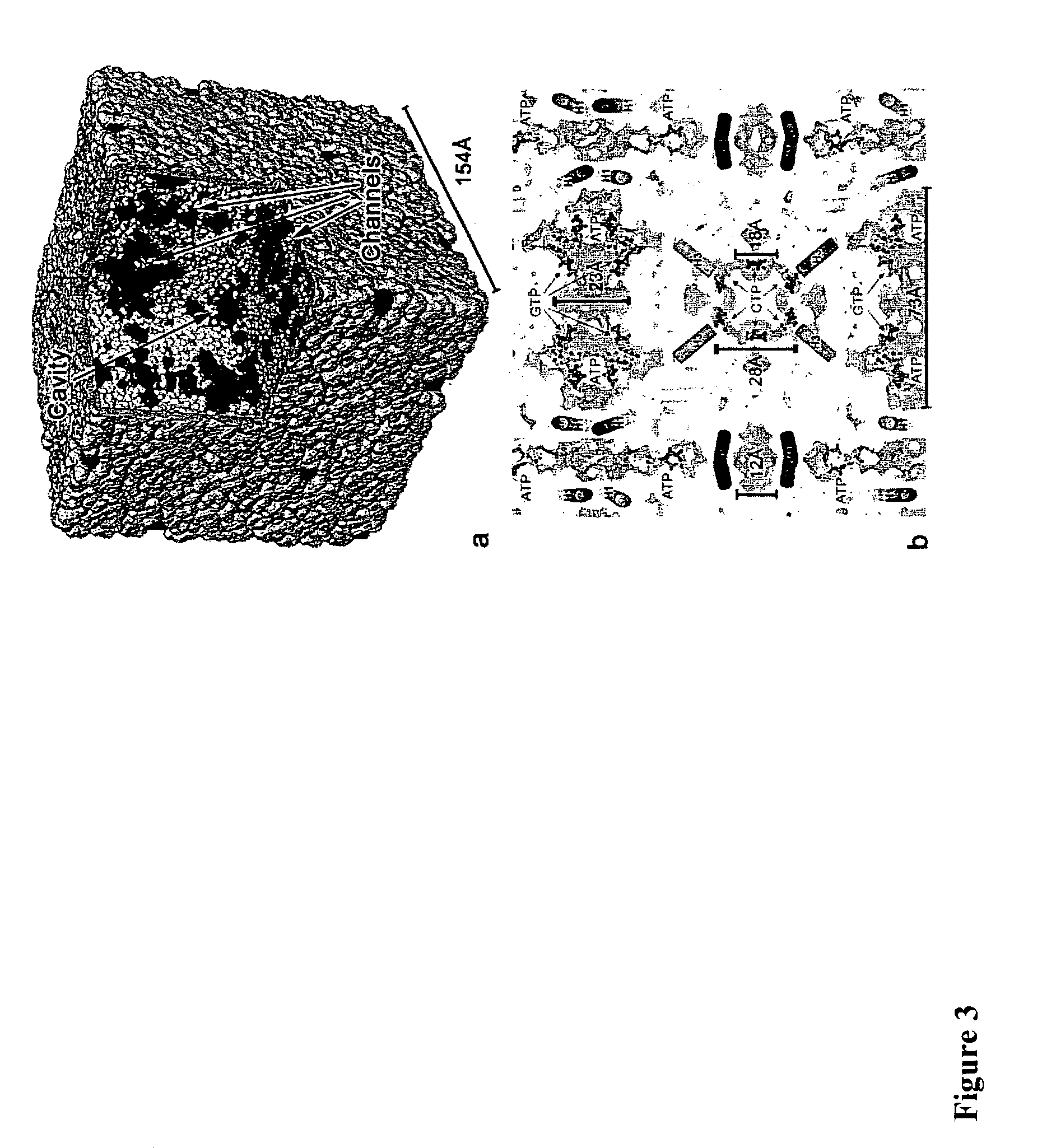
[0106]Crystals were spread on micromesh mounts (Thorne et al 2003) leaving a thin film of 50% ethylene glycol and data were collected at 120K on the MD2 diffractometer of the X06SA beamline (Swiss Light Source) with a strongly attenuated beam focused onto the crystal. Data were processed with Denzo / Scalepack (Otwinowski et al 1997) in space group I23 and datasets from 2-4 crystals were merged because of severe radiation damage. Successful heavy atom soaks were performed on recombinant polyhedra at high concentrations, usually half saturation in appropriate buffer. Heavy atom sites for four isomorphous derivatives including selenomethionine-substituted crystals were found by SHELX24 and refined at 2.6 Å using SHARP25. The structures of recombinant and infectious polyhedra and of polyhedra containing the ZIP-kinase were refined at resolutions of 2.1 Å, 1.98 Å and 2.45 Å respectively using Refmac526. The final models are essentially identical. The structure of in...

example 3

Model Analysis and Illustrations

[0109]The oligomeric and crystal contact interfaces were characterized with AREAIMOL from CCP428 and the PISA29 server at the European Bioinformatics Institute. Solvent cavities and channels were analyzed with VOIDOO30 (Uppsala Software Factory). Illustrations were prepared with PyMOL v0.99 (DeLano Scientific, San Carlos, Calif., USA. http: / / www.pymol.org).


PUM

PropertyMeasurementUnit
diameteraaaaaaaaaa
pHaaaaaaaaaa
pHaaaaaaaaaa

Abstract

Cypoviruses and baculoviruses are notoriously difficult to eradicate because the virus particles are embedded in micron-sized protein crystals called polyhedra. The remarkable stability of polyhedra means that like bacterial spores these insect viruses remain infectious for years in soil. Although these unique in vivo protein crystals have been extensively characterized since the early 1900s, their atomic organization remains elusive. Here we describe the 2 crystal structure of both recombinant and infectious silkworm cypovirus polyhedra determined using 5-12 micron crystals purified from insect cells. These are the smallest crystals yet used for de novo X-ray protein structure determination. It was found that polyhedra are made of trimers of the viral polyhedrin protein and contain nucleotides. Although the shape of these building blocks is reminiscent of some capsid trimers, polyhedrin has a new fold and has evolved to assemble in vivo into 3-D cubic crystals rather than icosahedral shells. The polyhedrin trimers are extensively cross-linked in polyhedra by non-covalent interactions and pack with an exquisite molecular complementarity similar to that of antigen-antibody complexes. The resulting ultra-stable and sealed crystals shield the virus particles from environmental damage. The structure suggests that polyhedra can serve as the basis for the development of robust and versatile nanoparticles for biotechnological applications such as in cell culture systems, microarrays and biopesticides.

Description

TECHNICAL FIELD[0001]The present invention relates to a method of introducing target molecules into a polyhedron or part thereof. The invention also relates to complexes formed by introducing target molecules into polyhedron or part thereof and to polyhedrin protein variants that are useful in the methods of the invention. The invention also relates to the use of the methods, complexes and polyhedrin protein variants for the stabilization of proteins in particular, for cell culture, biosensors, microarrays, and assays.BACKGROUND[0002]Any discussion of the prior art throughout the specification should not be considered an admission that such prior art is widely known or forms part of common general knowledge in the field.[0003]Stable maintenance of molecules, and in particular functional molecules, on support surfaces can be critical for analytical and diagnostic tools—for example, in high throughput analysis and functional characterization of biomolecules. However, techniques for st...

Claims


Application Information

Patent Timeline
Patent Type & Authority Patents(United States)
IPC IPC(8): G06F19/00
CPCA01N25/34A01N63/00A61K47/48776A61K47/48961B82Y5/00C07K14/005C07K14/475C07K14/485C07K14/50A01N61/00A01N63/02C07K2319/735C12N2710/14043C12N2720/12022A01N2300/00A61K47/6901A61K47/6949A01N63/50A01N63/23A01N63/40
Inventor METCALF, PETERCOULIBALY, FASSELI JOSEPHMORI, HAJIMEHAMADA, NORIOIKEDA, KEIKOCHIU, YUI LAM ELAINEIJIRI, HIROSHI
Owner NAT UNIV KYOTO INST OF TECH
Why Eureka
Try Eureka