
Method and apparatus for fusing the bones of a joint

a joint and bone technology, applied in the field of surgical methods and equipment, can solve the problems of affecting the relatively few options for restoring the form and function of the affected joint, and complicating the task of restoring the affected joint to a fully functional, pain-free condition

Active Publication Date: 2014-07-22
SCHELAND JOHN
View PDF4 Cites 33 Cited by
  • Summary
  • Abstract
  • Description
  • Claims
  • Application Information

AI Technical Summary

Benefits of technology

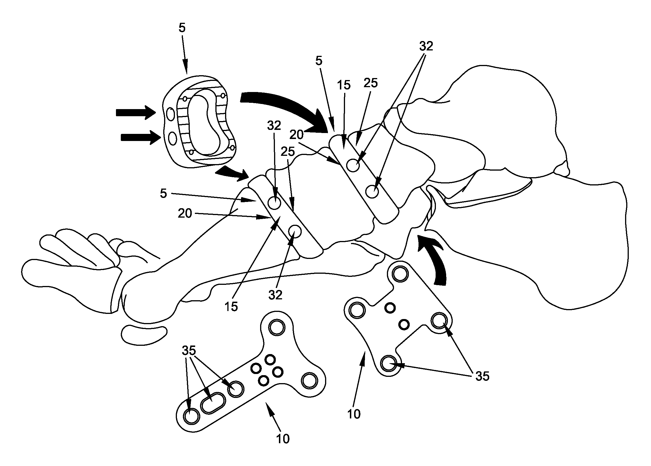
[0010]The present invention provides a new and improved method and apparatus for fusing the bones of a joint so as to provide relief to a patient.
[0012]The new and improved bone fusion system of the present invention is adapted to facilitate small bone arthrodesis, open-wedge osteotomies and bone defect healing, as well as other procedures, by maintaining bone position and stability, simplifying dissection and improving fixation at the fusion sites. The new and improved bone fusion system facilitates boney fusions and bone healing by delivering and incarcerating bone graft material at the fusion site. Furthermore, the present invention may be practiced using less invasive techniques so as to minimize soft tissue resection and / or other trauma, reduce vascular compromise and minimize the potential for developing post-operative neuromas.

Problems solved by technology

The joints in the limbs of a human are frequently subject to a loss of function due to trauma and / or disease.
As these joints become increasingly more painful and problematic for the patient, surgeons generally have relatively few options for restoring the form and function of the affected joints.
In addition, many clinical factors (including but not limited to patient age, patient weight and general patient health) further complicate the task of restoring the affected joints to a fully functional, pain-free condition.
In this respect it should also be appreciated that the smaller joints of the limbs are characterized by wide variations in topography, and impose substantial size constraints, which can make restoring an affected joint to pain-free mobility difficult or impossible.
Arthrodesis of this nature can be difficult to perform.
This is because the small bones in the foot and wrist are surrounded by many odd-shaped bones, with multiple ligamentous attachments, thus creating significant difficulty in achieving end-to-end contact.
To bring these bones together typically requires, among other things: extensive dissection of the surrounding ligaments to mobilize the bones; the use of high-angle interfragmentary screws, which often results in screw pull-out and unintended bone fracture; and plate fixation, which often results in increased unnecessary compressive loading of the screws, thereby resulting in hardware failure.
Furthermore, when arthrodesis is achieved without interpositional bone grafting, the area of fusion becomes shorter, resulting in iatrogenic deformity.
The first metatarsal open-wedge osteotomy is utilized to correct primus varus deformity when other options would result in an unacceptable shortening of the first metatarsal.
Present techniques require interpositional grafting where primary stabilization (by directly fixating the graft into the osteotomy) is difficult if not impossible to achieve.
This technique is inexact and often results in either over-correction or under-correction.
Recently, expensive Darco plates have been developed in an attempt to achieve this goal.
While these Darco plates have improved this procedure, the Darco plates are not completely satisfactory.
For one thing, the metal block spacer in the center of the Darco plates does not allow boney integration into the metal block spacer, thus requiring additional bone graft material to be placed into the remaining boney defect.
Furthermore, with the Darco plates, the bone graft material is not incarcerated into the boney defect and can migrate out of the osteotomy site during the post-operative period, thereby resulting in non-unions and repeat surgeries.
Also, the Darco plates are made out of stainless steel and titanium, which means that the Darco plates are not radiolucent, and hence it is difficult and / or impossible to perform radiographic studies (to evaluate bone healing) during the post-operative period.
These grafts are brittle and often fracture during placement and / or fixation.
If the graft is fractured during placement and / or fixation, it is no longer conducive to primary fixation and dorsal migration of the graft commonly occurs, resulting in loss of correction and pain from iatrogenic boney prominences in the foot.
However, these bone graft analogs are prohibitively expensive and have reduced bone healing properties when compared to autogenous bone.
Bone defects can occur as a result of the resection of bone lesions, the removal of non-healing fractures and the removal of small implants.
When bone defects occur, few options are available to the surgeon.
Most optimally, autogenous bone grafts are used, but this limits the surgeon's ability to stabilize the graft to the surrounding bone.
In many cases, external fixation is the best option, but this approach has a number of inherent complications (e.g., pin tract infection) which can cause patient morbidity.

Method used


Image

  • Method and apparatus for fusing the bones of a joint
  • Method and apparatus for fusing the bones of a joint
  • Method and apparatus for fusing the bones of a joint

Examples


Embodiment Construction

[0037]The present invention provides a new and improved method and apparatus for fusing the bones of a joint so as to provide relief to a patient.

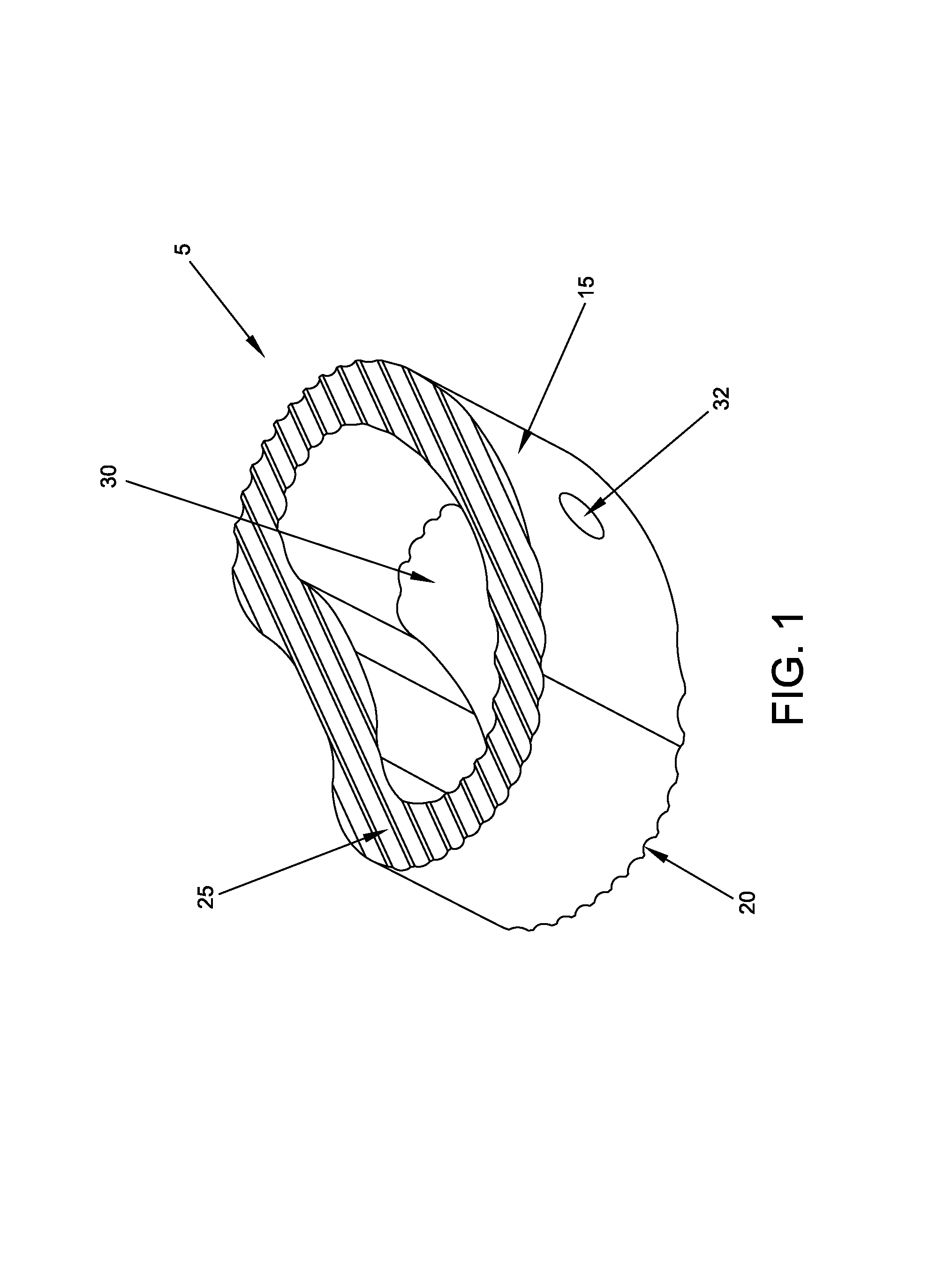
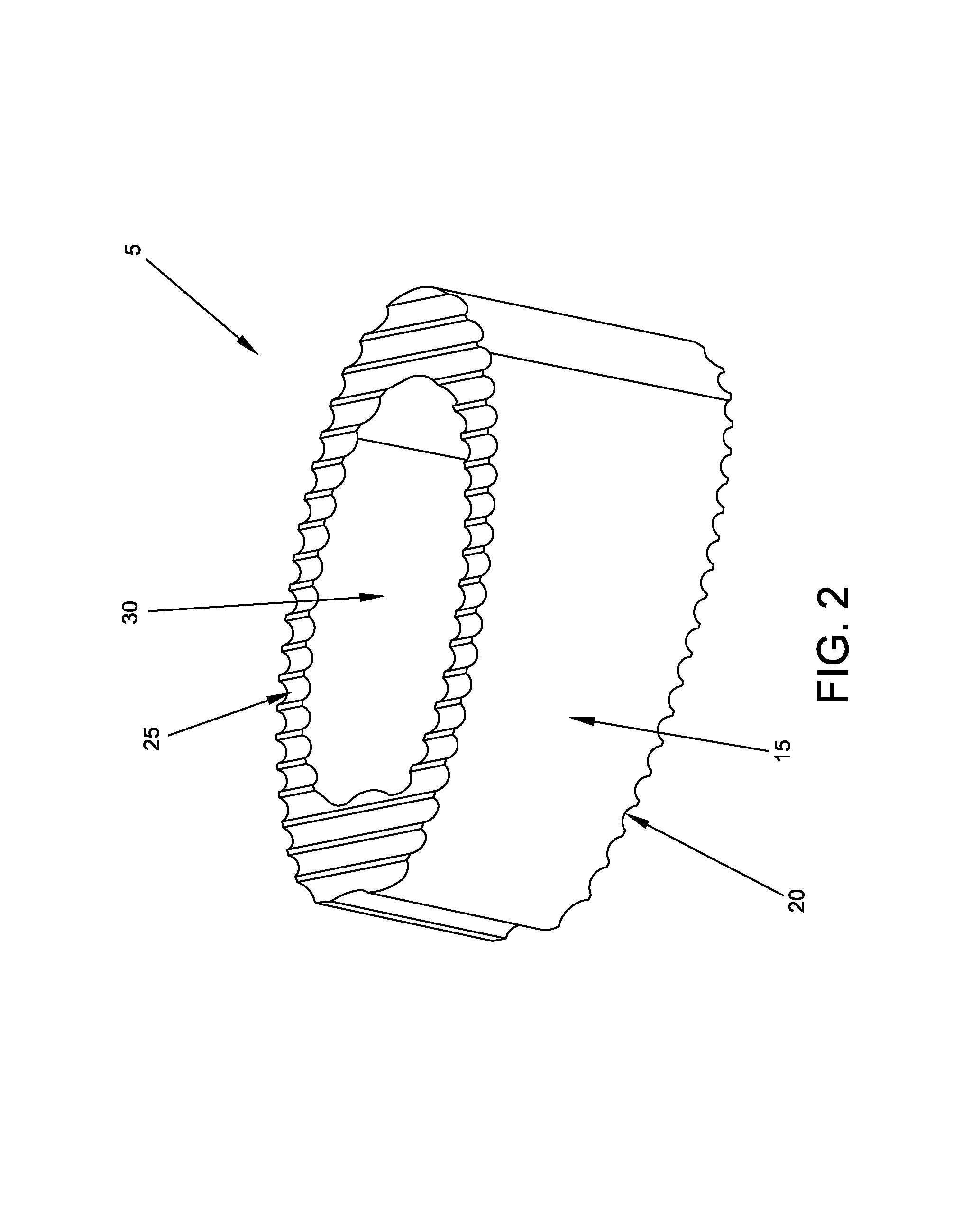
[0038]More particularly, the present invention comprises the provision and use of a new and improved bone fusion system for fusing the bones of a joint.

The New and Improved Bone Fusion System

[0039]The new and improved bone fusion system of the present invention is adapted to facilitate arthrodesis, open-wedge osteotomies and bone defect healing, as well as other procedures, by maintaining boney position and stability, simplifying dissection and improving fixation at the fusion sites. The new and improved bone fusion system facilitates boney fusions and bone healing by delivering and incarcerating bone graft material at the fusion site. Furthermore, the present invention may be practiced using less invasive techniques so as to minimize soft tissue resection and / or other trauma, reduce vascular compromise and minimize the potential for devel...


PUM


Abstract

Apparatus for fusing a first bone of a joint with a second bone of a joint, the apparatus comprising:a fusion block comprising a hollow tubular structure characterized by a first end, a second end and a lumen extending from said first end to said second end, the first end being configured to engage the first bone of the joint and the second end being configured to engage the second bone of the joint, the lumen being configured to span the distance from the first bone of the joint to the second bone of the joint and to receive and retain bone graft material therein, whereby to facilitate bone fusion across the fusion block; andat least one fusion plate for connecting the fusion block to the first bone of the joint and the second bone of the joint.

Description

REFERENCE TO PENDING PRIOR PATENT APPLICATION[0001]This patent application claims benefit of pending prior U.S. Provisional Patent Application Ser. No. 61 / 335,100, filed Dec. 31, 2009 by John Scheland et al. for BONE FUSION SYSTEM FOR EXTREMITIES, which patent application is hereby incorporated herein by reference.FIELD OF THE INVENTION[0002]This patent application relates to surgical methods and apparatus in general, and more particularly to surgical methods and apparatus for fusing the bones of a joint.BACKGROUND OF THE INVENTION[0003]The joints in the limbs of a human are frequently subject to a loss of function due to trauma and / or disease. As these joints become increasingly more painful and problematic for the patient, surgeons generally have relatively few options for restoring the form and function of the affected joints. In addition, many clinical factors (including but not limited to patient age, patient weight and general patient health) further complicate the task of res...

Claims


Application Information

Patent Timeline
Patent Type & Authority Patents(United States)
IPC IPC(8): A61F2/42
CPCA61F2/30A61F2002/30622A61F2/4225A61F2002/30578A61F2002/2835A61B17/8095A61B17/8061A61F2002/4223A61F2/4202
Inventor SCHELAND, JOHN
Owner SCHELAND JOHN
Learn More