Targeting method, targeting device, computer readable medium and program element

a targeting device and computer readable medium technology, applied in the field of targeting methods, can solve the problems of poor visualization of bony structures filled with air, poor visibility of bony anatomy, and low signal quality in obese patients (subcutaneous fat) and achieve the effect of simple, fast and effective targeting

Active Publication Date: 2014-08-05
KONINK PHILIPS ELECTRONICS NV
View PDF21 Cites 66 Cited by
  • Summary
  • Abstract
  • Description
  • Claims
  • Application Information

AI Technical Summary

Benefits of technology

[0007]It may be desirable to provide an efficient targeting method, a targeting device, a system, a computer readable medium and a program element usable in a targeting device. Thus, it is one intention of the present invention to present a simple, fast and effective targeting method, preferably, to approach a planning / preparation method for needle guided percutaneous interventions using C-arm based X-ray devices.
[0054]Using a three-dimensional image, e.g. a voxel representation, of a three-dimensional object as an initial image from which a two-dimensional image is selected, is an efficient way to ensure that at least one slice or slab of the three-dimensional image contains the target point. The voxel representation can be efficiently reconstructed out of data recorded in a computer tomography (CT) with well known methods of reconstruction.
[0058]In this context a graphic may mean a simplified image. Such a graphic can be easily overlaid on the two-dimensional image while still showing a sufficient contrast to be seen in this overlaying mode. Thus, it might be possible to display the target point on the overlaid image even in case in the image the first object or the target point is only depicted showing low contrast. The implementing of a scale in the graphic may be an efficient way to measure and / or to control the advancing of the first object in the second image. Alternatively, the target point may be added into the two-dimensional, i.e. the two-dimensional image may be altered by introducing the target point as a structure. This might be advantageous in case the target point is represented by a structure having low contrast in the image.
[0060]The rendering of the slices, volumes and / or graphics may be done with the same perspective as the corresponding images in order to achieve an improved tracking of the first object, e.g. a needle, in the object under examination. This may an efficient way to improve the targeting method by overcome a problem which might occur due to the fact that a first object, e.g. a needle, will be projected as a circle if the needle is pointed towards a focus position of the device used for taking the fluoroscopic images instead of the towards the endpoint (target point) of the planned path due to the perspective nature of the fluoroscopic images. This problem may be prevented by adapting the rotation of the C-arm in such a way that the focus position lays on the extension of the planned path line. That is, due to the perspective nature of the fluoroscopic images the object will not be projected as a circle if the detector plane is equal to the orthogonal plane of the planned path, to correct for this the orientation of the C-arm may be adapted in such a way that the focus point (e.g. X-ray source) is placed on the extension of the planned path, wherein the calculations are done from the volume coordinates to C-arm sensor coordinates.
[0062]A method according to an exemplary embodiment of the invention may have the advantages that an entrance point (puncturing spot) into a body as well as an entrance angle into the body may be determinable during a medical procedure. Further, the needle position in the body may be determinable during the procedure and shutter positions may be set to reduce the patient dose during a CT acquisitions to monitor the progress of the procedure.

Problems solved by technology

However, this technique suffers from multiple drawbacks, relatively poor visualization of bony structures filled with air, poor visibility when bony anatomy is superimposed on the structure of interest, low signal quality in obese patients (subcutaneous fat), for example.
The disadvantage of the technique is poor patient access and room sterility issues.
An intervention room equipped with a rotational X-ray device is much more suitable for such procedures, but for most of the procedures the contrast resolution of the X-ray device is not sufficient.
The problem with this process is that a tedious trial and error method must be followed to define / evaluate the resulting needle trajectory in the cross-sectional slices.
In the context of rotational X-ray, this process is hindered by the fact that sometimes the actual entry point is difficult to locate because the skin may not always be visible within the reconstructed region of interest.
Furthermore usually some global constraints must be fulfilled, such as entering a cavity or avoiding or passing in between bone or vessel material that is sometimes difficult to evaluate on cross sectional slices.

Method used


Image

  • Targeting method, targeting device, computer readable medium and program element
  • Targeting method, targeting device, computer readable medium and program element
  • Targeting method, targeting device, computer readable medium and program element

Examples


Embodiment Construction

[0075]The illustration in the drawings is schematically. In different drawings, similar or identical elements are provided with the similar or identical reference signs.

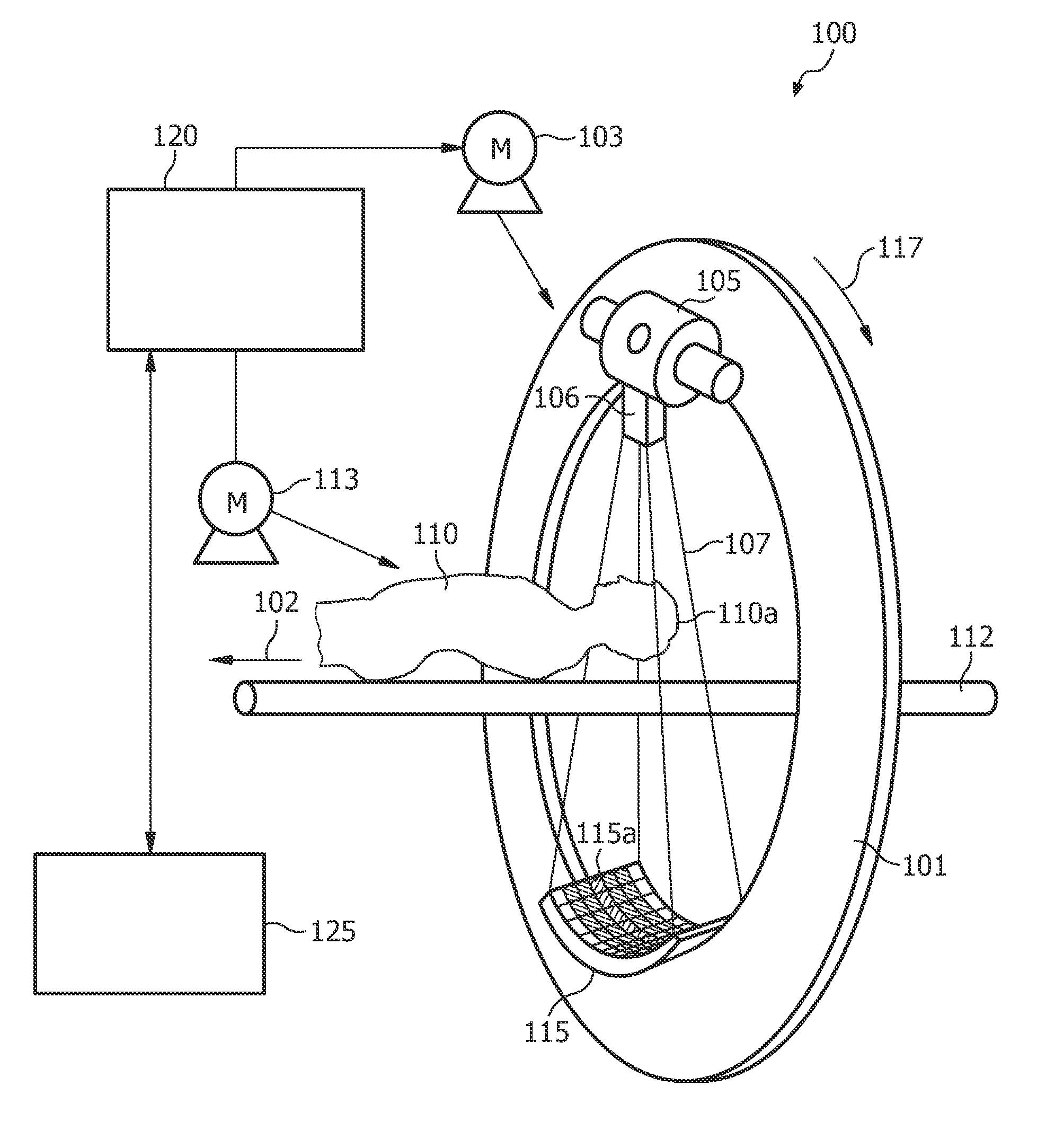
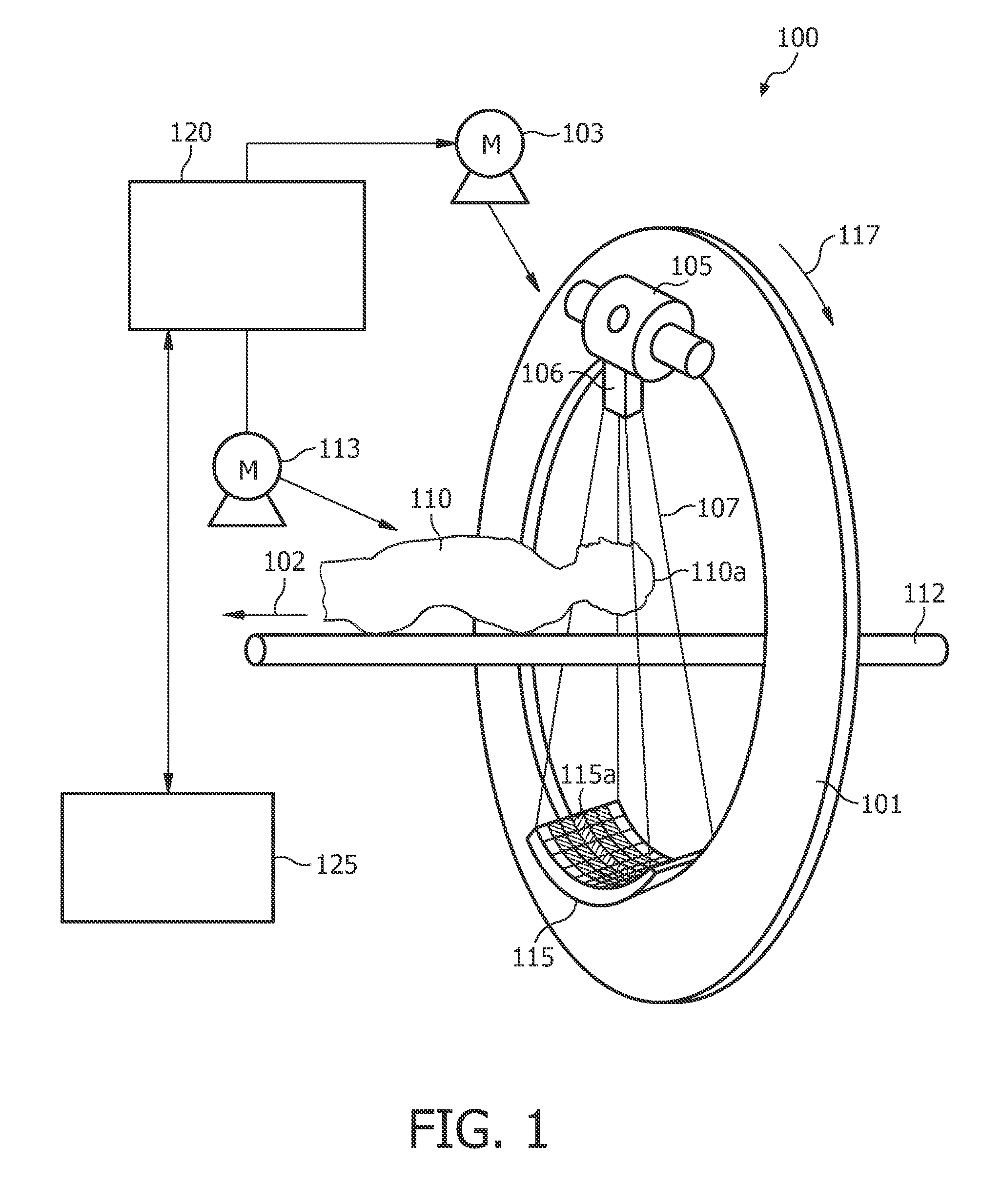
[0076]FIG. 1 shows an exemplary embodiment of a computed tomography scanner system which can be used in order to provide a three-dimensional image from which a slice can be selected. For the further targeting method a so-called C-arm device can be used which looks similar to the CT but comprises only a C-arm gantry, i.e. a gantry in form of a C, instead of a gantry, which is depicted in FIG. 1.

[0077]The computed tomography apparatus 100 depicted in FIG. 1 is a cone-beam CT scanner. The CT scanner depicted in FIG. 1 comprises a gantry 101, which is rotatable around a rotational axis 102. The gantry 101 is driven by means of a motor 103. Reference numeral 105 designates a source of radiation such as an X-ray source, which emits polychromatic or monochromatic radiation.

[0078]Reference numeral 106 designates an aperture ...


PUM


Abstract

This invention will introduce a fast and effective target approach planning method preferably for needle guided percutaneous interventions using a rotational X-ray device. According to an exemplary embodiment A targeting method for targeting a first object in an object under examination is provided, wherein the method comprises selecting a first two-dimensional image of an three-dimensional data volume representing the object under examination, determining a target point in the first two-dimensional image, displaying an image of the three-dimensional data volume with the selected target point. Furthermore, the method comprises positioning the said image of the three-dimensional data volume by scrolling and / or rotating such that a suitable path of approach crossing the target point has a first direction parallel to an actual viewing direction of the said image of the three-dimensional data volume and generating a second two-dimensional image out of the three-dimensional data volume, wherein a normal of the plane of the second two-dimensional image is oriented parallel to the first direction and crosses the target point.

Description

FIELD OF INVENTION[0001]The invention relates to a targeting method, a targeting device, a system, a computer readable medium and a program element, in particular to a method for targeting a first object in an object under examination.TECHNICAL BACKGROUND[0002]An important class of image guided interventional procedures are the so-called needle guided percutaneous interventions. The clinical procedures of interest are all needle guided embolisation, biopsies and ablation procedures in the domain of: vertrebroplasty, kyphoplasty, neuro biopsies in neoplastic cranial disease and percutaneous rhizotomy in trigeminal neuralgia.[0003]Two of the most frequently used imaging techniques for the image guidance are ultrasonic (US) based and computer tomography (CT) based. The US guided procedures are characterized by an interactive needle advancement monitoring and relatively good real time visualization of anatomical soft tissue structures. However, this technique suffers from multiple drawb...

Claims


Application Information

Patent Timeline
Patent Type & Authority Patents(United States)
IPC IPC(8): G06K9/00
CPCA61B17/3403A61B19/201A61B2019/507A61B2019/5242A61B34/10A61B90/11A61B2034/107A61B2090/3764
Inventor MIELEKAMP, PIETER MARIAHOMAN, ROBERT JOHANNES FREDERIKBABIC, DRAZENKO
Owner KONINK PHILIPS ELECTRONICS NV
Why Eureka
Try Eureka