Methods and system for the automated determination of immunofluorescent foci using a cell-based immunofluorescence assay using synthetic calibration particles

a cell-based immunofluorescence and automated determination technology, applied in the field of bioassays and diagnostics, can solve the problems of inability to describe reliable methods for standardising the assessment of the number of fluorescent foci, and achieve the effects of improving the accuracy of diagnosis of disease, increasing the number of dna double strand breaks, and accurate and reliable measurement of fluorescent foci

Active Publication Date: 2014-09-16
MEDIPAN +1
View PDF1 Cites 0 Cited by
  • Summary
  • Abstract
  • Description
  • Claims
  • Application Information

AI Technical Summary

Benefits of technology

[0020]By incorporating standardised reagents and methods an analytical platform can be established that is compatible with different technical setups throughout the world. Differences in technical setups relate to different microscope setups, different methods of generating fluorescence or different software setups for detecting fluorescent foci. A standardised approach overcomes these differences and ultimately enables accurate and reliable measurement of fluorescent foci in biological samples. This subsequently enables, in one aspect of the invention, an enhanced accuracy in diagnosis of disease, such as diseases represented by modified numbers of immunofluorescent foci, for example increased numbers of DNA double strand breaks.

Problems solved by technology

At the present time no reliable approaches for standardised assessment of fluorescent foci numbers have been described in the prior art, which are suitable for the analysis of biological samples as described herein.

Method used


Image

  • Methods and system for the automated determination of immunofluorescent foci using a cell-based immunofluorescence assay using synthetic calibration particles
  • Methods and system for the automated determination of immunofluorescent foci using a cell-based immunofluorescence assay using synthetic calibration particles
  • Methods and system for the automated determination of immunofluorescent foci using a cell-based immunofluorescence assay using synthetic calibration particles

Examples


example 1

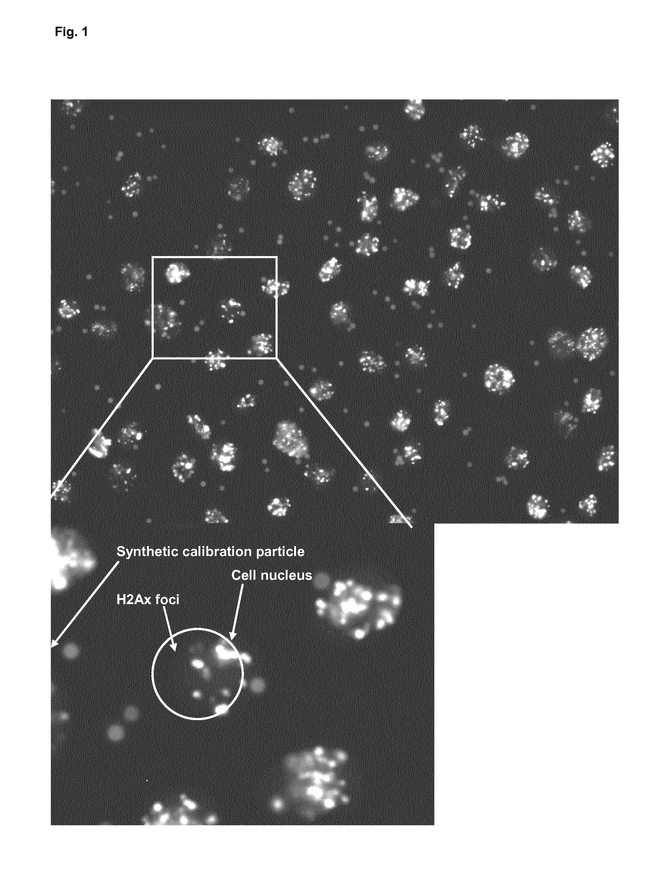
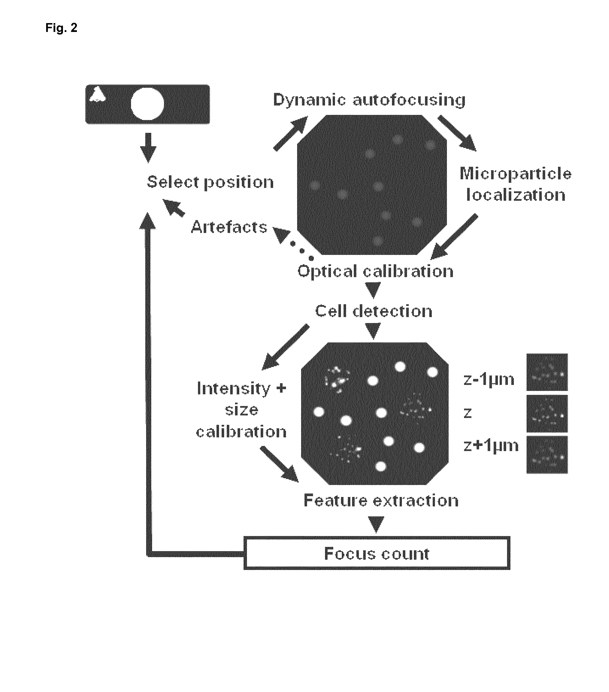
Demonstration of the Method Described Herein for Automated Determination of Immunofluorescent Foci by Means of an Immunofluorescence Assay Using Synthetic Calibration Particles

Immobilization of the Calibration Microparticles on Slides

[0073]Glass slides were coated with 10 μl per application spot of poly-lysine (Sigma, 5 μg / ml) were incubated for 4 hours in a moist incubation chamber. After rinsing the non bound poly-lysine from the slides using PBS, 10 μl of the calibration microparticle suspension (6000 calibration microparticles per application spot) were pipetted to the moist application spots. This suspension was subsequently dried under a sterile hood and allowed to rest on the slide overnight.

Preparation of Lymphocyte Suspension for the Detection of DSB in Peripheral Mononuclear Blood Cells

[0074]The blood sample of a test subject was inverted two times. 3 ml of uncoagulated blood (ratio 1:1) was pipetted onto the separation membrane of a centrifugation tube and subsequently ce...

example 2

Demonstration of the Use of the Method as Described Herein for Determining the Number of DNA Double Strand Breaks in Cells, for Determining DNA Damage or for Determining Radiation Exposure of a Cell and / or Cell Population to Ionizing or any Other Kind of Radiation

Radionuclide

[0085]188Re-perrhenate (188Re) was obtained by elution of a 40 GBq alumina based 188W / 188Re generator (ITG, München, Germany). The physical characteristics of 188Re are: physical half life 16.9 h, maximal beta energy 2.1 MeV, gamma emission 155 keV (15% abundance). The generator was eluted with 5 ml of 0.9% saline to achieve activity concentrations of about 1.0 GBq / ml of carrier free 188Re.

Cell Culture

[0086]The thyroid rat cell line PC CI3 (Clinical Cooperation Unit Nuclear Medicine, DKFZ, Heidelberg, Germany) is a sodium iodine symporter (NIS) positive subline of the FRTL5 cell line available in the American Type Culture Collection (ATCC). The cells were routinely grown as adherent monolayer in Ham's F12 medium...


PUM

PropertyMeasurementUnit
diameteraaaaaaaaaa
diameteraaaaaaaaaa
diameteraaaaaaaaaa

Abstract

The invention relates to a method for automated determination of immunofluorescent foci by means of an immunofluorescence assay using synthetic calibration particles, in addition to a system and kit for carrying out the method. In a preferred embodiment the method is characterized in that the immunofluorescent foci are gamma H2Ax foci.

Description

[0001]The invention relates to a method for automated determination of cellular immunofluorescent foci by means of an immunofluorescence assay, comprising providing a mixture of cells to be analysed and synthetic calibration particles, whereby cells and particles are fixed to a solid phase, detection of synthetic calibration particles, followed by calibration and focusing of a fluorescent microscopy device and automated evaluation system based on detection of the synthetic calibration particles, incubation of said mixture with one or more antibodies that bind to a target substrate of the cells, detection of antibodies bound to said substrates via said fluorescent microscopy device, and determination of immunofluorescent foci from image data generated by said fluorescent microscopy device using said automated evaluation system. In a preferred embodiment the method is characterized in that the immunofluorescent foci are gamma H2Ax foci.BACKGROUND OF THE INVENTION[0002]Standardization ...

Claims


Application Information

Patent Timeline
Patent Type & Authority Patents(United States)
IPC IPC(8): G01N33/53G01N31/00G01N33/543G01N33/569
CPCG01N33/56972G01N33/54346G01N33/5308
Inventor ROGGENBUCK, DIRK
Owner MEDIPAN
Why Eureka
Try Eureka