Method of identifying fetal antigens or cell surface markers using phage display technology
a phage and display technology, applied in the field of identifying fetal antigens or cell surface markers using phage display technology, can solve the problem of not preventing access
- Summary
- Abstract
- Description
- Claims
- Application Information
AI Technical Summary
Benefits of technology
Problems solved by technology
Method used
Image
Examples
example 1
Preparation of Peripheral Blood from Pregnant Women
[0256]The maternal blood sample (usually 3 ml) is divided into aliquots of 1 ml. Each aliquot is diluted 1:14 with 0.15 M NaCl, and the cells are allowed to sediment over night at 4° C. After sedimentation, the upper 12 ml of the supernatants are carefully removed and pooled.
[0257]The sedimented cells (total volume 2 ml / tube) are divided into aliquots of 0.5 ml.
Pre-Fixation without Erythrocyte Lysis
[0258]Cells of the supernatant is pre-fixed in a paraformaldehyde solution in PBS, with a final concentration of 0.5% of PFA for 10 minutes.
Recovering and Mounting on Slides
[0259]The cells are recovered by centrifugation for 10 minutes at 500 g. After centrifugation, the supernatant is discarded and the cell pellet is re-suspended in 0.15 M NaCl (usually 20 μl) and smeared onto poly-L-lysine coated slides (usually 2 slides).
Pre-Fixation with NH4Cl Mediated Erythrocyte Lysis
[0260]Each aliquot of the sedimented cells is diluted with 10 ml o...
example 2
Identification of Male Fetal Cells by Reverse Color Fish and Automated Scanning
[0264]Slides are recovered from the freezer and the airtight plastic back is removed. Before hybridization, slides are fixed and permeabilised 10 minutes in −20° C. cold methanol, 10 minute in −20° C. cold acetone and rinsed in phosphate buffered saline (PBS). Slides are then fixed again for 10 minutes in 2% PBS buffered paraformaldehyde (PFA) and rinsed for 5 minutes in PBS before they are dehydrated 3 minutes each in 60%, 80% and 99.9% ethanol and air-dried.
[0265]Chromosome-specific repeat probes, DXZ1 labeled with spectrum green and DYZ1 Labeled with spectrum orange (Vysis) are used for the first hybridization. Hybridization mixture containing both probes is prepared by mixing 1 part of the X-probe, 1 part of the Y-probe, 1 part of distilled water, and 7 parts of hybridization buffer (Vysis). For whole slide FISH, 28 μl of the hybridization mixture is added and covered by a 22×50 mm cover slips. Cover ...
example 3
Generation of Foetal Cell Specific Antibody Fragments
[0275]Foetal cell specific antibody fragments against cell markers are generated by use of phage display.
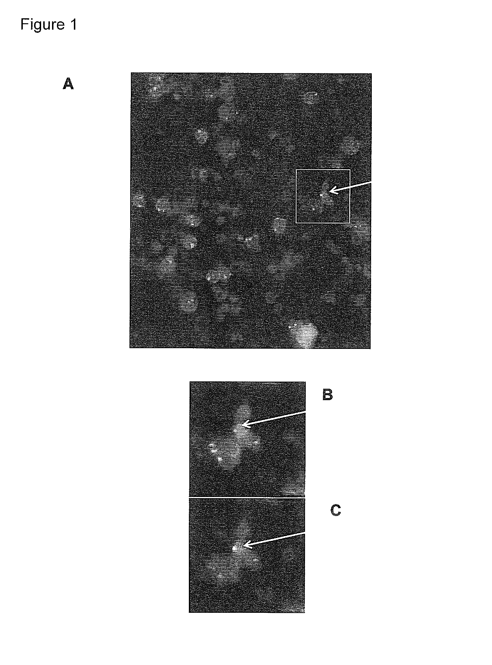
[0276]Based on the identification of foetal cells by FISH, the localization of the foetal cell on the slides is known. The identified foetal cell is subjected to biopanning whereby single chain fragment variable (scFv) antibody fragments are generated.
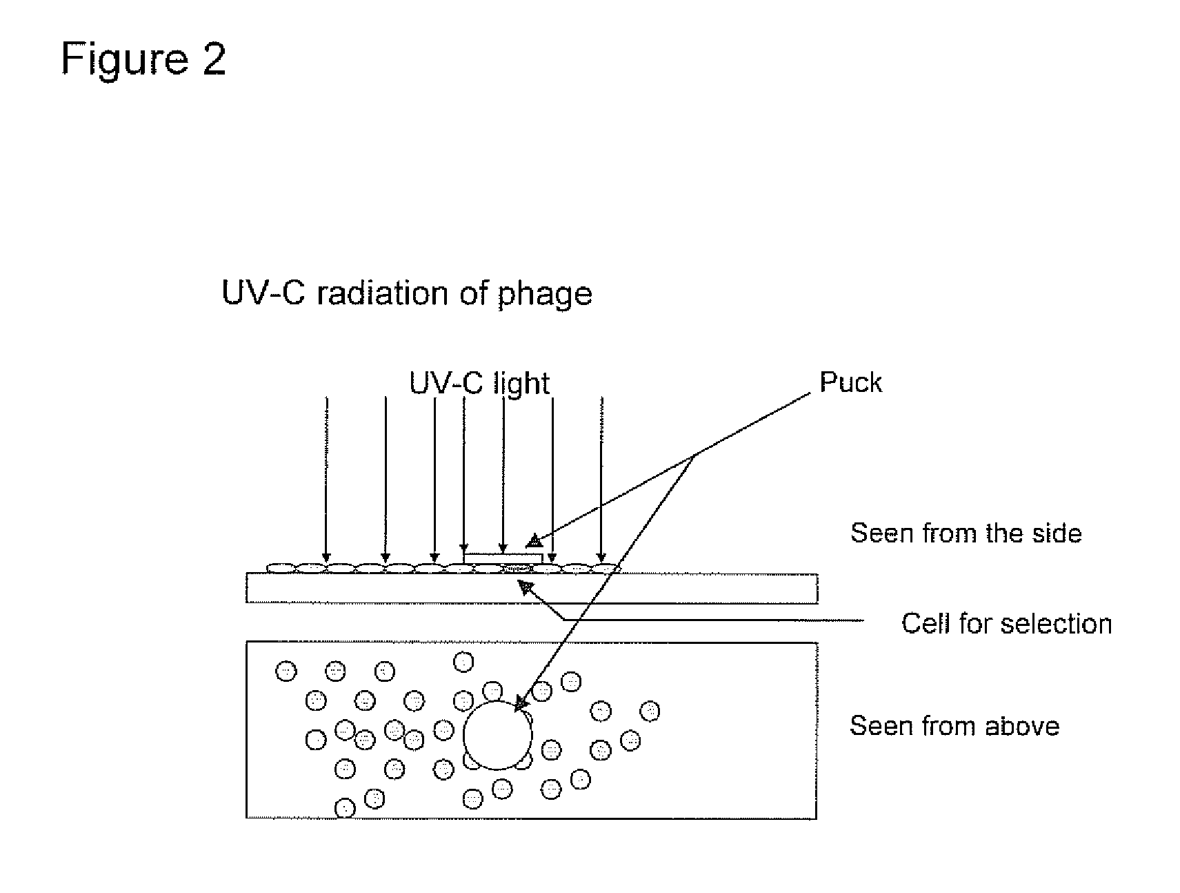
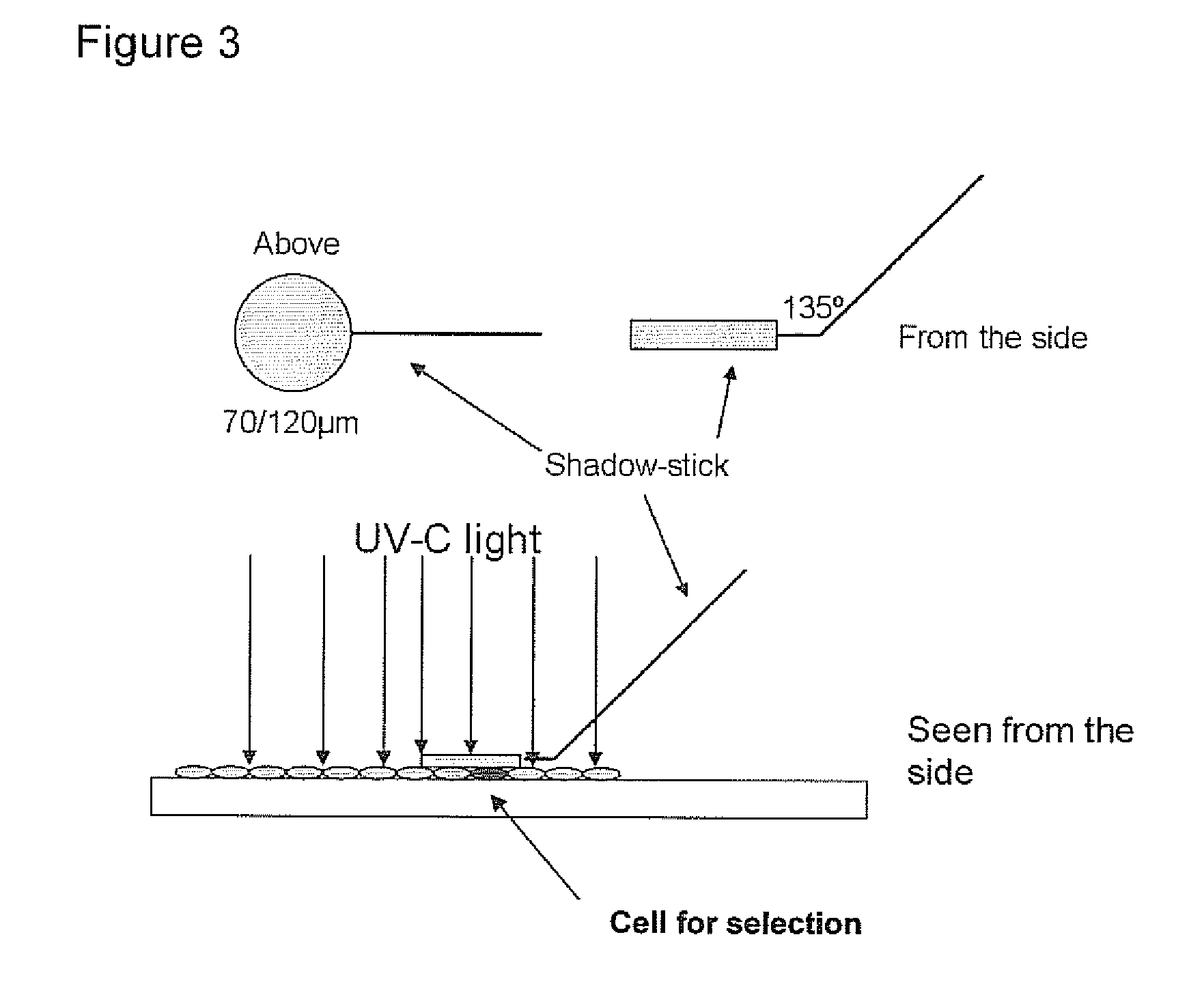
[0277]The selection of antibody fragments may be preformed as follows.[0278]1. The slide is blocked for 1 hour in 2% MPBS (skimmed milk powder in 1×PBS) at room temperature in order to reduce the amount of unspecific binding. This is done by subjecting the slide into MPBS in a holder for slides.[0279]2. The slide is washed one time in 1×PBS[0280]3. Biopanning: the scFv phage library (McCafferty et al. 1990) is incubated with the slide in 2% MPBS for one hour, with gentle shaking. A suitable library may have a semi-synthetic VH+VL repertoire and two single framework repertoires. An...
PUM
 Abstract
Description
Claims
Application Information
 - R&D
- Intellectual Property
- Life Sciences
- Materials
- Tech Scout
- Unparalleled Data Quality
- Higher Quality Content
- 60% Fewer Hallucinations
Browse by: Latest US Patents, China's latest patents, Technical Efficacy Thesaurus, Application Domain, Technology Topic, Popular Technical Reports.
© 2025 PatSnap. All rights reserved.Legal|Privacy policy|Modern Slavery Act Transparency Statement|Sitemap|About US| Contact US: help@patsnap.com


