Stabilization method for biological samples by combination of heating and chemical fixation

a biological sample and chemical fixation technology, applied in the field of biological sample stabilization, can solve the problems of difficult detection, difficult detection, difficult detection, etc., and achieve the effect of avoiding degradation of other constituents of samples, shortening the time needed to obtain blocking of enzymatic processes, and facilitating effective heating

Active Publication Date: 2015-03-24
DENATOR +1
View PDF2 Cites 3 Cited by
  • Summary
  • Abstract
  • Description
  • Claims
  • Application Information

AI Technical Summary

Benefits of technology

[0016]The present inventors have solved this problem by combining heat treatment with chemical fixation, providing an effective and reliable method for stabilizing biological samples.
[0017]Accordingly, the present invention provides a method for stabilizing a biological sample for analysis.
[0026]In an embodiment, the biological sample is given a shape that facilitates an effective heating in terms of the heating being uniform and fast. This helps to shorten the time needed to obtain blocking of enzymatic processes. By blocking certain enzymatic processes driven by proteins, degradation of other constituents of the sample is avoided. Because the time between taking the biological sample and performing a biological analysis has a large impact on the level of degradation, even after a short time, e.g., after as little as 1-3 minutes, it is important that heating takes place immediately after taking the sample. By heating the sample, proteins that function as enzymes lose their secondary and tertiary structure, and thereby their activity, minimizing degradation of the sample.
[0027]The methods according to the invention preserve the primary structure and post-translational modifications of proteins and peptides but simultaneously disrupts their original secondary, tertiary structures, and, where applicable, quaternary structures. The heating of the sample therefore has several advantages, including enabling species such as the relatively low-abundant neuropeptides and the more common post-translational modification of proteins that would otherwise be degraded to remain intact. In addition, the method minimizes degradation of neuropeptides and proteins in a reproducible manner. This method also makes it possible to compare the content and levels of proteins and peptides from different samples.

Problems solved by technology

Sample degradation, however, is both hard to impede, and hard to detect.
The result is that many analyses are unable to detect the presence of species that have degraded long before the analysis is carried out; correspondingly, such analyses may in fact identify degradation products of critical components in place of the original components.
Despite their ubiquity, proteins are extremely sensitive to their environments and thus are not always easy to detect and to identify because they can degrade very quickly.
Once an organism dies, or once a sample of tissue is extracted from a living organism, the regulatory balance of the organism or in the sample is lost and key proteins start to break down.
For example, some proteins whose natural role is to digest other proteins (a “proteolytic” function), and whose natural levels and activities are regulated while an organism is alive, may go out of control after death.
However, the study of tissue samples from patients or model organisms usually exposes the samples to a certain period of oxygen and nutrient depletion before homogenization and protease inactivation occurs.
Use of aldehyde solutions is problematic because it penetrates tissue slowly, ˜0.5-2 allowing for degradation of macromolecules prior to complete fixation and consequently does not arrest natural degradation of proteins (Fox et al., “Formaldehyde fixation” J. Histochem. Cytochem. 33:845-853, 1985).
Microwave irradiation is problematic because it is generally non-uniform, that is, some parts of the sample reach a temperature that is high enough to cause sample breakdown.
Furthermore, parts of the sample can reach temperatures above 100° C. creating small holes due to steam eruptions.
Furthermore, microwave irradiation has formerly been applied to living (non-human) subjects as part of a sacrificial protocol and thus has yet to be established as a tool for analyzing samples that have been extracted from subjects, both human and non-human.
This method does not address the problems related to rapid post-sampling degradation of the sample.
However, full benefit of the application of this technology e.g. in immunohistochemical studies is limited by the rapid loss of phosphorylation in samples before complete stabilization of the sample can be achieved (Mandell, “Phosphorylation state-specific antibodies.

Method used


Image

  • Stabilization method for biological samples by combination of heating and chemical fixation
  • Stabilization method for biological samples by combination of heating and chemical fixation

Examples


example 2

Stabilization of Samples from Mice Brain

Experimental

Biological Samples and Treatments

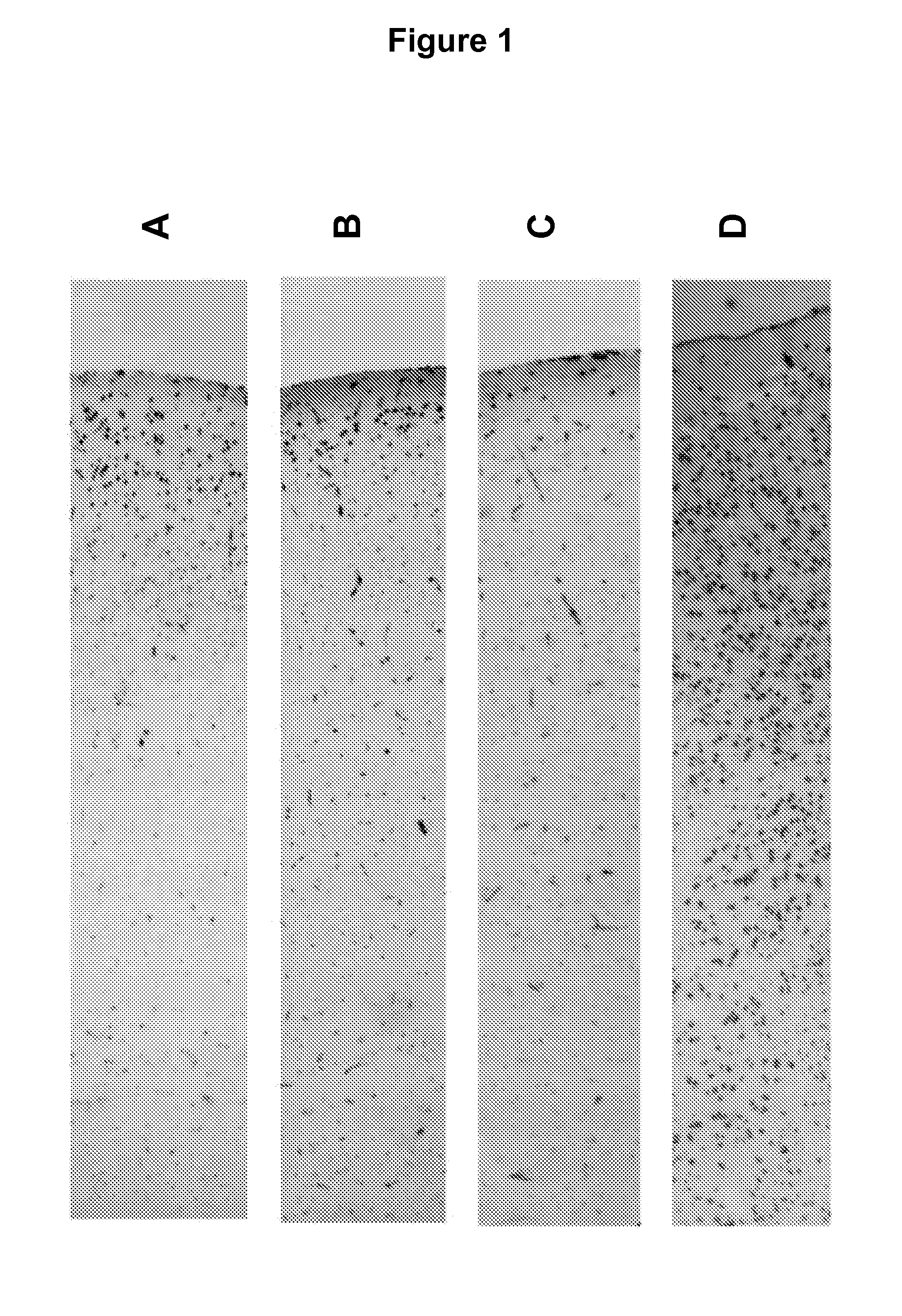
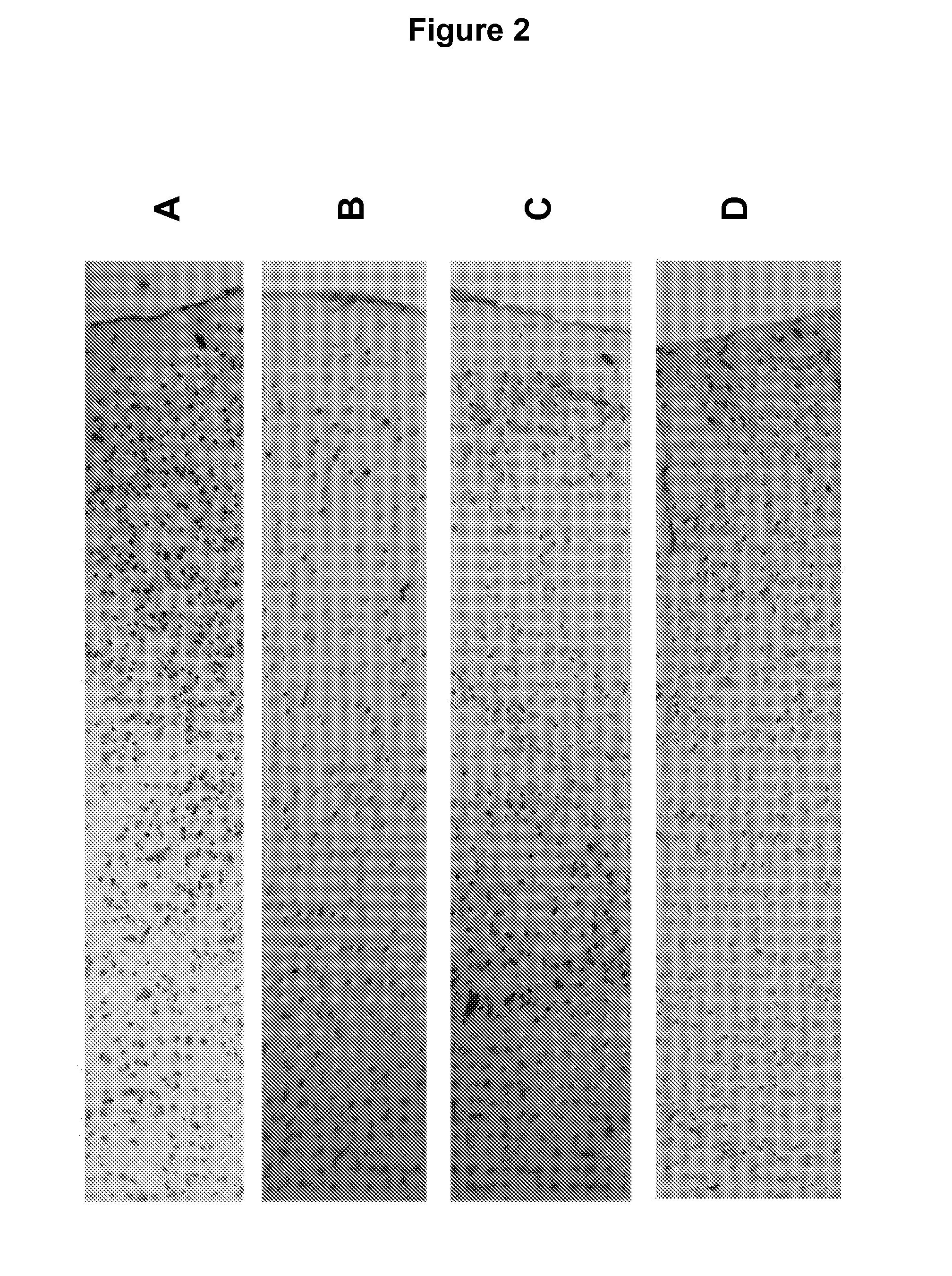
[0076]Mice were sacrificed by cervical dislocation and sample collected immediately. Whole brains were excised immediately following sacrifice and stabilized by heat treatment in the Stabilizor T1 instrument (Denator AB, Sweden) at auto settings for fresh tissue and subsequently incubated in room temperature for the following periods of time, A: 0 min, B: 15 min, C: 6 h, and D: 24 h prior to being submerged in NBF.

[0077]All samples were kept for 24 h in formalin solution at room temperature prior to paraffin imbedding. All samples were incubated without further sub dissection in the NFB. Prior to paraffin embedding samples were cut into halves and embedded such that sections could be cut through the centre of the original sample.

[0078]Histochemistry, Immunohistochemistry, and Image capture and processing were performed as described in Example 1.

Results

[0079]FIG. 2 describes the results of Immunohist...


PUM

PropertyMeasurementUnit
temperatureaaaaaaaaaa

Abstract

The present invention provides methods for stabilizing a biological sample for analysis. The invention more particularly provides methods combining heat treatment and chemical fixation of biological samples in order to maintain protein primary structure and post-translational modifications, such as protein phosphorylations.

Description

FIELD OF THE INVENTION[0001]The present invention relates to methods for stabilizing a biological sample for analysis. The invention more particularly relates to methods combining heating and chemical fixation of biological samples in order to maintain protein primary structure and post-translational modifications, such as protein phosphorylations.BACKGROUND TO THE INVENTION[0002]It is key to any analysis of a biological sample that the integrity of its constituents is conserved between the time that the sample is extracted from a living organism and the time that the analysis is carried out. Sample degradation, however, is both hard to impede, and hard to detect. The result is that many analyses are unable to detect the presence of species that have degraded long before the analysis is carried out; correspondingly, such analyses may in fact identify degradation products of critical components in place of the original components.[0003]The assemblage of proteins in an organism is the...

Claims


Application Information

Patent Timeline
Patent Type & Authority Patents(United States)
IPC IPC(8): G01N1/30G01N1/44
CPCG01N1/30G01N1/44G01N2001/305
Inventor BOREN, MATSSKOLD, KARLSODERQUIST, MARCUS
Owner DENATOR
Why Eureka
Try Eureka