Protection of virus particles from phagocytosis by expression of CD47
a virus particle and phagocytosis technology, applied in the field of phagocytosis protection of virus particles, can solve the problem that cd47, the self-signal, is not understood in targets less than 500 nm in size, and achieve the effect of increasing the life of the particle in vivo
- Summary
- Abstract
- Description
- Claims
- Application Information
AI Technical Summary
Benefits of technology
Problems solved by technology
Method used
Image
Examples
example 1
CD47 Inhibits Myosin Localization In Phagocytosis
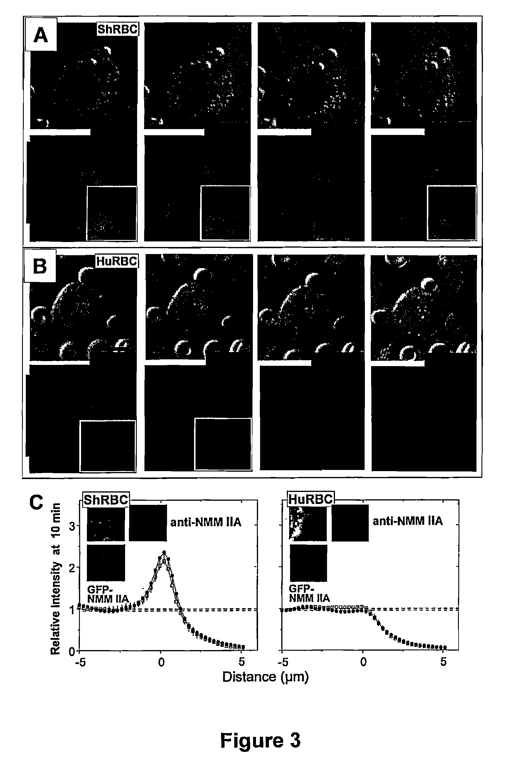
[0187]Pseudopod extension and phagocytic cup formation around the target involves extensive remodeling of the actin cytoskeleton. As explained previously, a hallmark of phagocytosis and endocytosis is the actin recruitment to the target cells (Allison et al., 1971, Nat. New Biol. 232(31):153-5; Newman et al., 1991, J. Immunol. 146(3):967-74), but to FcR-mediated phagocytosis, it is the involvement of myosin IIA (Araki, 2006, Front. Biosci. 11:1479-90; Tsai et al., 2008, J. Cell Biol. 180(5):989-1003). When using IgG-opsonized targets of 1.1 μm and 100 nm streptavidin polystyrene particles incubated with human-derived THP-1 macrophages, localization of F-actin to the synapse of the targeted particles was observed. Surprisingly, as shown in FIGS. 11B and 11C, the motor protein non-muscle myosin IIA (NMMIIA) localized during the phagocytosis of both the 1.1 μm particles and the 100 nm particles. As shown in FIGS. 11D and 11E, quantificat...
example 2
CD47 Inhibits Phagocytosis of Nano-scale Particles
[0188]In order to assess if CD47 is important in inhibiting FcR-mediated phagocytosis at nano-length scales, the uptake of different-sized streptavidin polystyrene particles having the same physical characteristics was studied to accurately compare the size ranges of interest. The extracellular immunoglobulin-like domains of human CD47 (hCD47) was recombinantly expressed with a spacer domain (Brown et al., 1994, Protein Eng 7(4):515-21; Brown et al., 1998, J. Exp. Med. 188(11): 2083-90) plus a C-terminal biotinylation site. Biotinylation allowed attachment of soluble hCD47 to streptavidin-coated beads of sizes ranging from 100 nm to 3 μm, with densities adjustable to levels previously measured for normal and diseased RBCs (Dahl et al., 2004, Blood 103(3):1131-6; Subramanian et al., 2006, Blood Cells Mol. & Dis. 36(3):364-372).
[0189]As shown in FIG. 12, particles were opsonized by pretreatment with anti-streptavidin antibody to induce...
example 3
SHP-1 and Myosin Inhibition Effects CD47 Potential
[0191]As explained previously, interaction of CD47 on the target and SIRPα on the macrophages leads to a number of downstream processes. This occurs through the receptor-ligation interactions of CD47-SIRPα leading to the phosphorylation SIRPα immunoreceptor tyrosine inhibitory motif (ITIM) (Kharitonenkov et al., 1997, Nature 386(6621):181-6) and subsequent SHP-1 phosphatase induction (Tsuda et al., 1998, J. Biol. Chem. 273(21):13223-9; Veillette et al., 1998, J. Biol. Chem. 273(35):22719-28; Vernon-Wilson et al., 2000, Eur. J. Immunol. 30(8):2130-7). In order to confirm that the interaction of hCD47 coupled to both the nano- and micro-scale streptavidin particles interacts specifically with SIRPα found on THP-1 human macrophages, FACS analysis was performed. As shown in FIG. 15, an antibody against SIRPα known to obstruct hCD47-SIRPα interaction (Subramanian et al., 2006, Blood 107(6):2548-2556) was pre-incubated on THP-1 macrophages...
PUM
| Property | Measurement | Unit |
|---|---|---|
| radius | aaaaa | aaaaa |
| radius | aaaaa | aaaaa |
| size | aaaaa | aaaaa |
Abstract
Description
Claims
Application Information
 - R&D
- Intellectual Property
- Life Sciences
- Materials
- Tech Scout
- Unparalleled Data Quality
- Higher Quality Content
- 60% Fewer Hallucinations
Browse by: Latest US Patents, China's latest patents, Technical Efficacy Thesaurus, Application Domain, Technology Topic, Popular Technical Reports.
© 2025 PatSnap. All rights reserved.Legal|Privacy policy|Modern Slavery Act Transparency Statement|Sitemap|About US| Contact US: help@patsnap.com


