Biological information imaging apparatus and method for analyzing biological information

a biological information and imaging apparatus technology, applied in the field of biological information imaging apparatus and a method for analyzing biological information, can solve the problem of limited regions in the living body that can be imaged, and achieve the effect of increasing the imaging range of biological information and high sensitivity

Inactive Publication Date: 2015-09-15
CANON KK
View PDF22 Cites 24 Cited by
  • Summary
  • Abstract
  • Description
  • Claims
  • Application Information

AI Technical Summary

Benefits of technology

[0023]The present invention can realize a biological information imaging apparatus that can measure the ultrasound generated from the optical absorber in the deep part of the subject living body with high sensitivity, and can increase an imaging range of biological information, and a method for analyzing biological information using the biological information imaging apparatus.

Problems solved by technology

Thus, a photoacoustic signal that can be observed by a sound wave detector placed on a surface of the living body cannot be obtained from the deep part of the subject, and the region in the living body that can be imaged is limited.

Method used


Image

  • Biological information imaging apparatus and method for analyzing biological information
  • Biological information imaging apparatus and method for analyzing biological information
  • Biological information imaging apparatus and method for analyzing biological information

Examples


embodiment 1

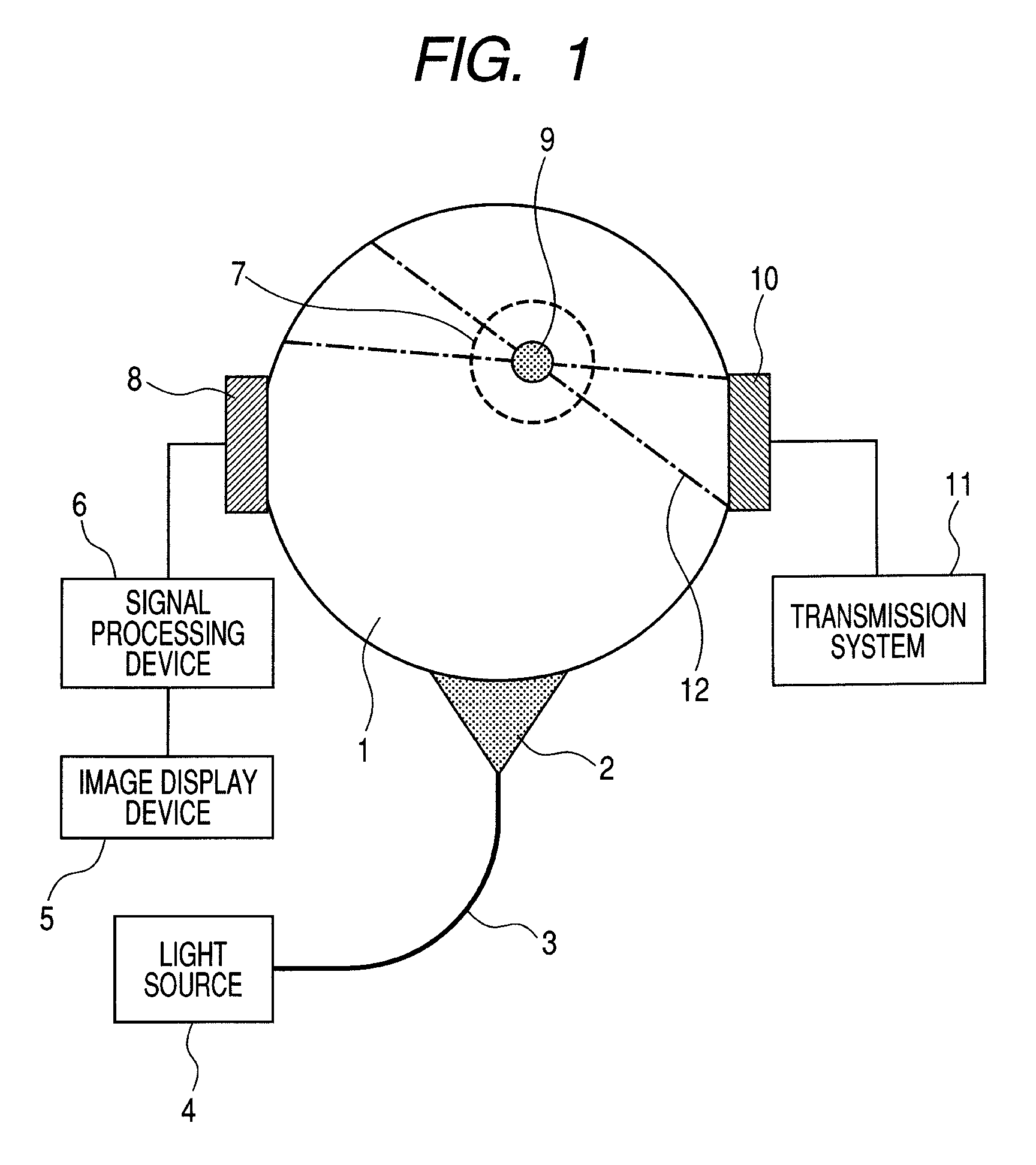
[0032]First, a biological information imaging apparatus according to Embodiment 1 of the present invention will be described.

[0033]FIG. 1 illustrates an exemplary construction of a biological information imaging apparatus according to the embodiment.

[0034]In FIG. 1, reference numeral 1 denotes a living body as a subject, 2; pulsed light, 3; an optical waveguide, 4; a light source, 5; an image display device, 6; a signal processing device, and 7; ultrasound generated from an optical absorber (sometimes referred to herein as “second ultrasound”).

[0035]Reference numeral 8 denotes an ultrasound detector (ultrasound detection unit), 9; an optical absorber, 10; an ultrasound transmission device (ultrasound transmission unit), 11; an ultrasound transmission control system, and 12; focused ultrasound (sometimes referred to herein as “first ultrasound”).

[0036]The biological information imaging apparatus of the embodiment can image information as described below for diagnosing tumor or vascul...

embodiment 2

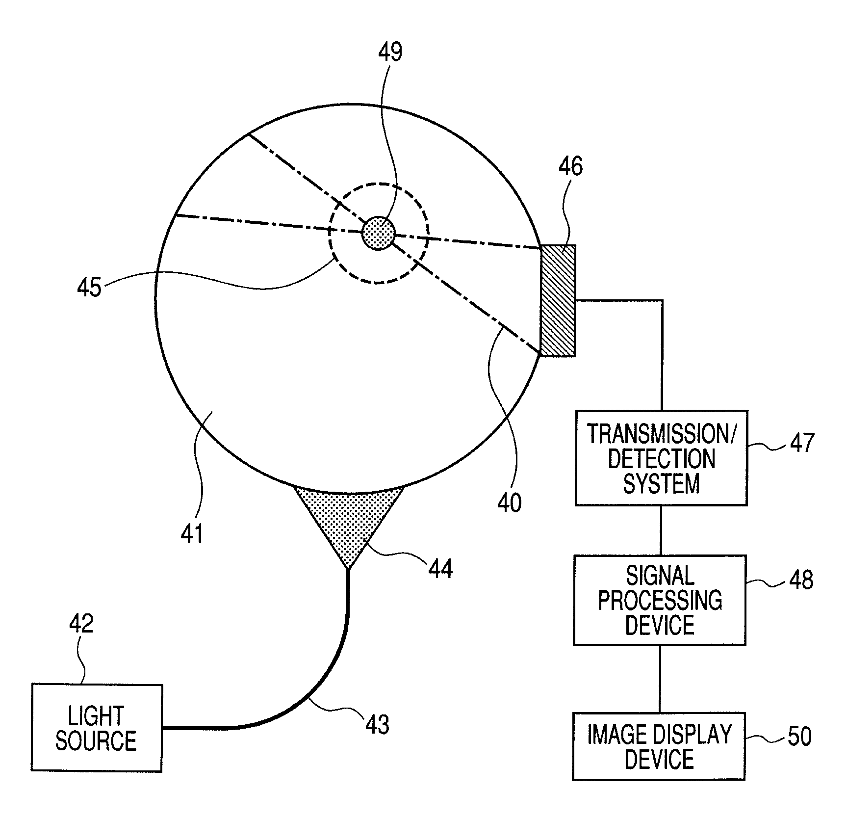
[0107]Next, a biological information imaging apparatus according to Embodiment 2 of the present invention will be described.

[0108]FIG. 4 illustrates an exemplary construction of the biological information imaging apparatus according to the embodiment.

[0109]The biological information imaging apparatus of the embodiment can image distribution information of a molecular probe introduced into a living body for diagnosing tumor or various diseases labeled by the molecular probe such as Alzheimer's Disease or carotid plaque.

[0110]The biological information imaging apparatus of the embodiment includes a light source 42 that irradiates a living body 41 with pulsed light 44 as illustrated in FIG. 4.

[0111]The light source 42 emits pulsed light, and includes an optical waveguide 43 for guiding light to a surface of the living body.

[0112]The biological information imaging apparatus of the embodiment also includes an ultrasound transmission / detection device 46 having both an ultrasound detection...

example

[0145]Next, an exemplary construction of the biological information imaging apparatus according to the example of the present invention will be described.

[0146]An exemplary construction of the biological information imaging apparatus that obtains information image relating to distribution of an optical absorber in a living body will be described with reference to FIG. 1.

[0147]Since imaging of the actual living body is difficult, the case of imaging a phantom that mimics the living body will be described. The phantom used is 1% of intralipid solidified with agar into a square shape, into which India ink solidified with agar into a spherical shape is inserted as an optical absorber.

[0148]As a light source 4, a Q switch Nd:YAG laser that can emit nanosecond pulsed light of 1064 nm is used.

[0149]A pulsed width is about 5 nanoseconds, and a repetition speed is 10 Hz. For example, energy of 1 pulsed light is 120 mJ.

[0150]The pulsed light is guided to a surface of a living body phantom usi...


PUM

PropertyMeasurementUnit
wavelengthaaaaaaaaaa
wavelength rangeaaaaaaaaaa

Abstract

There are provided a biological information imaging apparatus that can measure ultrasound generated from an optical absorber in a deep part of a subject living body with high sensitivity, and a method for analyzing biological information using the biological information imaging apparatus. The biological information imaging apparatus that detects ultrasound and images biological information, includes: a light source that irradiates the subject with light for generating ultrasound from an optical absorber existing in the subject; an ultrasound transmission unit that transmits focused ultrasound to a specific region where the optical absorber exists; and an ultrasound detection unit that detects an ultrasound synthesized signal due to interaction between ultrasound generated from the optical absorber that absorbs the light and the focused ultrasound transmitted to the specific region.

Description

BACKGROUND OF THE INVENTION[0001]1. Field of the Invention[0002]The present invention relates to a biological information imaging apparatus and a method for analyzing biological information.[0003]2. Description of the Related Art[0004]Imaging apparatuses using X-ray (mammography), ultrasound or MRI (magnetic resonance imaging) have been often used for diagnosing breast cancer.[0005]In recent years, a photoacoustic imaging apparatus has been noted, in which the imaging apparatus propagates pulsed light from a light source such as a laser into a living body, detects a photoacoustic signal generated by absorption of the propagated light into the living body, and images an initial pressure distribution or absorption coefficient distribution in the living body. This technique is referred to as PAT (PhotoAcoustic Tomography).[0006]Generally, photoacoustic tomography has the following advantages. First, photoacoustic tomography can perform functional imaging of oxygen metabolism and brain ...

Claims


Application Information

Patent Timeline
Patent Type & Authority Patents(United States)
IPC IPC(8): A61B5/00A61B8/08G01N29/24
CPCA61B5/0095A61B5/0091A61B5/4312A61B8/0825A61B5/489A61B5/4887A61B8/5223G01N29/2418A61B8/4416
Inventor FUKUTANI, KAZUHIKONAKAJIMA, TAKAOASAO, YASUFUMI
Owner CANON KK
Why Eureka
Try Eureka