Computerized image reconstruction method and apparatus

a computerized image and reconstruction method technology, applied in the field of computerized image reconstruction, can solve the problems of limiting the accuracy of proton therapy, unable to unable to achieve the direct visualization of the three-dimensional anatomy of the patient in the treatment room, so as to improve the convenience of treatment and improve the accuracy of patient treatment. , the effect of cost effectiveness

Active Publication Date: 2016-02-02
THE RES FOUND OF STATE UNIV OF NEW YORK
View PDF12 Cites 2 Cited by
  • Summary
  • Abstract
  • Description
  • Claims
  • Application Information

AI Technical Summary

Benefits of technology

[0013]Accordingly, the present invention provides an image reconstruction method for curved paths applicable to heavy particles, in addition to particles following straight line paths. Also provided is a method of use of a proton beam for dose calculation and patient anatomy positioning that provides improved convenience, cost effectiveness and achieves more precise patient treatment.
[0014]The present invention also provides improved image reconstruction of particle beams, resulting in improved treatment planning schemes and overcomes the above shortcomings, by minimizing the uncertainty from image reconstruction techniques. The present invention further provides more accurate, efficient reconstruction of pCT images for use in proton therapy and other diagnostic and therapeutic applications.

Problems solved by technology

However, travel paths of heavy particles, such as proton, muon, neutron, carbon, alpha particle, do not follow a straight line within the body, and present image reconstruction difficulties for proton Computed Tomography (pCT).
However, existing proton treatments have several problems associated with radiation dose calculations due to varied positioning of patient anatomy, and are currently performed based on X-ray computed tomography or xCT, with the patient positioned with the help of X-ray radiographs, hence direct visualization of the three-dimensional (3D) patient anatomy in the treatment room is presently impossible, limiting the accuracy of proton therapy.
Further, conventional practices for estimating the proton path rely on single line-type curve estimations and fail to consider proton deflection and scattering within the body.
Because of the difference of X-ray and proton interaction with the body tissues, the xCT attenuation map introduces an uncertainty into proton therapy treatment planning.
Since the Bragg peak releases a large amount of energy locally, a minor error on the targeted cancerous volume could cause significant harm to the surrounding normal tissues.
Thus, the accuracy of xCT for proton treatment planning is limited due to the difference in physical interactions between X-ray photons and particle protons, which partially obviates the advantage of proton therapy.
Further, conventional methods do not accurately modify xCT in the treatment room based on patient position.

Method used


Image

  • Computerized image reconstruction method and apparatus
  • Computerized image reconstruction method and apparatus
  • Computerized image reconstruction method and apparatus

Examples


Embodiment Construction

[0023]The following detailed description of various embodiments of the present invention will be made in reference to the accompanying drawings. In describing the present invention, explanation about related functions or constructions known in the art are omitted to avoid obscuring the present invention with unnecessary detail.

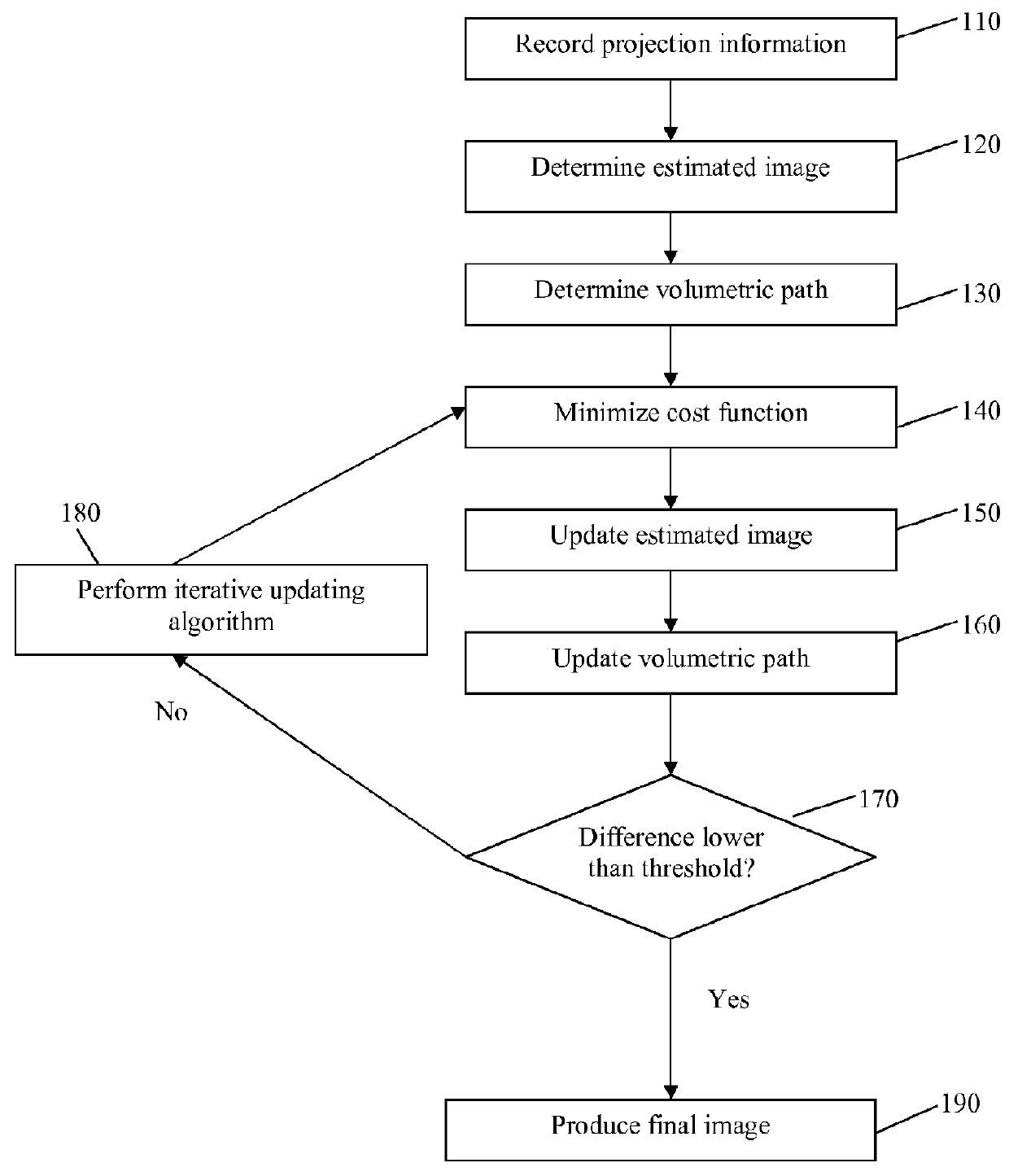
[0024]The image formation principles of proton beams for pCT rely on hardware configuration and data acquisition to reconstruct an image from projected data along proton trajectories through the body. Image formation for pCT relies on the interaction of incident energy with the tissues inside the body. The knowledge of the interaction and the accuracy of measuring the difference of the exit energy from the incident energy determine the quality of the reconstructed image about the body internals.

[0025]A single-proton-tracking pCT scanner tracks individual protons pre and post patient with two-dimensional (2D) sensitive Silicon Strip Detectors (SSDs), providing ...


PUM


Abstract

Provided herein is a computerized image reconstruction apparatus and method that includes recording projection path information and energy loss information of a plurality of particles traversing an object being imaged and determining an estimated image of the object based on the projection path information and the energy loss information sampled into a projection format. The estimated image includes an active volume defined by an enclosure border. Cost function minimization uses an Adaptive-weighted Total Variation cost function, a Penalized Weighted Least-Squares cost function, or an Alpha-Divergence cost function to update the estimated image. An iterative updating algorithm corresponding to the cost function updates the estimated image and produces a final image based on the estimated image according to a predetermined threshold.

Description

PRIORITY[0001]This application claims priority to U.S. Provisional Application No. 61 / 593,634, filed with the U.S. Patent and Trademark Office on Feb. 1, 2012, the entire content of which is incorporated herein by reference.BACKGROUND OF THE INVENTION[0002]1. Field of the Invention[0003]The present invention generally relates to computerized image reconstruction and, more particularly, to image reconstruction from projected data along curved paths of proton particles through an object being imaged.[0004]2. Description of the Related Art[0005]Computed tomographic image reconstruction has typically focused on X-rays or photons that pass through the body in a straight line. Any path deviation from an original direction is considered as the path of scattered particles and may be ignored. Such imaging modalities, such as X-ray Computed Tomography (xCT) or simple CT, Single-Photon Emission Computed Tomography (SPECT) and Positron Emission Tomography (PET), provide high energy X-rays or Ga...

Claims


Application Information

Patent Timeline
Patent Type & Authority Patents(United States)
IPC IPC(8): G06K9/00G06T11/00A61B6/03
CPCG06T11/005A61B6/032G06T11/006G06T2207/10124G06T2207/30096G06T2211/424G06T2207/10081
Inventor LIANG, ZHENGRONGMA, JIANHUALIU, YAN
Owner THE RES FOUND OF STATE UNIV OF NEW YORK
Why Eureka
Try Eureka