
Treatment of leaky or damaged tight junctions and enhancing extracellular matrix

a tight junction, leaky or damaged technology, applied in the field of treatment, can solve the problems of loss of blood pressure, organ dysfunction or failure, death, global tissue hypoxia, etc., and achieve the effect of enhancing growth and repair of surrounding tissues

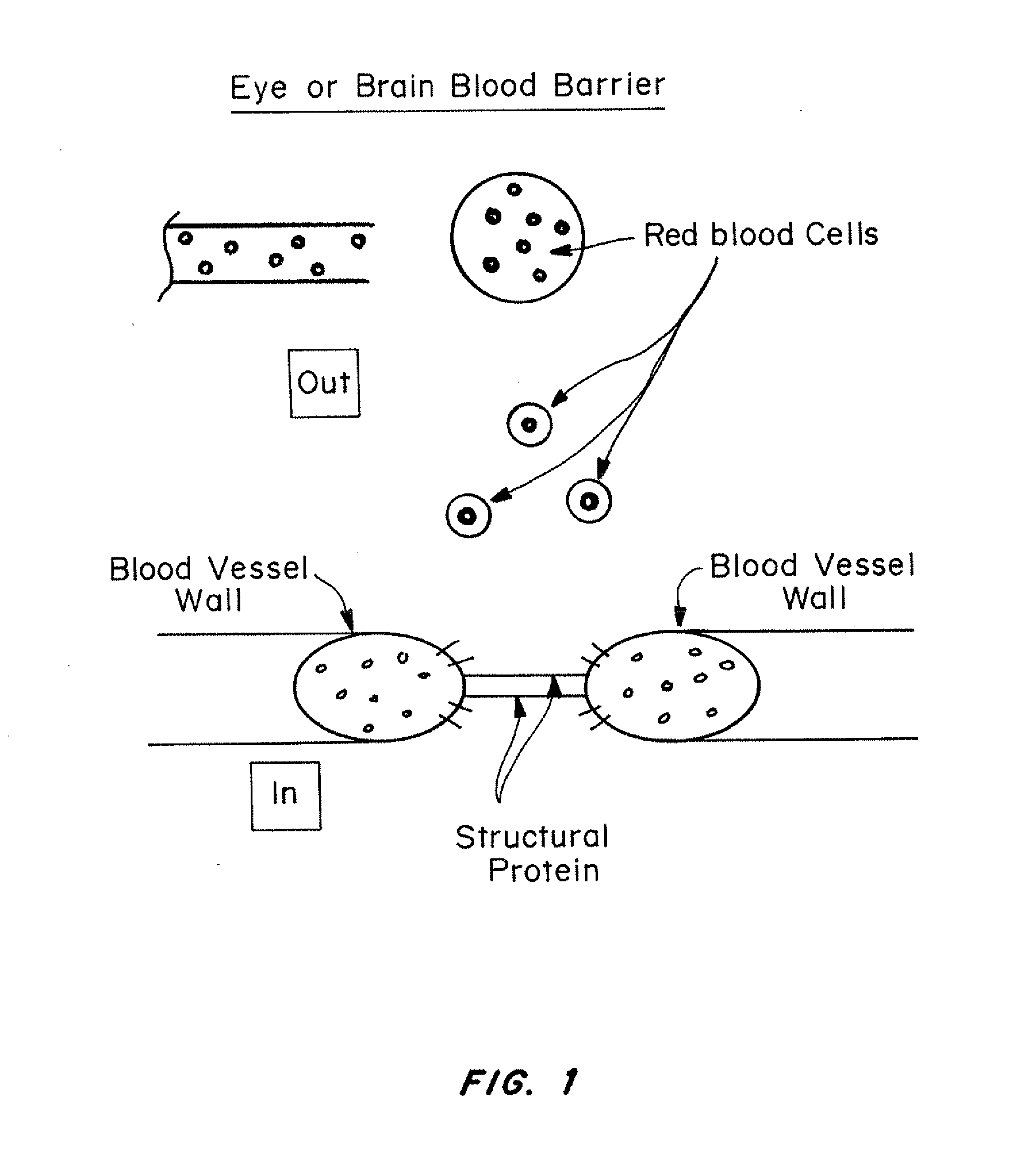
Active Publication Date: 2016-08-16
ARCH BIOSURGERY
View PDF63 Cites 24 Cited by
  • Summary
  • Abstract
  • Description
  • Claims
  • Application Information

AI Technical Summary

Benefits of technology

The patent describes materials that can be used to create, for example, scaffolds for growing new tissues or skin. These materials are designed to break down in the body naturally and safely, and can even improve the growth and repair of surrounding tissues. This is important because traditional scaffolds can be toxic and cause secondary harm to the body.

Problems solved by technology

Such leakage can lead to fluid invading the tissues, causing a loss in blood pressure, organ dysfunction or failure, and death.
This over-response disrupts homeostasis through an uncontrolled cascade of inflammation, coagulation, and impaired fibrinolysis.
Deranged micro-circulatory function leads to global tissue hypoxia and direct tissue damage.
This ultimately results in organ failure, and often, death.
Anti-infectives, resuscitation, and supportive care do not necessarily prevent the progressive organ dysfunction that occurs in many patients.
Microcirculatory dysfunction may persist despite adequate global values of oxygen delivery, making resuscitation procedures ineffective.
One third of women undergoing mastectomy with axillary evacuation for primary breast cancer suffer from postoperative seromas leading to unnecessary costs and complications such as infections and new operations.
Another condition for which there are insufficient treatments is the loss of fluid which occurs due to sepsis or burns, where the junctions between cells begin to leak interstitial fluid, causing dehydration, electrolyte imbalance, and cell death.
These disorders can lead to further complications when the release of inflammatory molecules from the damaged tissue elicits further tissue damage.
However, Zhang does not disclose the use of such materials for treatment and support of disorders associated with leaky, damaged tight junction and weak, diseased, or injured extracellular matrix.
However, these applications do not describe the use of such materials for treatment and support of disorders associated with leaky, damaged tight junction and weak, diseased, or injured extracellular matrix.

Method used


Image

  • Treatment of leaky or damaged tight junctions and enhancing extracellular matrix
  • Treatment of leaky or damaged tight junctions and enhancing extracellular matrix
  • Treatment of leaky or damaged tight junctions and enhancing extracellular matrix

Examples


example 1

Hemostasis in a Brain Injury

Methods and Materials

[0182]Adult Syrian hamsters were anesthetized with an intraperitoneal injection of sodium pentobarbital (50 mg / kg) and adult rats were anesthetized with an intraperitoneal injection of ketamine (50 mg / kg). The experimental procedures adhered strictly to the protocol approved by the Department of Health and endorsed by the Committee on the Use of Laboratory Animals for Teaching and Research of the University of Hong Kong and the Massachusetts Institute of Technology Committee on Animal Care.

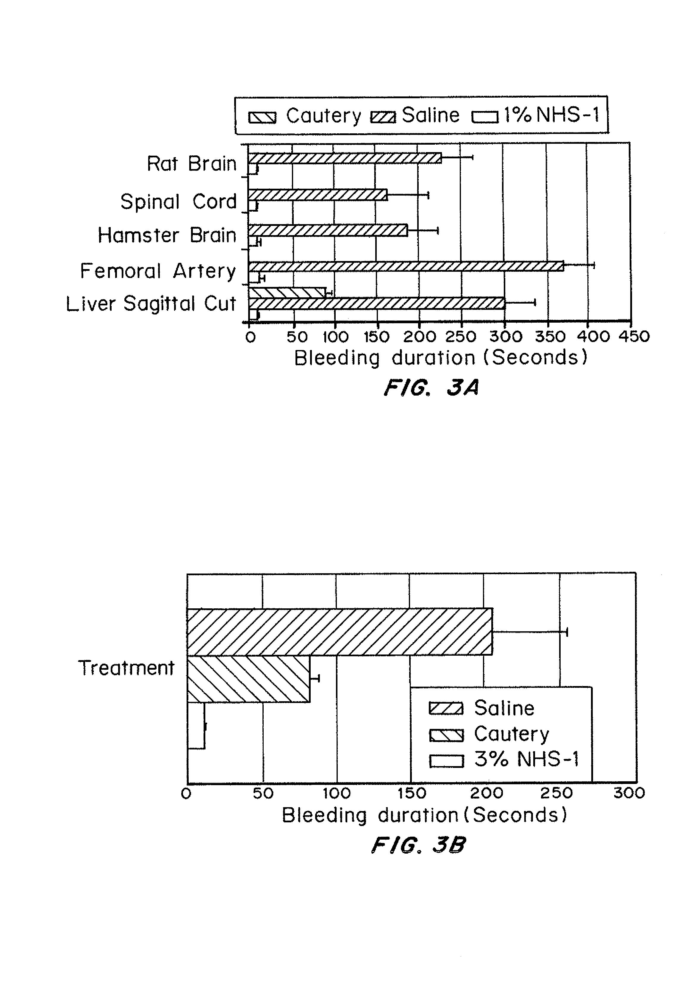
[0183]The animals were fitted in a head holder. The left lateral part of the cortex was exposed. Each animal received a transection of a blood vessel leading to the superior sagittal sinus (FIG. 4a). With the aid of a sterile glass micropipette, 20 μl of 1% NHS-1 solution was applied to the site of injury or iced saline in the control cases. The animals were allowed to survive for up to six months.

Results

[0184]Initial experiments in the brain includ...

example 2

Hemostasis in a Spinal Cord Injury

Methods and Materials

[0186]Under an operating microscope, the second thoracic spinal cord segment (T2) was identified before performing a dorsal laminectomy in anesthetized adult rats. After opening the dura mater, a right hemisection was performed using a ceramic knife. Immediately after the cord hemisection 20 μl of 1% solution of RADA16-I (SEQ ID NO: 60) (NHS-1) was applied to the area of the cut for bleeding control. The controls received a saline treatment. The animals were allowed to survive for up to 8 weeks as part of another experiment.

Results

[0187]The spinal environment and the brain environment were investigated for similarities or differences. Secondary damage caused by surgery can be reduced by quickly bringing bleeding under control. After laminectomy and removal of the dura, the spinal cord was hemisected at T2, from the dorsal to ventral aspect and treated (N=5) with 20 μl of 1% NHS-1. Hemostasis was achieved in 7.6 seconds. In the s...

example 3

Hemostasis in a High Pressure Femoral Artery Wound

Methods and Materials

[0188]Rats were placed on their backs and the hind limb was extended to expose the medial aspect of the thigh (FIG. 4b). The skin was removed and the overlying muscles were cut to expose the femoral artery and sciatic nerve. The femoral artery was cut to produce a high pressure bleeder. With a 27 gauge needle, 200 μl of 1% RADA16-I (SEQ ID NO: 60) (NHS-1) solution was applied over the site of injury. In two cases powder was applied to the injury site which also worked. (Data not shown and was not included in the analysis). Controls were treated with a combination of saline and pressure with a gauge. All animals were sacrificed four hours after the experiment.

Results

[0189]The femoral artery of 14 adult rats was surgically exposed, transected and then treated with 200 μl of 1% solution of NHS-1 or iced saline and packing. The treated cases (n=10) took about 10 seconds to achieve hemostasis (FIG. 3a). The controls (...


PUM

PropertyMeasurementUnit
sizeaaaaaaaaaa
distanceaaaaaaaaaa
distanceaaaaaaaaaa

Abstract

Self assembling peptides and peptidomimetics can be utilized for the treatment and support of disorders associated with leaky or damaged tight junction and weak, diseased, or injured extracellular matrix. The self-assembling materials generally have alternating hydrophilic or hydrophobic residues or hydrophobic and / or hydrophilic sections which allow the material to react or interact with the glycoproteins found in the ECM. Diseases in which treatment with these materials applied to or near the site in need of treatment include diabetic retinopathy, sepsis, burns, and certain neurodegenerative diseases such as Parkinson's and Alzheimer's. The formulations can be administered by injection, spraying, topically or by catheter or via a wound dressing or other material to which it is applied and then applied to the site in need of treatment.

Description

CROSS-REFERENCE TO RELATED APPLICATIONS[0001]This application claims priority to U.S. Ser. No. 60 / 894,872, filed on Mar. 14, 2007.FIELD OF THE INVENTION[0002]The present application is in the field of materials to alter or enhance cell matrix deposition around tissues, especially blood vessels, connective tissue and tissues with tight junctions.BACKGROUND OF THE INVENTION[0003]There are a number of disorders associated with leakage around blood vessels and within the tight junctions between cells. Such leakage can lead to fluid invading the tissues, causing a loss in blood pressure, organ dysfunction or failure, and death. Many of these disorders are a result of massive cell death, such as that associated with chemotherapy of a large tumor, sepsis following systemic bacterial infection, inflammation associated with burns, and large surgical resections.[0004]Severe sepsis results from the body's systemic over-response to infection. This over-response disrupts homeostasis through an u...

Claims


Application Information

Patent Timeline
Patent Type & Authority Patents(United States)
IPC IPC(8): A61K38/10A61K38/08C07K7/06A61K38/07C07K5/103
CPCA61K38/07A61K38/08A61K38/10C07K5/1008C07K7/06A61P1/00A61P1/04A61P1/16A61P13/02A61P17/02A61P25/00A61P25/28A61P27/02A61P29/00A61P3/10A61P31/04A61P43/00A61P7/00A61P9/00A61P9/10A61P9/14A61K45/06
Inventor ELLIS-BEHNKE, RUTLEDGENORCHI, TERRENCE W.KELLY, STEPHEN RICHARD
Owner ARCH BIOSURGERY
Learn More