Endotracheal tube coupling adapters

a technology of endotracheal tube and adapter, which is applied in the field of endotracheal tube coupling adapters, can solve the problems of requiring a relatively long time to be obtained and interpreted, and affecting the survival of acutely ill patients

Active Publication Date: 2016-09-20
AVENT INC
View PDF362 Cites 20 Cited by
  • Summary
  • Abstract
  • Description
  • Claims
  • Application Information

AI Technical Summary

Benefits of technology

[0084]For purposes of summarizing the disclosure, certain aspects, advantages and novel features of embodiments of the inventions have been described herein. It is to be understood that not necessarily all such advantages can be achieved in accordance with any particular embodiment of the inventions disclosed herein. Thus, the embodiments disclosed herein can be embodied or carried out in a manner that achieves or optimizes one advantage or group of advantages as taught herein without necessarily achieving other advantages as can be taught or suggested herein.

Problems solved by technology

Depending on how busy the x-ray department is at the time and other unpredictable factors, such as time of day and number of personnel available, a confirmatory chest x-ray can take a relatively long time to be obtained and interpreted, which can seriously threaten the survival of an acutely ill patient.
In addition, chest x-rays are relatively expensive and expose the patient to unnecessary or undesirable radiation.

Method used


Image

  • Endotracheal tube coupling adapters
  • Endotracheal tube coupling adapters
  • Endotracheal tube coupling adapters

Examples


example 1

[0548]A feasibility test was performed on an endotracheal tube removed from a patient that was extubated after five days. The endotracheal tube was freshly blind suctioned just prior to extubation. Biofilm was visible from the outside of the endotracheal tube before cleaning. An embodiment of the endotracheal tube cleaning device was inserted within the endotracheal tube and a cleaning procedure was performed as described herein. Two ccs of biofilm was removed. Microbiology testing was performed on the biofilm and the following types of bacteria were found: staph aureus, pseudomonas, streptococcus and candida. After cleaning with the endotracheal tube cleaning device, 99% of the bacteria was removed from the endotracheal tube (as determined by the change in colony counts).

example 2

[0549]A feasibility test was performed on an endotracheal tube removed from a patient that was extubated after four days. The endotracheal tube was freshly blind suctioned just prior to extubation. Biofilm was not visible from the outside of the endotracheal tube before cleaning. An embodiment of the endotracheal tube cleaning device was inserted within the endotracheal tube and a cleaning procedure was performed as described herein. 1.5 ccs of biofilm was removed. Microbiology testing was performed on the biofilm and the following types of bacteria were found: staph aureus, pseudomonas and streptococcus. After cleaning with the endotracheal tube cleaning device, 99% of the bacteria was removed from the endotracheal tube (as determined by the change in colony counts).

example 3

[0550]Endotracheal tube resistance testing was performed on an endotracheal tube removed from a patient. The testing was performed at a ventilation flow rate of 60 liters per minute. Bloody secretions and visible accumulation was present in the distal half of the endotracheal tube. In some embodiments, the secretions and biofilm collect at the “bottom” or distal half of the endotracheal tube. The endotracheal tube was an 8 mm endotracheal tube. The endotracheal tube showed resistance to flow similar to a new 7 mm tube. In one embodiment, an 8 mm to 7 mm reduction in effective internal diameter results in a relative increase in resistance of about 70%. Generally, a 20% increase in resistance is enough to impede flow and be detrimental. An embodiment of the endotracheal tube cleaning device was inserted within the endotracheal tube and a cleaning procedure was performed as described herein. After cleaning, the endotracheal tube resistance was reduced by about 90%. The 8 mm endotrachea...


PUM


Abstract

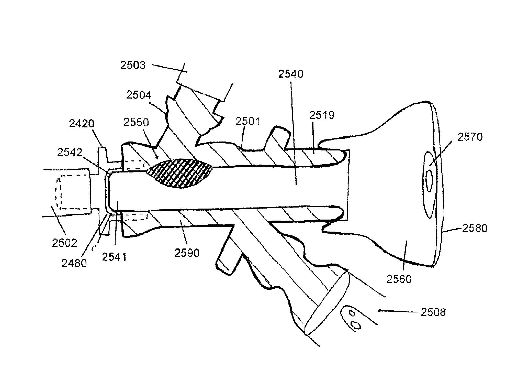
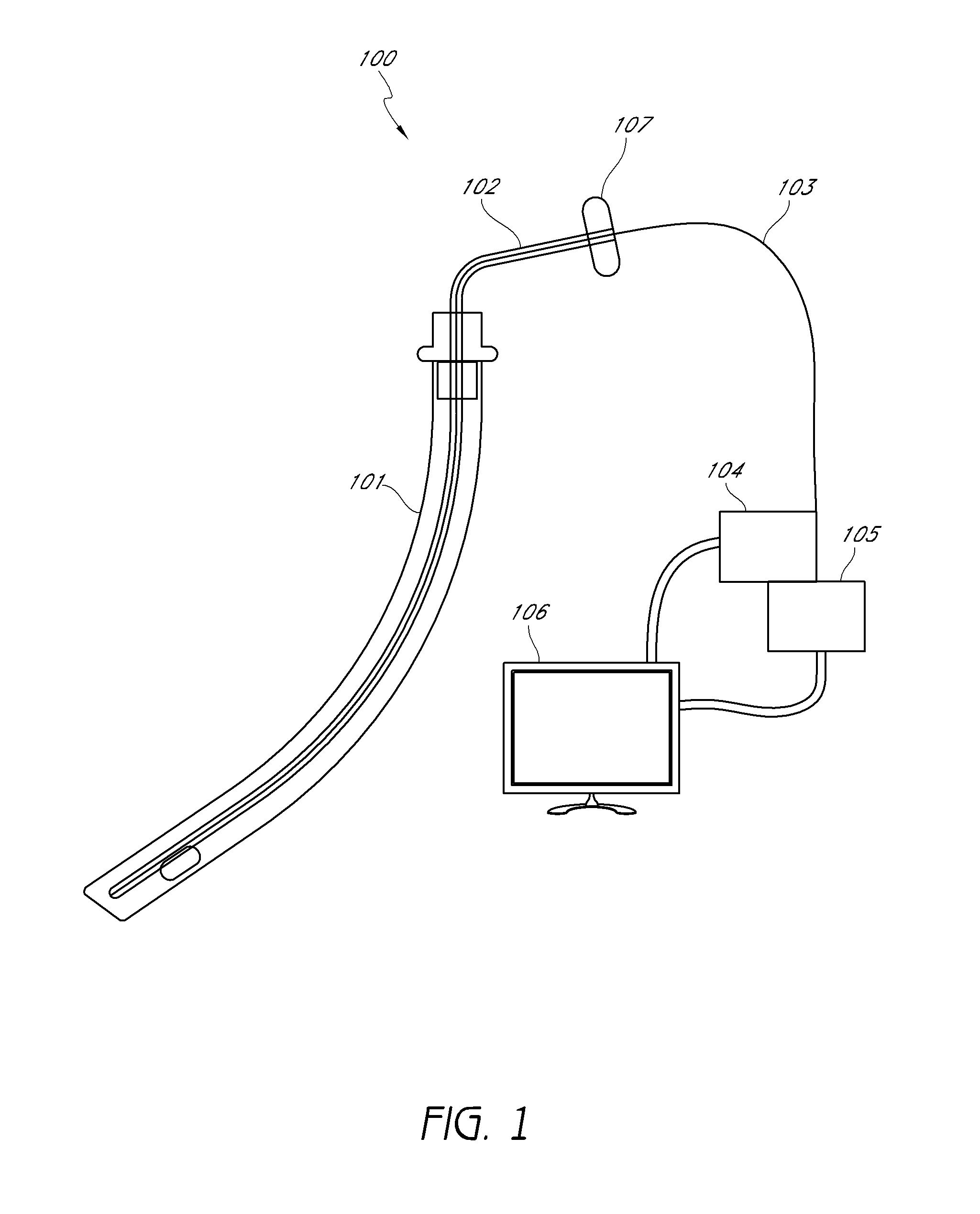
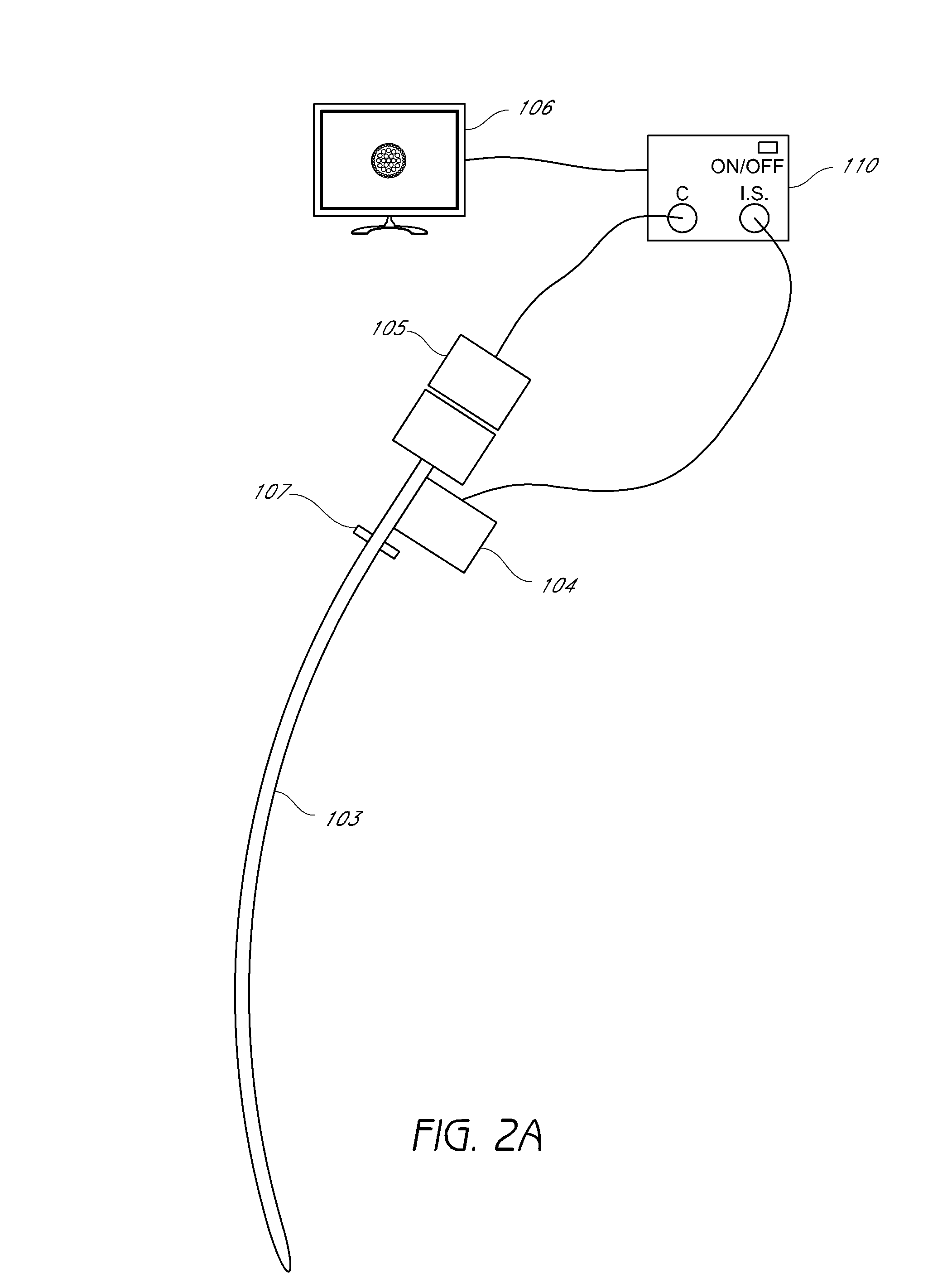
Endotracheal tube coupling adapters for facilitating collection and removal of biofilm or any debris from endotracheal tubes for disposal or laboratory testing are disclosed. The endotracheal tube coupling adapters may be adapted for use in closed suction systems. The endotracheal tube coupling adapters may be configured to remain coupled to an endotracheal tube from the time of intubation until the time of removal and to receive visualization, cleaning, and/or biofilm collection devices or members without being removed from the endotracheal tube.

Description

CROSS-REFERENCE TO RELATED APPLICATIONS[0001]This application claims priority under 35 U.S.C. §119(e) to U.S. Provisional Application No. 61 / 552,417 filed Oct. 27, 2011, the entire content of which is hereby incorporated by reference herein. This application is related to U.S. application Ser. No. 13 / 638,567, filed Sep. 28, 2012, which is a U.S. national phase application filed under 35 U.S.C. §371 of International Application No. PCT / 2011 / 030229 filed Mar. 28, 2011, which claims priority under 35 U.S.C. §119(e) to U.S. Provisional Application No. 61 / 318,761 filed Mar. 29, 2010 and to U.S. Provisional No. 61 / 370,425 filed Aug. 3, 2010, and which is a continuation-in-part of U.S. application Ser. No. 12 / 849,672 filed Aug. 3, 2010, the entire content of each of which is hereby incorporated by reference herein.FIELD[0002]Embodiments disclosed herein relate generally to cleaning and / or visualization of native airways and other anatomical passages, and more specifically, to removal of po...

Claims


Application Information

Patent Timeline
Patent Type & Authority Patents(United States)
IPC IPC(8): A61B1/267A61M16/04A61M16/08
CPCA61B1/267A61M16/0463A61M16/0486A61M16/0816A61M2209/10A61M16/1055A61M2205/7536A61M16/106A61M16/0833A61M16/0434A61M2016/0413A61M16/0418A61M16/0431A61M16/0427A61M16/0484
Inventor VAZALES, BRAD EUGENECHERSKY, DAVID MARK
Owner AVENT INC
Why Eureka
Try Eureka