Methods of amplifying whole genome of a single cell

a single cell, whole genome technology, applied in the direction of dna preparation, biochemistry apparatus and processes, fertilization, etc., can solve the problems of inability to amplify the genome in its entirety, short amplification products, and common formation of primer dimers

Active Publication Date: 2017-04-11
PRESIDENT & FELLOWS OF HARVARD COLLEGE
View PDF9 Cites 2 Cited by
  • Summary
  • Abstract
  • Description
  • Claims
  • Application Information

AI Technical Summary

Benefits of technology

[0008]Embodiments of the present disclosure are directed to a method of amplifying DNA such as a small amount of genomic DNA or a limited amount of DNA such as a genomic sequence or genomic sequences obtained from a single cell or a plurality of cells of the same cell type or from a tissue, fluid or blood sample obtained from an individual or a substrate. According to certain aspects of the present disclosure, the methods described herein can be performed in a single tube with a single reaction mixture and programmable thermocycles. The methods described herein can provide substantial coverage of the entire genome of a single cell producing amplified DNA for high-throughput sequencing.
[0009]According to an additional aspect, methods are provided herein for performing whole genome amplification of single cells with high fidelity and amplification uniformity or coverage across different loci in the genome which is useful for further sequencing or analysis using high throughput sequencing platforms known to those of skill in the art. Methods provided herein minimize amplification bias and provide substantially complete or complete genome coverage of DNA sequencing of genomic DNA from a single cell. Methods described herein can amplify greater than 90 percent of genomic DNA from a single cell while greater than 70 percent or 75 percent of the genomic DNA can be sequenced with a sequencing depth of 7× or 10× or 15× with little, substantially few or no chimera sequences. Methods described herein reduce or eliminate creation of sequencing artifacts and facilitate advanced genomic analysis of single nucleotide polymorphisms, copy number variations and structural variations. Methods described herein have particular application in biological systems or tissue samples characterized by highly heterogenous cell populations such as tumor and neural masses. Methods described herein to amplify genomic DNA facilitate the analysis of such amplified DNA using next generation sequencing techniques known to those of skill in the art and described herein.
[0011]The DNA amplification methods of the present disclosure will be useful for amplifying small or limited amounts of DNA, which will allow multiple sites in the DNA sample to be genotyped for high-throughput screening. Additionally, the present method will allow for the rapid construction of band specific painting probes for any chromosomal region, and can also be used to microdissect and amplify unidentifiable chromosomal regions or marker chromosomes in abnormal karyotypes. The presently disclosed method will also allow for the rapid cloning of amplified DNA for sequencing or generating DNA libraries. Thus, the method will not only be a valuable tool for genotype analysis and high-throughput screening, it should also be a valuable tool in cytogenetic diagnosis.
[0012]Embodiments described herein utilize non-standard PCR cycles to perform whole genome amplification from a single mammalian cell. Certain polymerases provide exemplary results. A step may be provided during cycling to anneal a free primer to a complementary primer sequence on an amplicon, such as at the 3′ end of the amplicon, thereby preventing hybridization within the amplicon, i.e. hybridizing with itself, or between amplicons, such as to form chimeras. Procedures are described for removing primer sequences on either ends of the amplicons generated using the methods described herein.

Problems solved by technology

Formation of primer dimers is a common problem in existing methods for DNA or RNA amplification using random primers.
Other problems known in the art when using random primers to amplify DNA include an inability to amplify the genome in its entirety due to locus dropout (loss), generation of short amplification products, and in some cases, the inability to amplify degraded or artificially fragmented DNA.
PCR-based methods such as GenomePlex and Picoplex can induce significant amplification bias across different loci in the genome which results in significant amplification loci dropout.
Multiple displacement amplification utilizes low temperature amplification which leads to large amounts of chimera sequences that do not belong to the original genome resulting in significant false discovery rate of one out of several kilobases and introduces artifacts into the whole genome sequencing analysis.
While methods for whole genome amplification have been proposed, such methods can generally be inefficient, complex and expensive.

Method used


Image

  • Methods of amplifying whole genome of a single cell
  • Methods of amplifying whole genome of a single cell
  • Methods of amplifying whole genome of a single cell

Examples


example i

Single Cell DNA Amplification

Cell Selection by Laser Dissection Microscopy

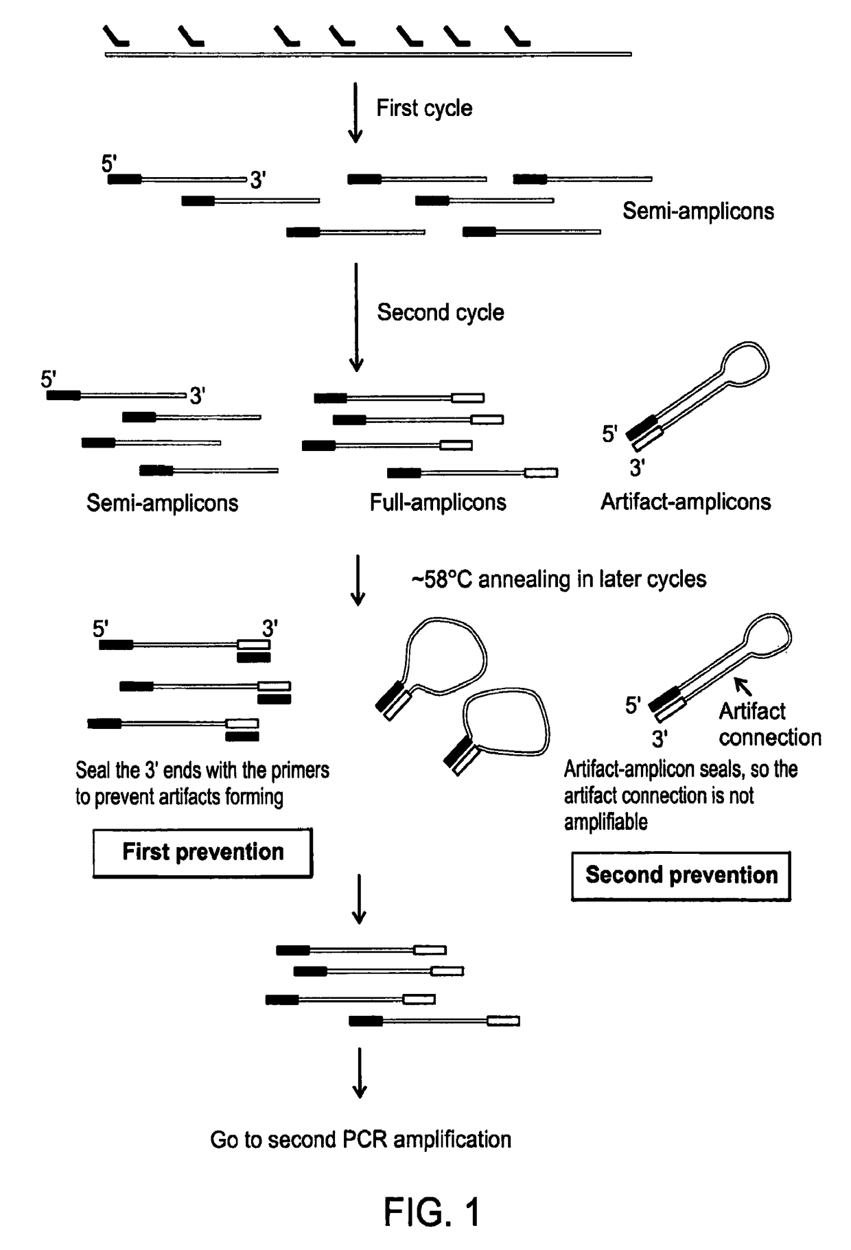
[0108]A cell is selected, cut from a culture dish, and dispensed in a tube using a laser dissection microscope (LMD-6500, Leica) as follows. The cells are plated onto a membrane-coated culture dish and observed using bright field microscopy with a 10× objective (Leica). A UV laser is then used to cut the membrane around an individually selected cell such that it falls into the cap of a PCR tube. The tube is briefly centrifuged to bring the cell down to the bottom of the tube. 5 μl lysis buffer (30 mM Tris-Cl PH 7.8, 2 mM EDTA, 20 mM KCl, 0.2% Triton X-100, 12.5 μg / ml Qiagen Protease) is added to the side of the PCR tube and span down. The captured cell is then thermally lysed using the using following temperature schedule on PCR machine: 50° C. 3 hours, 75° C. 20 minutes, 80° C. 5 minutes.

Linear Pre-Amplification

[0109]In the linear pre-amplification, a pair of quasi-degenerated primers is used to initiate over...

example ii

Further Amplification of Amplicons of Example I Using Standard Methods

[0115]Standard PCR amplification is used to exponentially amplify the amplicons produced in Example I as follows. The following reaction buffer is prepared and added to the PCR tube which is being maintained on ice.

3.0 μl ThermoPol Buffer (NEB)

1.0 μl dNTP (10 mM)

26 μl H2O (Ambion)

0.1 μl primer (100 μM) (5′-GT GAG TGA TGG TTG AGG TAG TGT GGA G-3′) (SEQ ID NO:3)

1.0 μl DeepVentR (exo-) (New England Biolabs)

[0116]Amplification is performed with standard PCR procedures as follows to generate 1-2 μg of DNA material.

94° C.—20 seconds

59° C.—20 seconds

72° C.—3 minutes

Repeat the above cycles 17×

72° C.—5 minutes

4° C.∞

[0117]After exponential amplification, DNA can be purified using a Qiagen column and stored for a next procedure to remove the primer end of the DNA amplicons.

[0118]Additional methods of amplification known to those of skill in the art can be used as follows.

[0119]One of the best known amplification methods is P...

example iii

Removing Priming Sequences from PCR Amplicons

[0134]A phosphorothioate nucleotide strategy is used to remove the priming sequences of the amplicon product. The DNA amplicons are re-amplified using dA, dT, dC and phosphorothioated dG. Since the primer sequence does not include the dC nucleotide, the complementary sequence will not contain the dG nucleotide. The product is digested from the 3′ end using exonuclease III. However as the phosphorothioate bond cannot be cleaved by exonuclease III, the 3′ end can be digested up to the phosphorothioated dG. In the same enzyme mix, Mung Bean exonuclease is also added, which will remove the protruding 5′ end formed from exo III cleavage.

[0135]The reaction buffer and conditions are as following: 1× Buffer 1 from New England Biolabs, 5 U ExoIII and 0.5 U Mung Bean exonuclease, both enzymes are available from New England Biolabs. The reaction takes 2 minutes at room temperature and can be stopped by adding 5 μl 0.5M EDTA.

[0136]After removing prim...


PUM

PropertyMeasurementUnit
temperatureaaaaaaaaaa

Abstract

Methods and compositions for amplifying the whole genome of a single cell are provided.

Description

RELATED APPLICATION DATA[0001]This application is a National Stage Application under 35 U.S.C. 371 of co-pending PCT application PCT / US2012 / 038930 designating the United States and filed May 22, 2012; which claims the benefit of provisional application No. 61 / 621,271 and filed Apr. 6, 2012; which claims the benefit of provisional application No. 61 / 550,677 and filed Oct. 24, 2011; which claims the benefit of provisional application No. 61 / 510,539 and filed Jul. 22, 2011; which claims the benefit of provisional application No. 61 / 490,790 and filed May 27, 2011 each of which are hereby incorporated by reference in their entireties.STATEMENT OF GOVERNMENT INTERESTS[0002]This invention was made with government support under 3R01HG005097-02 and 3R01HG005097-02S1 from the National Institute of Health. The Government has certain rights in the invention.BACKGROUND[0003]Field of the Invention[0004]Embodiments of the present invention relate in general to methods and compositions for amplifyi...

Claims


Application Information

Patent Timeline
Patent Type & Authority Patents(United States)
IPC IPC(8): C12Q1/68C12N15/10C12P19/34
CPCC12Q1/6883C12N15/1093C12Q1/6848C12Q1/6886C12Q2525/155C12Q2525/161C12Q2549/119
Inventor XIE, XIAOLIANG SUNNEYZONG, CHENGHANGLU, SIJIA
Owner PRESIDENT & FELLOWS OF HARVARD COLLEGE
Why Eureka
Try Eureka