Balloon immobilization device for radiation treatment

a radiation treatment and balloon technology, applied in the field of balloon immobilization device for radiation treatment, can solve the problems of difficult radiation therapy for bladder cancer patients, unreliable radiation treatment effect, and exposure of normal healthy organs to unnecessary radiation

Active Publication Date: 2018-02-20
GIAP HUAN +2
View PDF10 Cites 5 Cited by
  • Summary
  • Abstract
  • Description
  • Claims
  • Application Information

AI Technical Summary

Benefits of technology

[0012]The inflatable balloon is coupled to the flexible tube at the proximal end. The shape of the balloon can be elliptical, pyramidal, cylindrical, or spherical, and the size or volume can be small, medium, or large, depending on the volume of the bladder and organs. As stated previously, the inflatable balloon can have varying degrees of radiopacity, depending on its purpose and location. For example, depending on the location of the cancerous cells within the bladder and the orientation of the linear accelerator beam, it may be beneficial to shield specific areas from unnecessary radiation exposure that can be harmful to healthy cells by making parts of the balloon more or less radiopaque than others.
[0016]Another embodiment of the present invention can be used for limiting the movement of the rectum during radiation treatment of rectal cancer. Rectal cancer occurs when there is cancerous or abnormal cell growth in the lining of the rectum. Like the bladder, the rectal walls expand and contract as the organ fills and empties with fecal matter from the digestive process. Also like the bladder, the movement can make precise radiation therapy difficult. There are circumstances where only part of the rectal circumference is needed to be irradiated while sparing the uninvolved rectal areas from the radiation treatment portal. The movement and deformation of the rectum makes the precise delivery of radiation to these particular areas even more difficult.
[0017]In this embodiment, the inflatable balloon of the present invention would be oblong to fit the shape of the rectum and the size would vary depending on the volume of the rectum of the patient. One or more lumen would be used for the inflation of the inflatable balloon and for possible irrigation or introducing radio protector agents into the rectum. In addition, the configuration of radiopaque markers in the inflatable balloon could be used for multiple reasons, including information on the location of the cancerous cells in the lining of the rectum. In addition, the inflatable balloon can have varying degrees of radiopacity depending on the purpose of the device and can therefore shield untreated areas from any radiation.
[0020]Depending on the purpose and circumstances of the case, one or more lumina could be used. The first lumen will inflate the inflatable balloon and another may be used for possible irrigation or introducing radio protector agents into the vagina. The radiopaque markers can be used to aid in finding the location of the cancerous cells, without the use of invasive surgery to implant markers. In addition, the varying radiopacity of the inflatable balloon can shield healthy areas of the vagina from harmful radiation, while still exposing the area needing treatment.
[0022]Depending on the purpose and circumstances of the case, one or more lumina could be used. The first lumen will inflate the inflatable balloon and another lumen may be used for possible irrigation or introducing radio protector agents into the uterus. Radiopaque markers can be used to aid healthcare professionals in locating the position of the cancerous cells, without the use of invasive surgery to implant markers. In addition, the varying radiopacity of the inflatable balloon can shield healthy areas of the uterus from harmful radiation, while still exposing the area or areas needing treatment.

Problems solved by technology

However, some tumors do not have a consistent, precise position because of the nature and location of the cancer in the internal organs.
For example, radiation therapy for patients with bladder cancer is difficult due to the anatomy of the bladder.
Organ motion and deformation (change in shape) prevent the accurate localization of the radiation beam to the tumor and as a result exposes normal healthy organs to unnecessary radiation.
Therefore, the three-dimensional image acquired prior to starting a radiation therapy plan may not be useful for subsequent radiation appointments if, in comparison to when the image was produced, the bladder has expanded or deflated with urine.
Consequently, if the beam is not in the precise position, it could cause damage to healthy cells.
Clinical studies show that bladder patients undergoing chemo-radiation therapy have about a 50% survival rate (similar to surgery); however, about 20-30% of these patients surviving the chemo-radiation treatment have significant radiation damage to their bladder making it useless (incontinence).
This serious side effect is due to the unnecessary exposure of the normal bladder to high doses of radiation during radiation treatment as a result of the change in shape and size of the bladder during the radiation treatment.
Due to the inaccurate delivery of radiation during bladder cancer treatment and its negative side effects, the radiation dosage delivered to the bladder tumor cannot be given as high as needed.
Although the use of this technology would improve chances of bladder preservation, it is still difficult to immobilize the bladder during the radiation treatment to limit organ movement and deformation.
Moreover, it is also difficult to place markers within the bladder as a guide for radiation treatment without the use of invasive surgery.
However, the Foley catheter is primarily used for irrigation of the bladder and it is not designed to maintain the bladder in a constant shape and volume.
The Foley catheter balloon is much smaller than the interior of the bladder and thus cannot control the expansion or contraction of the bladder.

Method used


Image

  • Balloon immobilization device for radiation treatment
  • Balloon immobilization device for radiation treatment
  • Balloon immobilization device for radiation treatment

Examples


Embodiment Construction

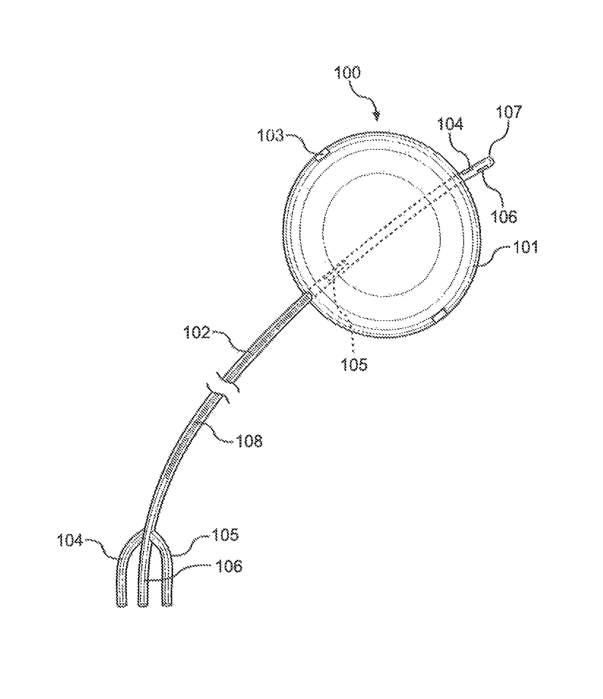
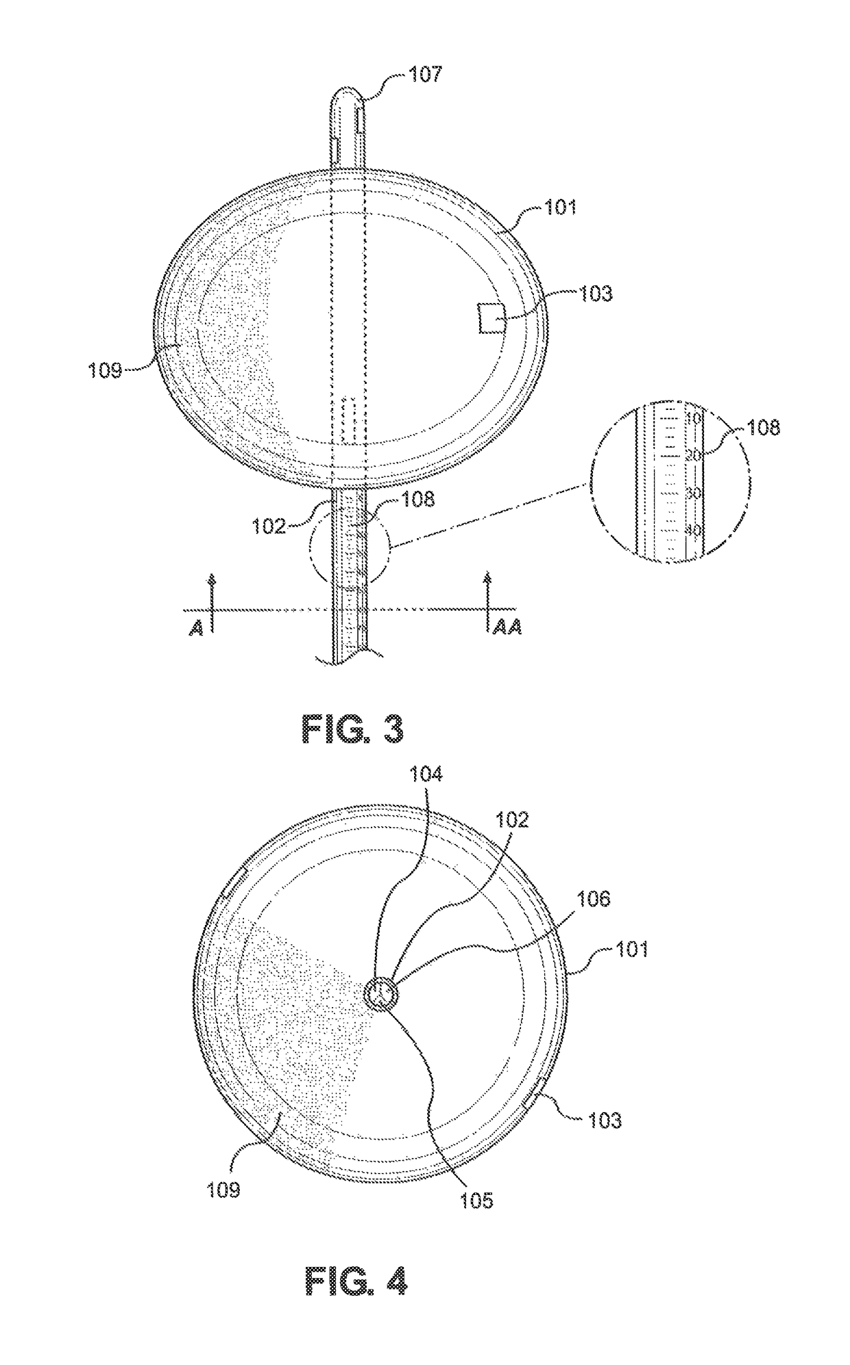
[0047]Referring initially to FIG. 1, the preferred embodiment of the Balloon Immobilization Device for Radiation Treatment for bladder cancer of the present invention is shown and generally designated 100. The Balloon Immobilization Device for Radiation Treatment 100 consists of a flexible tube 102, a catheter tip 107, an inflatable balloon 101, and two radiopaque markers 103. The inflatable balloon 101 is made of an expandable membrane incorporated with slightly radiopaque materials. As shown, the inflatable balloon 101, when deflated, is oblong and thin enough to allow insertion into the urethra (not shown) or other orifices subsequently discussed.

[0048]The flexible tube 102 can be of any length depending on the use of the Balloon Immobilization Device for Radiation Treatment 100 of the present invention. Within the flexible tube 102 are three (3) separate lumina, first lumen 104, second lumen 105, and third lumen 106. The flexible tube 102 distally trifurcates into three (3) dist...


PUM


Abstract

The Balloon Immobilization Device for Radiation Treatment of the present invention includes a catheter assembly with radiopaque markers and one or more lumina. The catheter assembly consists of a flexible tube and inflatable balloon. The flexible tube has distal and proximal ends, with one or more separate lumina within and along the length of the flexible tube. At least one lumen is in fluid communication with the exterior of the flexible tube at the distal end and the interior of the inflatable balloon and at least one lumen in fluid communication with the exterior of the flexible tube at the distal end and the exterior of the flexible tube at the proximal end. The inflatable balloon varies in shape and size and is made of a radiopaque material having varying degrees of radiopacity. Radiopaque markers are located at different locations on the balloon and flexible tube.

Description

FIELD OF THE INVENTION[0001]The present invention pertains generally to a device for use in the radiation treatment of cancers. More specifically, the present invention pertains to a device that can immobilize internal organs and provide spatial orientation information to healthcare providers during radiation therapy treatment for various cancers. The present invention is particularly, but not exclusively, useful as a device for the radiation therapy treatment of cancers of the bladder, vagina, rectum, and uterus.BACKGROUND OF THE INVENTION[0002]Radiation therapy is the medical use of high doses of ionizing radiation and typically used in the treatment of cancer to kill malignant cells. Radiation therapy can be used as a primary treatment or used in conjunction with other treatments, such as chemotherapy or surgery. In addition to cancerous cells, radiation therapy can damage healthy cells, making it imperative that treatment is carefully planned to minimize side effects. The type o...

Claims


Application Information

Patent Timeline
Patent Type & Authority Patents(United States)
IPC IPC(8): A61M29/02A61F5/37A61N5/10A61B17/42A61B17/22
CPCA61F5/37A61N5/10A61N2005/1097A61B2017/4216A61B2017/22069
Inventor GIAP, HUANGIAP, FANTINEGIAP, BOSCO
Owner GIAP HUAN
Why Eureka
Try Eureka