Method for preparing acellular matrix

A technology for decellularized matrix and tissue organs, which is applied in the field of preparing acellular matrix, can solve the problems of destroying the ultrastructure of extracellular matrix and excessive molecular weight, and achieve the effect of promoting hydrolysis reaction, strong specificity, and promoting decellularization effect

Inactive Publication Date: 2008-10-01
ZHONGSHAN OPHTHALMIC CENT SUN YAT SEN UNIV
View PDF0 Cites 31 Cited by
  • Summary
  • Abstract
  • Description
  • Claims
  • Application Information

AI Technical Summary

Problems solved by technology

The physical method refers to the destruction of cells by physical methods such as freezing, high pressure, ultrasonic waves, and osmotic pressure changes. It also destroys the ultrastructure of the extracellular matrix to varying degrees. At the same time, the broken cell debris must be removed by other means. This removal process will further damage the ultrastructure of the extracellular matrix.
Proteases can effectively remove cellular components, but trypsin and neutral protease have a wide range of substrates, causing a large amount of structural and functional proteins in the extracellular matrix to degrade
Nucleases selectively degrade nucleic acids without causing damage to extracellular matrix components, but their molecular weight is too large, which can easily cause immune reactions

Method used


Image

  • Method for preparing acellular matrix
  • Method for preparing acellular matrix
  • Method for preparing acellular matrix

Examples


Embodiment 1

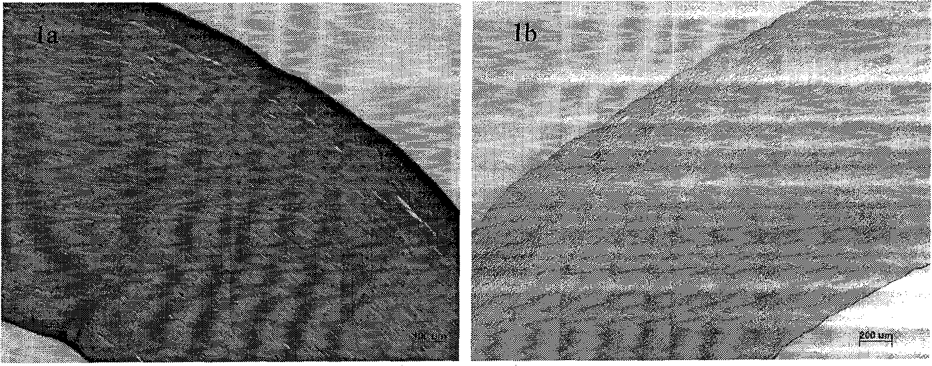
[0048] The purpose of this example is to prepare porcine corneal decellularized matrix with porcine cornea. (note: the phospholipase solution used in this embodiment can contain phospholipase A 1 , A 2 , B 1 , B 2 , any one or more of C, D. The surfactants used can be ingredients with surface activity that can be produced in vivo or in the human body, such as: cholate, deoxycholate, chenodeoxycholate, glycocholate, glycochenodeoxychole Any one or more of salt, taurocholate, taurochenodeoxycholate, lipopolysaccharide, lipoprotein, lysolecithin, or polyethylene glycol, TritonX-100, its effect Same as below).

[0049] 1. Under normal aseptic operation at room temperature, use a 10.0mm trephine to remove fresh pig eye corneal slices. Soak the obtained porcine corneal slices in carbonate buffer solution containing antibiotics (100 U / ml penicillin G, 100 μg / ml streptomycin sulfate) for 2-5 times, each time for 2-10 minutes.

[0050] 2. Put the porcine corneal piece into 10ml ...

Embodiment 2

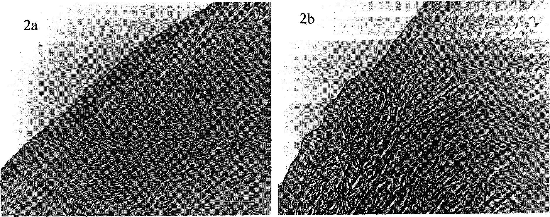
[0057] The purpose of this example is to prepare porcine limbal acellular matrix with porcine limbal tissue. (note: the phospholipase solution used in this embodiment can contain phospholipase A 1 , A 2 , B 1 , B 2 , any one or more of C, D, the effect is the same as the following).

[0058] 1. At room temperature, under routine aseptic operation, remove fresh porcine corneal limbal tissue of 2 mm each area inside and outside, and use antibiotics (100 U / ml penicillin G, 100 μg / ml streptomycin sulfate) on the pig corneal limbal tissue. Soak in carbonate buffer solution 2-5 times, 2-10 minutes each time.

[0059] 2. Put the porcine limbal tissue into 10ml of sterile pure water and soak in a water bath at 4°C for 10-60 minutes.

[0060] 3. Put the porcine limbal tissue into 10 ml of sterile phospholipase solution, and shake it in a water bath at 4°C for 6-24 hours. (Sterile phospholipase solution: prepared using carbonate buffer, pH range 8-12, phospholipase A 1 +A 2 =50~...

Embodiment 3

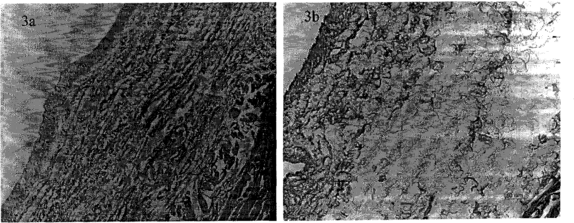
[0064] The purpose of this example is to use porcine conjunctiva to prepare porcine conjunctival acellular matrix. (note: the phospholipase solution used in this embodiment can contain phospholipase A 1 , A 2 , B 1 , B 2 , any one or more of C, D. The surfactants used can be ingredients with surface activity that can be produced in vivo or in the human body, such as: cholate, deoxycholate, chenodeoxycholate, glycocholate, glycochenodeoxychole Any one or more of salt, taurocholate, taurochenodeoxycholate, lipopolysaccharide, lipoprotein, lysolecithin, or polyethylene glycol, TritonX-100, its effect Same as below).

[0065] 1. At room temperature, under routine aseptic operation, remove the fresh porcine conjunctiva tissue within a range of 1 × 1 cm, and use carbon dioxide containing antibiotics (100 U / ml penicillin G, 100 μg / ml streptomycin sulfate) on the obtained porcine conjunctiva tissue. Soak in salt buffer solution 2-5 times, 2-10 minutes each time.

[0066] 2. Put...


PUM


Abstract

The invention discloses a method of preparing acellular matrixes by using phospholipase. The method of the invention is characterized in that stand-by organ tissue is first pre-treated and then added into solution containing the phospholipase to prepare the acellular matrixes under a controlled condition; the prepared acellular matrixes are then washed. By adopting the preparation method of the invention, the obtained acellular matrix can have good physical property and biological function. Therefore, the preparation method in the invention is not only a great breakthrough in the tissue engineering, but also opens a new way for clinical treatment of diseases. The preparation method of the invention has the advantages of reliable theory, simple and flexible process technique, good product reproducibility and is very easy to be industrialized.

Description

technical field [0001] The invention belongs to the field of tissue engineering, and in particular relates to a method for preparing an acellular matrix. Background technique [0002] Various bioscaffold materials derived from acellular matrices have achieved success in preclinical studies in animal experiments and clinical applications in human diseases. By removing heterogeneous or allogeneic cells in various tissues and organs and retaining the complex structural and functional proteins, the acellular matrix of the corresponding tissues and organs can be obtained. Different decellularization methods directly affect the composition and ultrastructure of the resulting acellular matrix, and ultimately affect the host's response to the acellular matrix after transplantation. [0003] The current preparation methods of acellular matrix mainly include: 1 physical method; 2 chemical method; 3 enzymatic method. The physical method refers to the destruction of cells by physical ...

Claims


Application Information

Patent Timeline
Patent Type & Authority Applications(China)
IPC IPC(8): A61L27/36
CPCA61L27/3687A61L27/3604A61L2430/40
Inventor 王智崇陈冬武征
Owner ZHONGSHAN OPHTHALMIC CENT SUN YAT SEN UNIV
Why Eureka
Try Eureka