A Method for the Quantification of Membrane Proteins Using Mass Spectrometry

A technology for proteins and proteolytic enzymes, which is applied in the field of quantitative determination of membrane proteins existing in cell membranes, and can solve problems such as the reduction of quantitative accuracy.

Inactive Publication Date: 2011-12-21
TOHOKU UNIV
View PDF4 Cites 0 Cited by
  • Summary
  • Abstract
  • Description
  • Claims
  • Application Information

AI Technical Summary

Problems solved by technology

Furthermore, peptides with different sequences detected by the same channel may be detected a

Method used


Image

  • A Method for the Quantification of Membrane Proteins Using Mass Spectrometry
  • A Method for the Quantification of Membrane Proteins Using Mass Spectrometry
  • A Method for the Quantification of Membrane Proteins Using Mass Spectrometry

Examples


Embodiment 1

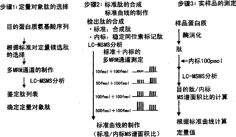
[0431] Peptide sample preparation

[0432] Human ABCG2 (human ATP binding cassette transporter G2) was selected as a cell membrane protein, and samples were prepared in order to select detectable proteins by LC-MSMS. First, using the cell membrane of ABCG2-expressing cells as a peptide supply source, it was prepared as follows. That is, the ABCG2 expressed cell membrane protein was denatured in a pH 8.5 buffer solution of 7M guanidine hydrochloride, 0.1MTris-HCl, and 10mM EDTA, and it was reduced with DTT in order to protect the SH group of the cysteine ​​residue. and carbamidomethylation with iodoacetamide. Dialyze in 50 mM ammonium bicarbonate, add trypsin in an amount of 1 / 100 of the protein weight, and enzymatically digest at 37° C. for 16 hours to prepare peptide samples.

[0433] Preparation of quantitative target peptides and stable isotope-labeled peptides

[0434] Human ABCG2 expressed cell membrane peptide samples were determined by LC-MSMS. From the measurement ...

Embodiment 2

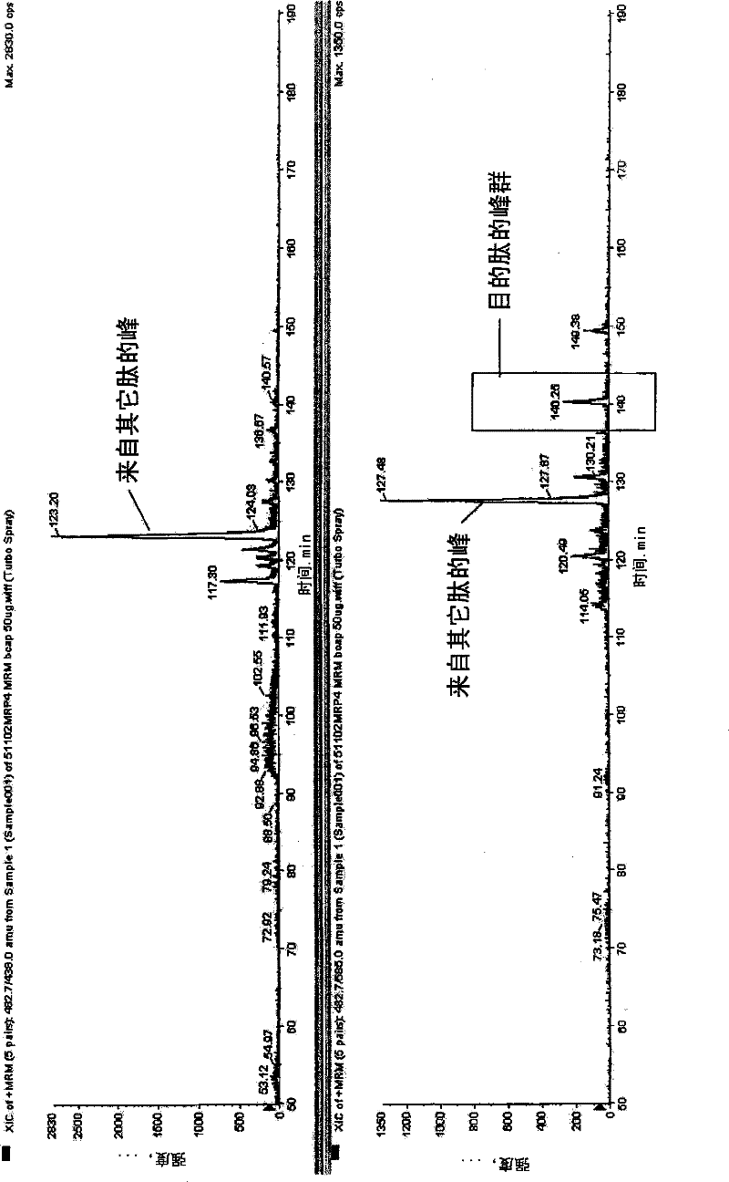
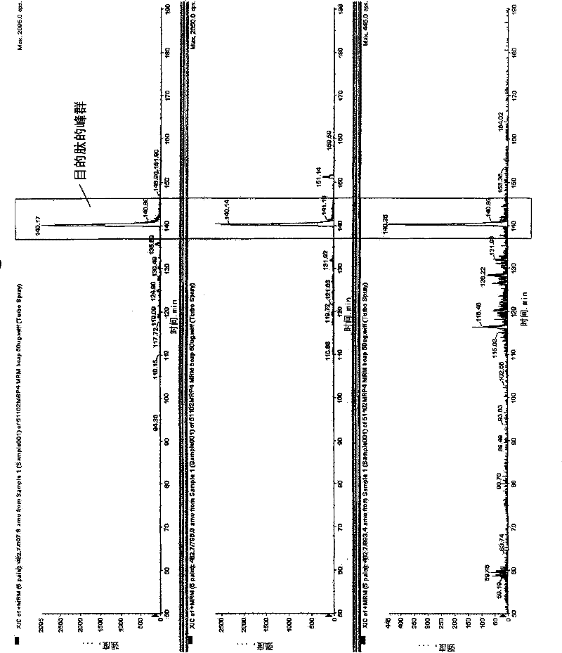
[0441] As the criteria for selecting peptides to be quantified, (1), (2), (3), (4), (5), (6), (7), (8), (9) and (10) are used, and necessary When using (11), similarly to Example 1, a standard curve was prepared for the target peptides for protein synthesis and quantification described in Table 1 below, and its linearity was discussed. To 10 fmol, 50 fmol, 100 fmol, 500 fmol, and 1000 fmol of non-labeled peptides, 500 fmol of stable isotope-labeled peptide was added, respectively, and measured by LC-MSMS. Any of the above-mentioned peptides all show the linearity of the standard curve in the range of 10fmol-1000fmol, and it can be confirmed that the target protein can be quantified in this range ( Figure 6 ). In addition, as a transporter protein supply source, a cell membrane protein of a human leukemia cell line was selected for quantification. Table 1 shows the quantitative limit value of the obtained standard curve and the quantitative value of the leukemia cell membran...

Embodiment 3

[0446] As the criteria for selecting peptides to be quantified, (1), (2), (3), (4), (5), (6), (7), (8), (9) and (10) are used, and necessary When using (11), similarly to Example 1, for the proteins described in Table 2 below, peptides to be quantified were synthesized, a standard curve was prepared, and its linearity was discussed. 100 fmol of stable isotope-labeled peptide was added to 1 fmol, 5 fmol, 10 fmol, 50 fmol, 100 fmol, 500 fmol, and 1000 fmol of non-labeled peptide, respectively, and measured by LC-MSMS. Any of the above-mentioned peptides all showed the linearity of the standard curve, and it can be confirmed that the target protein can be quantified within this range ( Figure 6 ). Table 2 shows the quantitative limit value of the obtained standard curve and the quantitative value of the leukemia cell membrane.

[0447] Table 2

[0448] protein sequence stable isotope sequence Limit of quantitation (fmol) ...


PUM


Abstract

The invention provides a method for quantitatively measuring the membrane protein existing on the cell membrane simply, quickly and with high precision by using the stable isotope-labeled peptide as a probe through mass analysis. Prepare oligopeptide fragments by fragmenting cell membrane proteins, identify them by LC-MSMS, use peptides obtained by proteolytic enzymes and peptides specific to target molecules as necessary standards, set the content of hydrophobic amino acids and sequence conditions, and the number of amino acid residues , various scoring criteria for specific amino acid sequence conditions, etc., preferably select peptide fragments with high total scores, and use ESI method to select ionizable peptide fragments according to the quantitative object peptide selection criteria, and use the quantitative object peptide and stable isotope labeling Peptides, high-precision quantification of cell membrane proteins by mass analysis.

Description

technical field [0001] The present invention relates to a method for quantitative determination of cell membrane protein, specifically, a stable isotope-labeled peptide as a probe, using liquid chromatography-mass spectrometry (LC-MSMS) for mass analysis, thereby quantitatively determining the presence of Methods for Membrane Proteins in Cell Membranes. Background technique [0002] The proteins that make up the biomembrane include cell membrane protein (plasma membrane protein: also called membrane protein). Cell membrane proteins have functions such as receptors for enzymes, peptide hormones, growth factors, and endocrine substances (autacoids), transporters for sugars, etc., ion channels, and cell membrane antigens. activity function. Cell membrane proteins are proteins or glycoproteins integrated in the lipid bilayer of the cell membrane. They exist in various states throughout the cell membrane (membrane-penetrating protein), located on the surface of the cell membran...

Claims


Application Information

Patent Timeline
IPC IPC(8): G01N27/62C12Q1/37G01N30/72
CPCH01J49/00G01N2030/8813G01N2333/705G01N2030/8831G01N33/6848G01N30/88G01N30/7266G01N2030/8818G01N30/8679G01N2030/8827
Inventor 上家润一大槻纯男寺崎哲也
Owner TOHOKU UNIV
Why Eureka
Try Eureka